Telangana TSBIE TS Inter 2nd Year Zoology Study Material 6th Lesson Genetics Textbook Questions and Answers.
TS Inter 2nd Year Zoology Study Material 6th Lesson Genetics
Very Short Answer Type Questions
Question 1.
What is pleiotropy?
Answer:
The phenomenon of multiple effects of a single gene is called pleiotropy i.e., the same gene is activated in several different tissues producing different phenotypic effects.
Question 2.
What are the antigens causing ‘ABO’ blood grouping? Where are they present?
Answer:
Antigens are present on the plasma membrane of the RBCs. They are also called iso agglutinogens. They are antigen A antigen B are responsible for ABO blood grouping.
Question 3.
What are the antibodies of ‘ABO’ blood grouping? Where are they present?
Answer:
Isoagglutinins (or) Antibodies are present in the blood plasm. Iso aggulutinin A and Iso agglutinin B are the antibodies of ABO blood grouping.
Question 4.
What are multiple alleles?
Answer:
When more than two allelic forms occur at a same locus on the homologous chromosomes of an organism, they are called multiple alleles. When more than two alleles exist in a population of specific organism, the phenomenon is called multiple allelism.
Question 5.
What is erythroblastosis foetalis?
Answer:
When father is Rh+ and mother is Rh– in the second Rh+ baby onwards, due to immunological in compatibility between mother and growing foetus, the RBC of Rh+ foetus are destroyed. This is called erythroblastosis foetalis.

Question 6.
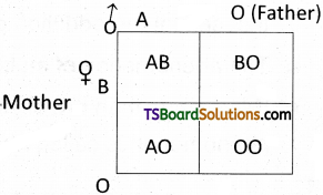
A child has blood group ‘O’. If the father has blood group ‘A’ and mother blood group ‘B’. Work out the genotypes of the parents and the possible genotypes of the other offspring.
Answer:
Child blood group is ‘O’ i.e., the genotype will be ‘OO’. Father blood group phenotype A hence it must AO genotype. Mother blood group phenotype B hence it must BO genotype. Possible genotypes of other offsprings are AB, BO, AO phenotypes are AB, B, A.
Question 7.
What is the genetic basis of blood types in ABO system in man?
Answer:
ABO blood group phenotypes are specified by Isoagglutinogen (I) gene with three alleles IA, IB and I° located on chromosome 9 (nine). IA IA and IA I° genotypes specify group. IB IB and IB I° genotype specify B group. IA IB genotype specified AB group and I°I° specifies ‘O’ group phenotypes.
Question 8.
What is polygenic inheritance?
Answer:
Many characters, such as human skin colour and height, an either – or classification is impossible because the characters vary in the population in gradations along a continuum. These are called quantitative characters. Quantitative variation usually indicates polygenic inheritance, an additive/ cumulative effect of two or more genes on a single phenotypic character.
Question 9.
Compare the importance of Y – chromosome in human being and Drosophila.
Answer:
- In human beings XX – XY type is seen. A pair of X chromosomes is present in female and XY is present in male. During spermatogenesis among males, two types of gametes 50% sperms with X and 50% with ‘Y’- chromosomes. It is evident that Y – chromosome decides the sex of child (if XX female if Xy male).
- In sex determination of Drosophila the sex of an individual is determined by the ratio of number of its chromosomes and that of its autosomal sets, the ‘Y’ chromosome taking no part in the determination of sex. The ratio is termed as sex index.
Question 10.
Distinguish between heterogametic and homogametic sex determination systems.
Answer:
If the two sex chromosomes / allosomes are different (XY or ZW) or it contains only one allosome (XO or ZO) the individual is heterogametic and if the two sex chromosomes / allosomes are similar (XX or ZZ) the individual is homogametic.
Question 11.
What is haplo – diploidy?
Answer:
In Hymenopterous insects like honey bees, sex is determined by the number of sets of chromosomes (haploid or diploid) in a bee, the fertilized eggs (diploid) develop into females and the unfertilized eggs (haploid) develop into males. This method of sex determination is haplodiploidy.
Question 12.
What are Barr bodies?
Answer:
Darkly stained, highly condensed and heterochromatinized X-chromosome present in the somatic cells like buccal mucosa, fibroblasts of female human beings is called Barrbody or Sex chromatin body.
Question 13.
What is Klinefelter’s syndrome?
Answer:
This genetic disorder is caused by trisomy of 23rd pair. The karyotype is 47, XXY. A Klinefelter male possesses an additional X – chromosome along with normal XY. At the same time, feminine sexual development is not entirely suppressed. Slight enlargement of breast (gynecomastia) and hips are often rounded.
Question 14.
What is Turner’s syndrome?
Answer:
The karyotype is 45, X is due to monosomy 23rd pair where one X – chromosome is last. A turner female does not show Barr bodies in her somatic cells. The symptoms are short stature, gonadal dysgenesis, webbed neck and broad shield like chest with widely spaced nipples.
Question 15.
What is Down syndrome?
Answer:
Down syndrome is a genetic condition that causes delays in physical and intellectual development. The cause of this genetic disorder is the presence of an additional copy of the chromosome numbered 21 (Trisomy of 21st set). The Karyotype is designated as TRISOMY 21 (47, XX, + 21). Characters are short statured, round head, furrowed tongue and partially opened mouth. Mental development and physical development is retarded.
Question 16.
What is Lyonisation?
Answer:
X – chromosome inactivation is also called Lyonisation. (It is proposed by Mary Lyon and Liane Russell). It is a process by which one of the two copies of the X – chromosome present in the body cells of female mammals is activated.
Question 17.
What is sex – linked inheritance?
Answer:
The inheritance of a trait that is determined by a gene located on one of the sex chromosomes is called sex – linked inheritance.
Question 18.
Define hemizygous condition.
Answer:
Genes that are present on the X or Y chromosome are called sex linked genes. The genes located on X – chromosome, whose alleles are absent on the Y – chromosome are called X – linked genes. Male human beings are hemizygons that is genes alleles are not present on Y-chromosome. Sometimes alleles are absent on X – chromosome of male. They are called holandric genes or Y – linked genes.
Question 19.
What is crisscross inheritance?
Answer:
The criss cross pattern or inheritance (skip gene – ration in heritance) in which a gene responsible for the white eyes is transmitted from a male parent to a male grandchild through carrier female of the first generation.
Question 20.
Why are sex – linked recessive characters more common in the male human beings?
Answer:
Sex linked recessive characters more common in male human beings because a mutation in a gene on the chromosome causes the phenotype to be expressed in males who are necessarily hemizygous for the recessive allele and they have only one X – chromosomes.
Question 21.
Why are sex – linked dominant characters more common in the female human beings ?
Answer:
X – linked dominant inheritance is a mode of genetic inheritance by which a dominant gene is carried on the X – chromosome. X linked dominant inheritance indicates that a gene responsible for a genetic disorder is located on the chromosome, and only one copy of the allele is sufficient to cause the disorder when inherited from a parent who has the disorder X – linked dominant traits are more prominent in woman.
Question 22.
What are sex limited characters?
Answer:
Sex limited genes are autosomal genes present in both males and female. Their phenotypic expression is limited to only one sex due to internal hormonal environment, e.g.: Beard in man, development of breast and secretion of milk in woman etc., are sex limited traits.
Question 23.
What are sex influenced characters?
Answer:
Sex influenced genes are the autosomal genes present in both males and females. In sex influenced inheritance, the genes behave differently in the two sexes, probably because of sex hormones providing different cellular environments in males and females. Cases of sex influenced inheritance include pattern baldness in humans, horn formation in certain breeds of sheep. (Dorset Horn Sheep)
Question 24.
How many base pairs are observed in human genome? What is the average number of base pairs in a human gene?
Answer:
The human genome contains 3164.7 million nucleotide bases. The average gene consists of 3000 bases, but sizes vary greatly with the largest known human gene being the one that codes for the protein called dystrophin.
Question 25.
What is ‘junk DNA’?
Answer:
Some DNA is involved in regulating the expressions of the genes that code for specific proteins. The remaining non-functional DNA is called Junk DNA.
Question 26.
What are VNTRs?
Answer:
No two people (other than identical twins) have exactly the same sequence of bases in their DNA. Restriction Fragment Length Polymorphism RFLPs-(pronounced riflips) are characteristic to every person’s DNA. They are called Variable Number Tandem Repeats (VNTRs) and are useful as Genetic markers.
Question 27.
List out any two applications of DNA finger printing technology.
Answer:
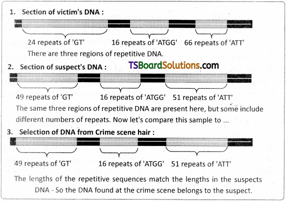
- DNA finger printing is a technique by which the DNA of an individual can be compared with that found in a sample or another individual (a suspect in a crime)
- DNA finger printing is used particularly paternity / maternity testing and for forensic work.
- Taxonomical applications – to study of phylogeny.
Short Answer Type Questions
Question 1.
Briefly mention the contribution of T.H. Morgan to genetics.
Answer:
Experimental verification of the “Chromosomal theory of inheritance” by Thomas Hunt Morgan and his colleagues, led to discovering the basis for the variation that sexual reproduction produced. For his work, Morgan selected a species for fruit fly, Drosophila melanogaster, which can be grown on simple synthetic medium in the laboratory. It completes its life cycle in about two weeks, and a single mating could produce a large number of progeny.
Further, it has many types of morphological, hereditary variations that can be seen under a low power microscope. Another advantage of the fruit fly is that it has only four pairs of chromosomes, which are easily distinguishable under a light microscope. There are three pairs of autosomes and one pair of sex chromosomes. Female fruit flies have a pair of homologous X – chromosomes, and males have one X – chromosome and one Y – chromosome.
Question 2.
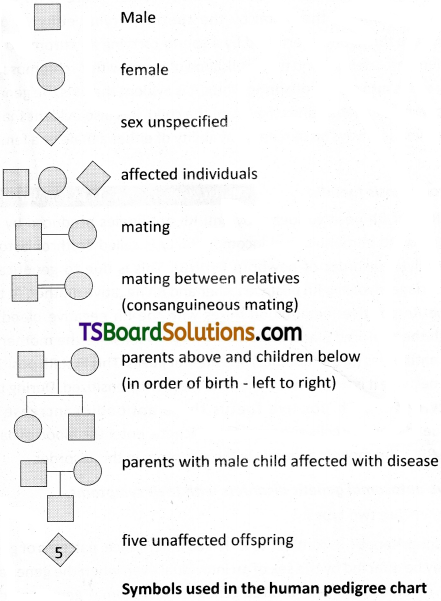
What is pedigree analysis? Suggest how such can analysis, can be useful.
Answer:
Pedigree analysis is useful in many ways like it helps to work out the possible genotypes from the knowledge of the respective phenotypes. It helps to study the pattern of inheritance of a dominant or a recessive trait. The possible genetic makeup of a person for a trait can also be known with the help of the pedigree chart. Some of the important standard symbols used in the pedigree analysis are shown in the figure.
Question 3.
How is sex determined in human beings? [March 2020, 2018 (A.P.); March 2015 (T.S.)]
Answer:
Sex determination in Humans : It has already been mentioned that the sex determining mechanism in case of humans is XX – XY type. Out of 23 pairs of chromosomes present, 22 pairs are exactly same in both males and females; these are the autosomes. A pair of X- chromosomes is present in the female, where as the presence of an X and Y – chromosome are determinant of the male characteristic. During spermatogenesis among males, two types of gametes are produced. 50 percent of the total sperm produced carry the X – chromosome and the rest 50 percent has Y – chromosome besides the autosomes. Females, however, produce only one type of ovum with an X – chromosome.
There is an equal probability of fertilisation of the ovum by the sperm carrying either X or Y chromosome. In case the ovum is fertilised by a sperm carrying X – chromosome, the zygote develops into a female and the fertilisation of ovum with Y – chromosome carrying sperm results into a male offspring. Thus, it is evident that it is the genetic makeup of the sperm that determines the sex of the child. It is also evident that in each pregnancy there is always 50 percent probability of either a male or a female child.
Question 4.
Describe erythroblastosis foetolis. [March 2019, ’17, May ’17 (A.P.); Mar. ’14]
Answer:
Destruction of RBC of Rh positive foetus by anti Rh antibodies produced by Rh negative mother due to immunological incompatibility is called Erythroblastosis foetalis or Haemolytic disorder of newborn (HDNB). This is due to genetically incompatible marriage involving Rh positive father and Rh negative mother. At the time of birth the Rh positive foetal blood mixes with the Rh negative blood of mother, through the ruptured placenta.
The Rh antigens sensitize the mother to produce anti Rh antibodies (IgG antibodies) and memory cells. This first Rh positive by is unaffected because it is delivered by the time mother is sensitized. During the next pregnancy bearing Rh positive foetus these antibodies increase in concentration due to memory cells and cross the placenta, enter the blood of baby and destroy the RBC. Haemolytic anaemia is the symptom in this disorder.
Question 5.
Mention any two autosomal genetic disorders with their symptoms.
Answer:
Autosomal disorders are two types.
1) Sex – Limited inheritance :
in contrast to X – linked inheritance, patterns of gene expression may be affected by the sex of an individual even when the genes are not on the X – chromosome. Sex – limited genes are autosomal genes present in both males and females. Their phenotypic expression is limited to only one sex due to internal hormonal environment, e.g. beard in man, development of breast and secretion of milk in woman etc., are sex limited traits.
2) Sex – influenced Inheritance :
Sex – influenced genes are the autosomal genes present in both males and females. In sex -influenced inheritance, the genes behave differently in the two sexes, probably because the sex hormones provide different cellular environments in males and females. Thus, the heterozygous genotype may exhibit one phenotype in males and the contrasting one in females. Cases of sex – influenced inheritance include pattern baldness in humans, horn formation in certain breeds of sheep (e.g. Dorset Horn sheep).
Question 6.
Describe the genetic basis of ABO blood grouping.
Answer:
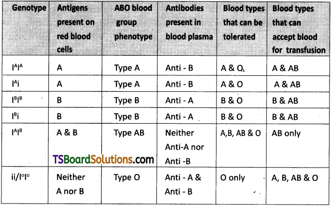
Bernstein discovered that these blood group phenotypes were inherited by the interaction of three ‘autosomal alleles’ of the gene named I, located on chromosome 9. IA, IB and i (or I°) are the three alleles of the gene I. The antibodies ‘anti – A’ and ‘anti – B’ are called isoagglutinins (also called isohaemagglutinins) which are usually IgM type. The isoagglutinins of an individual cause agglutination reactions with the antigens of another individual. The alleles IA and IB are responsible for the production of the respective antigens ‘A’ and ‘B’. The allele i does not produce any antigen. The alleles lA and lB are dominant to the allele i, but co-dominant to each other (IA = IB > i). A child receives one of the three alleles from each parent, giving rise to six possible genotypes
Table : Genetic control of the human ABO blood groups
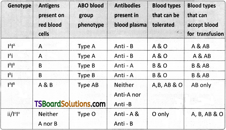
and four possible blood types (phenotypes). The genotypes are IAIA, IAi, IBIB, IBi, IAIB and ii. The phenotypic expressions of IAIA and IAi are ‘A’ – type blood, the phenotypic expressions of IAIA and IBi are ‘B’ – type blood, and that of IAIB is ‘AB’ – type blood. The phenotype of ii (I°I°) is ‘O’ – type blood.
Question 7.
Describe male heterogamety.
Answer:
Male Heterogamety : In this method of sex determination, the males (heterogametic) produce dissimilar gametes while females (homogametic) produce similar gametes. Male heterogamety is of two kinds, XX – XO type and XX – XY type.
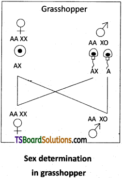
i) XX – XO type :
In some insects such as bugs, grasshoppers and cockroaches, females are with two X – chromosomes and males are with one X – chromosome in each somatic cell. McClung discovered this type in grasshoppers. The unpaired X – chromosome determines the male sex. The karyotype of the female (homogametic) is AAXX and that of the male (heterogametic) is AAXO. All the ova contain ‘AX’ complement of chromosomes and the sperms are of two types. One half of the sperms have ‘AX’ complement and the other half have ‘A1 complement of chromosomes. The sex of the offspring depends on the type of sperm that fertilizes the ovum.
ii) XX – XY type :
In human beings and some insects such as Drosophila, both females and males have the same number of chromosomes. The karyotype of the female is AAXX and that of the male is AAXY. Females are ‘homogametic’ with ‘XX’ chromosomes.
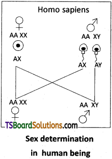
They produce similar ova having one X – chromosome each. Males are ‘heterogametic’ with X and Y – chromosomes. They produce two kinds of sperms; one half of them with X – chromosome and tbe other half with Y – chromosome. The sex of the offspring depends on the fertilizing sperm. The XX – XY type is also found in most other mammals.
Question 8.
Describe female heterogamety.
Answer:
Female Heterogamety :
In this method of sex determination, the males produce ‘similar gametes’ while females produce ‘dissimilar gametes’. Female heterogamety is of two kinds, ZO – ZZ type and ZW – ZZ type.
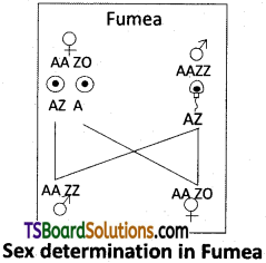
i) ZO – ZZ type : In moths and some butterflies, female is heterogametic with one z – chromosome (ZO) and male is homogametic with two Z – chromosomes (ZZ). The karyotype of female is AAZO and male is AAZZ. Females produce two kinds of ova, half of them with a Z – chromosome and the other half with no sex chromosome. Males produce similar type of sperms. The sex of the offspring depends on the type of ovum that is fertilized.
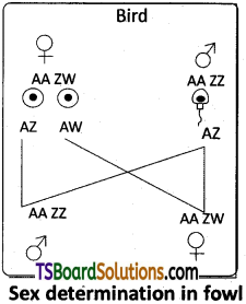
ZW – ZZ type :
In birds, reptiles, some fishes, etc., the females are heterogametic with ZW – allosomes and males are homogametic with ZZ – allosomes. The karyotype of the female is AAZW and that of the male is AAZZ. All sperms are similar with the allosome – Z. Ova are of two different kinds; one half of the ova are with the allosome – Z and the other half with the allosome – W. The sex of the offspring depends on the typ£of ovum that is fertilized.
Question 9.
Describe the Genic Balance Theory of sex determination. [March 2015 (A.P.)]
Answer:
Genic balance theory of sex determination was proposed by Calvin Bridges with reference to sex determination in Drosophila. He proposed that both the X- chromosomes and autosomes together play a role in sex determination in Drosophila, where as Y-chromosome has no role. This theory explains that genes for maleness are located on autosome and for femaleness on X-chrombsorne in Drosophila. Y-chromosome in lacks male – determining factor but contains gametic information essential to male fertility. Sex in Drosophila is determined by ratio of X-chromosome to the number of haploid sets of autosomes.
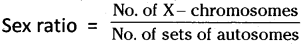
The chromosomal complement, X/A ratio and sex of Drosophila are tabulated below.
| Chromosomal complement | X/A ratio | Sex |
| AAX | 0.5 | Male |
| AAXX | 1.0 | Female |
| AA XXX | 1.5 | Metafemale |
| AAA XX | 0.67 | Intersex |
| AAA X | 0.33 | Metamale |
Question 10.
Explain the inheritance of sex linked recessive character in human being.
Answer:
Colour Blindness :
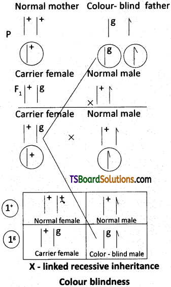
It is a sex -linked recessive disorder. Retina of the eye in man contains the cells sensitive to red and green colours, this phenotypic trait is genetically controlled. Its alleles are located on the X-chromosome. When a woman with normal vision (homozygous) marries a colour – blind man, all the sons and daughters are normal, but daughters are carriers (heterozygous),. If a carrier woman marries a man with normal vision, all the daughters and half of the sons have normal vision and another half of sons are colour – blind. Colour – blind trait is inherited from a male parent to his grand sons through carrier daughter, which is an example of crisscross pattern of inheritance.
Question 11.
Describe the experiment conducted by Morgan to explain sex linkage.
Answer:
Thomas H. Morgan (Father of Modern Genetics ) discovered sex linkage in Drosophila melanogaster. Morgan wanted to analyze the behavior of the two alleles of a fruit fly’s eye – colour gene. When he crossed a white eyed (mutant) male to a normal (wild) red eyed female, in the F1 generation all the males and females were red eyed. When the F1 generation ‘red eyed female’ was crossed to a ‘red eyed male’, in the F2 generation all the females were red eyed and 50 percent of the males were ‘white eyed’.
The white eyed trait from the male is inherited to the male of the F2 generation through the ‘carrier daughter’ of the F1 generation. This pattern of inheritance is called crisscross pattern of inheritance (skip generation inheritance) in which a gene responsible for the white eyes is transmitted from a male parent to a male grandchild through carrier female of the first generation.
In a reciprocal cross (to test the role of parental sex on inheritance pattern), in which a white eyed female was crossed to a red eyed male, the results were different. The first generation male offspring had white eyes while the female offspring had red eyes. The reason was that the allele responsible for the white eye is sex – linked (more specifically X – linked, as it occurs on the X – chromosome) and recessive. Males always inherit the X – linked recessive traits from the female parents.
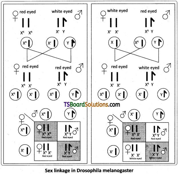
Morgan’s discovery that transmission of the X – chromosome in Drosophila correlates with the inheritance of an eye – colour trait was the first solid evidence indicating that a specific gene is associated with a specific chromosome.
Question 12.
Explain the inheritance of sex influenced characters in human beings.
Answer:
Sex influenced genes are autosomal genes in both males and females, whose phenotypic expression is different in different sexes, dominant in one sex and recessive in the other.
Pattern baldness is a sex influenced trait in human being, in which a fringe of hair is present low on the head. The gene for baldness ‘B’ is dominant in males and recessive in females in heterozygous condition.
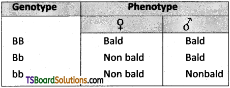
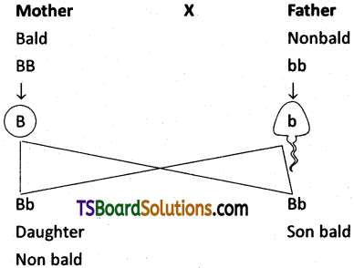
When mother is homozygous bald and father is homozygous nonbald in the progeny all the daughters are nonbald and all sons are bald.
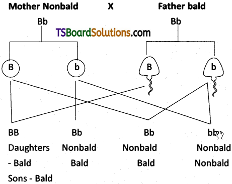
If both the parents are heterozygous bald, nonbald daughters are 1 : 3, and sons are 3 : 1. In the progeny bald, non-bald ratio is 1 : 1.
Question 13.
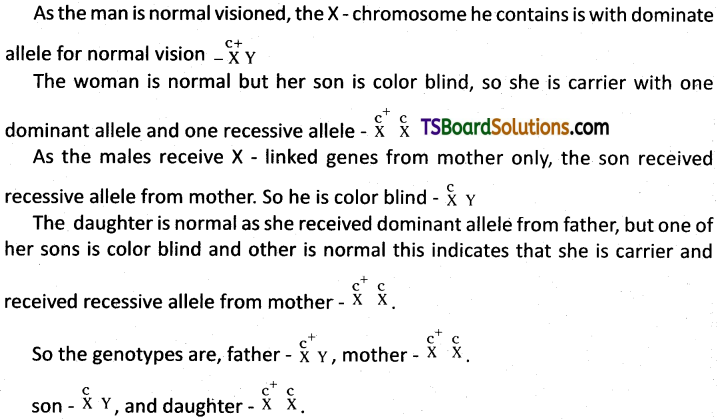
A man and woman of normal vision have one son and one daughter. Son is colour – blind and his son is with normal vision. Daughter is with normal vision, but one of her sons is colour – blind and the other is normal. What are the genotypes of the father, mother, son and daughter?
Answer:
Color blindness in human being is because of recessive X – linked gene, and show criss – cross inheritance.
Question 14.
A colour – blind man married a woman who is the daughter of a colour – blind father and mother homozygous normal vision. What is the probability of their daughters being colour – blind?
Answer:
The woman he married is a carrier (heterozygous) since her father is color blind but mother is homozygous normal. When a color-blind man marries a carrier woman in the progeny half daughters are carriers and half daughter are color blind.
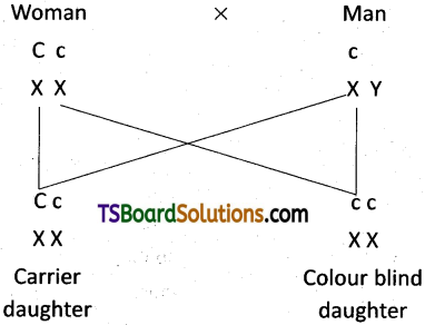
So the probability of their daughters being color blind is 50%.
Question 15.
A heterozygous bald man who is non – haemophilic, married a woman who is homozygous for the non – bald trait and is haemophilic. What is the probability of her male children becoming bald and haemophilic?
Answer:
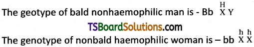
When these two marry they produce two kinds of sons, half heterozygous bald and haemophilic and half nonbald and haemophilic.
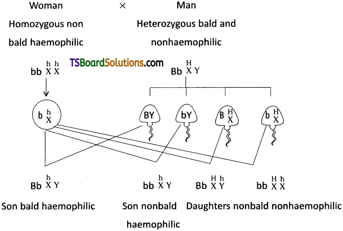
So the probability of their male children being bald and haemophilic is 50%.
Question 16.
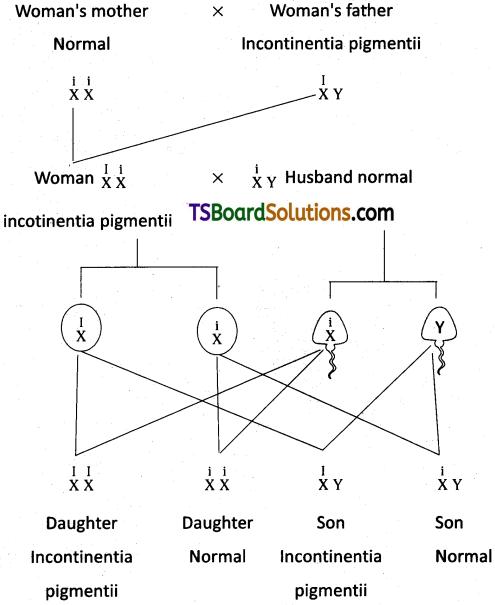
A woman’s father shows ‘IP’but her mother and husband are normally pigmented. What will be the phenotypic ratio of her children?
Answer:
In continentia pigmentii is ‘X’ linked dominant truit. It is more in females than in males, because they have two chances to inherit this allele.
As that womans, mother is normal and father is incontinentia pigmenti, she is heterozygous for incontinentia pigmentii. As her husband is normal in their progeny half daughters are heterozygous incontinentia pigmentii, half daughters are normal, half sons are incontinentia pigmentii and half sons are normal, so the phenotype ratio is 1 : 1 : 1 : 1.
Question 17.
Write the salient features of ‘HGP’.
Answer:
Salient Features of Human Genome: Some of the salient observations drawn from human genome project are as follows :
- The human genome contains 3164.7 million nucleotide bases.
- The average gene consists of 3000 bases, but sizes vary greatly, with the largest known human gene being the one that codes for the protein called dystrophin.
- The total number of genes is estimated at 30,000. Almost all (99.9%) nucleotide bases are exactly the same in the people.
- The functions are unknown for over 50% of the genes discovered.
- Less than 2 percent of the genome codes for proteins.
- Repeated sequences make up very large portion of the human genome.
- Repetitive sequences are stretches of DNA sequences that are repeated many times. They are thought to have no direct coding functions, but they shed light on chromosome structure, dynamics and evolution.
- Chromosome 1 has the highest number of genes (2,968), and the Y – chromosome has the fewest genes (231).
- Scientists have identified about 1.4 million locations where single base DNA differences (SNPs – single nucleotide polymorphism, pronounced as snips) occur in humans. This information promises to revolutionise the processes of finding chromosomal locations for disease – associated sequences and tracing human history.
Question 18.
Describe the steps involved in DNA finger printing technology.
Answer:
The following is the process (protocoJ) of DNA (genetic) fingerprinting.
- First step is obtaining DNA sample from substances like blood, semen, hair roots, bone or saliva.
- The DNA is cut at specific sites into small fragments using restriction enzymes and then amplified by rDNA or PCR methods.
- Double stranded DNA is split into single stranded DNA using alkaline chemicals.
- The DNA fragments are applied to one end of thin agarose gel and separated into individual bands, with fragments in each one progressively smaller in size, by electrophoresis.
- A thin dylon membrane covered by paper towels is placed over the gel, as the
paper towels absorb the moisture from gel, DNA is transferred into nylon membrane, this process is blotting. - A radio active DNA probe is introduced, which binds with specific complemental DNA sequences on nylon membrane. The excess DNA probe is washed away.
- When a photographic / X – ray film is placed on the nylon membrane radio active probes will expose the film producing a pattern of thick and thin bands. This pattern of bars is DNA (genetic) fingerprint.
Long Answer Type Questions
Question 1.
What are multiple alleles ? Describe multiple alleles with the help of ABO blood groups in man. [March 2020, 2018 (A.P.); March 2014; May/June ’14]
Answer:
Multiple alleles and human blood groups :
Generally a gene has two alternative forms / versions called alleles. They are present at the same locus in a pair of homologous chromosomes. Two alleles of a gene can form three genotypes in a diploid organism. Sometimes a gene may have more than two alleles. When more than two allelic forms occur at the same locus on the homologous chromosomes of an organism, they are called mutiple alleles when more than two alleles exist in a population of a specific organism, the phenomenon is called mutiple allelism.
As mentioned above ‘multiple alleles’ cannot be observed in the genotype of a diploid individual, but can be observed in a population. The number of genotypes that can occur for multiple alleles is given by the expression n (n + 1) /2 where, n = number of alleles. A well known example of multiple allelism in man is the expression of ABO blood types by three alleles of a single gene which can produce six genotypes.
ABO Blood Types :
The ABO blood group system was proposed by Karl Landsteiner. He was awarded the Nobel Prize in Physiology or Medicine in 1930 for his work. The phenotypes (blood types) A, B, AB and O types are characterized by the presence or absence of ‘antigens’ on the plasma membrane of the RBCs. The A and B antigens are actually carbohydrate groups (sugar polymers) that are bound to lipid molecules (fatty acids) protruding from the membrane of the red blood cell. They are also called isoagglutinogens because they cause blood cell agglutination in the case of incompatible blood transfusions.
‘Blood type A’ persons have antigen A on their RBCs and anti – B antibodies in the plasma. ‘Blood type B’ persons have antigen B on their RBCs and anti – A antibodies in the plasma. ‘Blood type AB’ person have antigens ‘A’ and ‘B’ on theRBCs and no antibodies in the plasma. ‘Blood type O’ persons have no antigens on their RBCs and both ‘anti – A, and ‘anti – B’ antibodies are present in the plasma.
Bernstein discovered that these phenotypes were inherited by the interaction of three ‘autosomal alleles’ of the gene named I, located on chromosome 9. IA, lB and i (or I°) are the three alleles of the gene I. The antibodies ‘anti – A’ and ‘anti – B’ are called isoagglutinins (also called isohaemagglutinins) which are usually IgM type. The isoagglutinins of an individual cause agglutination reactions with the antigens of another individual. The alleles lA and lB are responsible for the production of the respective antigens ‘A’ and’B’. The allele i does nto produce any antigen. The alleles lA and lB are dominant to the allele i, but co-dominant to each other (lA = lB > i). A child receives one of the three alleles from each parent, giving rise to six possible
Table: Genetic control of the human ABO blood groups
genotypes and four possible blood types (phenotypes). The genotypes are IAIA, IAi, IBIB, IBi, IAIB and ii. The phenotypic expressions of IAIA and IAi are ‘A’ – type blood, the phenotypic expressions of IBIb and IBi are ‘B’ – type blood, and that of IAIB is ‘AB’ – type blood. The phenotype of ii (I°I°) is ‘O’ – type blood.
Question 2.
Describe chromosomal theory of sex determination.
Answer:
Sex Determination :
The mechanism of sex determination has always been a puzzle to geneticists. In fact, the cytological observations made in a number of insects led to the development of the concept of genetic / chromosomal basis of sex determination.
Sex Chromosomes :
In most of the animals a pair of chromosomes is responsible for the determination of sex. These two chromosomes are called sex chromosomes or allosomes. The chromosomes other than the sex chromosomes are called autosomes. The first indication that sex chromosomes were distinct from the other chromosomes came from the experiments conducted by Henking. He could trace a specific nuclear structure all through spermatogenesis in wasps, and it was also observed by him that 50 percent of the spermatozoa received this structure after spermatogenesis, whereas the other 50 percent did not receive it.
Henking gave the name X – body to this structure, but he could not explain its significance. Further investigations by other scientists led to the conclusion that the X – body of Henking was in fact a chromosome and that is why it was given the name X – chromosome. Stevens and Wilson first identified Y – chromosome as a sex determining chromosome in the mealworm, Tenebrio molitor. The revealed that the chromosomal basis of sex depended on the presence or absence of the Y – chromosome.
Heterogametic Sex Determination :
Heterogametic sex refers to the sex of a species in which the sex chromosomes are not similar.’The process of sex determination by allosomes is called genetic or chromosomal sex determination. In the heterogametic sex determination, one of the sexes produces ‘similar’ gametes and the other sex (heterogametic sex) produces ‘dissimilar / unlike gametes. The sex of the young one is determined at the time of syngamy (fertilization). It depends on which gamete of the two dissimilar gametes unites with the other gamete produced by the ‘homogametic parent’.
Male Heterogamety :
In this method of sex determination, the males (heterogametic) produce dissimilar gametes while females (homogametic) produce similar gametes. Male heterogamety is of two kinds, XX – XO^type and XX – XY type.
XX – XO type :
In some insects such as bugs, grasshoppers and cockroaches, females are with two X – chromosomes and males are with one X – chromosome in each somatic cell. McClung discovered this type in grasshoppers. The unpaired X – chromosome determines the male sex. The karyotype of the female (homogametic) is AAXX and that of the male (heterogametic) is AAXO. All the ova contain ‘AX’ complement of chromosomes and the sperms are of two types. One half of the sperms have ‘AX’ complement and the other half have ‘A’ complement of chromosomes. The sex of the offspring depends on the types of sperm that fertilizes the ovum.
XX – XY type :
In human beings and some insects such as Drosophila, both females and males have the same number of chromosomes. The karyotype of the female is AAXX and that of the male is AAXY. Females are ‘homogametic’ with ‘XX’ chromosomes.
They produce similar ova having one X – chromosome each. Males are ‘heterogametic’ with X and Y – chromosomes. They produce two kinds of sperms; one half of them with X – chromosome and the other half with Y – chromosome. The sex of the offspring depends on the fertilizing sperm. The XX – XY type is also found in most other mammals.
Female Heterogamety :
In this method of sex determination, the males produce ‘similar gametes’ while females produce ‘dissimilar gametes’. Female heterogamety is of two kinds, ZO – ZZ type and ZW – ZZ type.
ZO – ZZ type :
In moths and some butterflies, female is heterogametic with one Z-chromosome (ZO) and male is homogametic with two Z – chromosomes (ZZ). The karyotype of female is AAZO and male is AAZZ. Females produce two kinds of ova, half of them with a Z – chromosome and the other half with no sex chromosome. Males produce similar type of sperms. The sex of he offspring depends on the type of ovum that is fertilized.
ZW – ZZ type :
In birds, reptiles, some fishes, etc., the females are heterogametic with ZW – allosomes and males are homogametic with ZZ – allosomes. The karyotype of the female is AAZW and that of the male is AAZZ. All sperms are similar with the allosome – Z. Ova are of two different kinds; one half of the ova are with the allosome – Z and the other half with the allosome – W. The sex of the offspring depends on the type of ovum that is fertilized.
Sex Determination in Humans :
It has already been mentioned that the sex determining mechanism in case of human is XX – XY type. Out of 23 pairs of chromosomes present, 22 pairs are exactly same in both males and females; these are the autosomes. A pair of X- chromosome is present in the female, whereas the presence of an X and Y – chromosome are determinant of the male characteristic. During spermatogenesis among males, two types of gametes are produced. 50 percent of the total sperm produced carry the X – chromosbme and the rest 50 percent has Y – chromosome besides the autosomes. Females, however, produce produce only one type of ovum with an X – chromosome.
There is an equal probability of fertilisation of the ovum by the sperm carrying either X or Y- chromosome. In case the ovum js fertilised by a sperm carrying X – chromosome, the zygote develops into a female and the fertilisation of ovum with Y – chromosome carrying sperm results into a male offspring. Thus, it is evident that it is the genetic makeup of the sperm that determines the sex of the child. It is also evident that in each pregnancy there is always 50 percent probability of either a male or a female child.
Question 3.
What is crisscross inheritance? Explain the inheritance of one sex linked recessive character in human beings. [Mar. ’19, 17, May ’17 (A.P.); Mar. ’15 (A.P. & T.S.)]
Answer:
The crisscross pattern of inheritance (skip generation inheritance) is one in which a gene responsible for the sex linked recessive character is transmitted from a male parent to a male grand child through a carrier female of the first generation. Colour blindness is the best example for criss – cross inheritance in human being.
Colour blindness :
It is a sex-linked recessive disorder. Retina of the eye in man contains the cells sensitive to red and green colours. This phenotypic trait is genetically controlled. Its alleles are located on the X – chromosome; When a woman with normal vision (homozygous) marries a colour – blind man, all the sons and daughters are normal, but daughters are carriers (heterozygous). If a carrier woman marries a man with normal vision, all the daughters and half of the sons have normal vision and another half of sons are colour – blind. Colour – blind trait is inherited from a male parent to his grandsons through carrier daughter, which is an example of crisscross pattern of inheritance.
Haemophilia :
Haemophilia A is recessive X – linked genetic disorder involving lack of the functional clotting Factor – VIII and represents 80% of haemophilia cases. Haemophilia B is also a recessive X – linked genetic disorder involving lack of the functional clotting Factor IX. When a person with hemophilia is injured, bleeding is prolonged because a firm clot is slow to form. Haemophilia follows the characteristic crisscross pattern of inheritance like that of colour – blindness.
Duchenne Muscular dystrophy :
Duchenne muscular dystrophy (DMD) is a recessive X – linked form of muscular dystrophy, affecting around 1 in 3,600 boys. The disease is characterized by a progressive weakening of the muscles and loss of coordination. Affected individuals rarely live past their early 20s. The disorder is caused by a mutation in the dystrophin gene (the largest known gene in humans) located on the X – chromosome, which codes for the protein dystrophin, an important structural component within muscle tissue (connects sarcolemma and the outer most layer of muscle filaments and supports muscle fiber strength).
If the mother is known to be a carrier of this gene, about X – linked recessive inheritance half of her male children are expected to Colour blindness be affected. All female children born to a carrier mother are expected to be normal, since the possibility of their being homozygous for this sex – linked recessive gene is virtually non – existent.
Question 4.
Write an essay on common genetic disorders.
Answer:
Genetic disorders :
A number of disorders in human beings have been found to be associated with the inheritance of changed “or altered genes or chromosomes. Genetic disorders may broadly be grouped into two categories – Mendelian disorders and Chromosomal disorders.
Mendelian disorders :
Medelian disorders are genetic diseases showing Mendelian pattern of inheritance, caused by a single mutation in the structure of DNA, which causes a single basic defect with pathologic consequences, in some cases. Mendelian disorders are also called monogenic diseases. Monogenic diseases run in families and can be dominant or recessive and autosomal or sex linked (allosomic). The pattern of inheritance of Mendelian disorders can be traced in a family with the help of pedigree charts and their analyses. The most common and prevalent Mendelian disorders are Haemophilia, Cystic fibrosis, Sickle – cell anaemia, Colour blindness, phenylketonuria, Thalassemia, DMD, Albinism, etc.
Haemophilia:
Haemophilia A (caused by deficiency of clotting factor VIII) and Haemophilia B (caused by deficiency of clotting factor IX) are X – linked recessive disorders that impair the body’s ability to control clotting or coagulation of blood. Haemophilia C is an autosomal recessive disorder involving lack of the functional clotting ‘factor XI’. Haemophilia is also called bleeder’s disease. Haemophilia A and B follow the characteristic crisscross pattern of inheritance like that of colour – blindness. In this disease, a single protein that is a part of the cascade of reactions involved in the clotting of blood is affected.
Haemophilia is more likely to occur in males than in females. This is because female have two X -chromosomes while males have only one, and so the defective gene on the X will certainly express in the male who carries it. As it is caused by a recessive allele on the X chromosome, a female human being has to be ‘double recessive’ to express haemophilia. Because the chance of a female having two defective copies of the gene (alleles) is very remote, the females are mostly asymptomatic carriers of the disorder. The ‘allele’ is typically passed on from an affected father to 50% of his grand sons through his ‘carrier daughters’. The family pedigree of Queen Victoria shows a number of haemophilic descendents, as she was a carrier for the disease.
Sickle – cell anaemia :
Sickle – cell anaemia is an autosomal recessive genetic blood disorder characterized by red blood cells that assume an abnormal, rigid, sickle-shape in hypoxia conditions (at high altitudes or under physical stress, for instance). Sickled cells may clump and clog small blood vessels, often leading to other symptoms throughout the body, including physical weakness, pain, organ damage, and even paralysis.
This disease is controlled by a single pair of alleles, HbA and Hbs found on the chromosome 11. The homozygous individuals for sickle – cell anaemia (HbA Hbs) express the diseased phenotype. Heterozygous individuals (HbA Hbs) appear ‘unaffected’ but they are still, carriers of the disease. Even though two sickle cell alleles are necessary to cause sickle cell anaemia, one dose can affect the phenotype. Persons ‘heterozygous’ to sickle cell trait can usually lead a healthy life but in prolonged periods of reduced oxygen content in the blood may suffer from symptoms of SCD as both normal and sickle cell haemoglobin are formed in them.
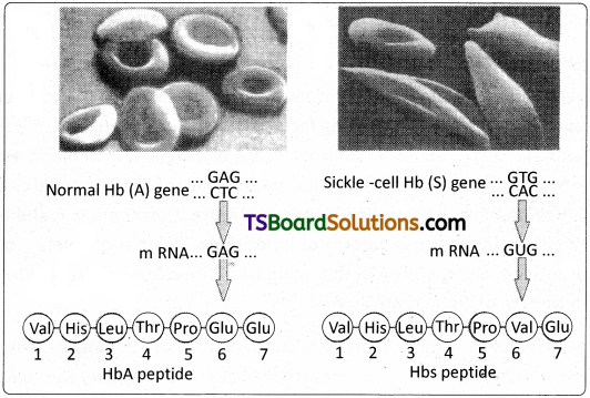
Micrograph of the red blood cells and the amino acid composition of the relevent portion of β – chain of haemoglobin; (a) From a normal individual; (b) From an individual with sickle – cell anaemia.
Sickle cell anaemia is caused by a point mutation in the DIMA that codes for the beta globin polypeptide chains of the haemoglobin molecule, causing the replacement of the glutamic acid in the sixth position by valine. The heterozygous individuals are relatively resistant to the most severe effects of malaria such as those of ‘falciparum malaria’ also (although they are not resistant to malaria infection) – an effect called heterozygote advantage. The heterozygous individuals carry the deleterious alleles in their genomes (genetic load).
Phenylketonuria (PKU) :
Phenylketonuria was discovered by A. Foiling. This is an autosomal recessive, metabolic genetic disorder caused by a mutation in the gene (PAH. Phenylalanine hydroxylase gene) located on the chromosome 12 for the hepatic enzyme ‘phenylalanine hydroxylase’, rendering it nonfunctional. The affected individual lacks the above mentioned enzyme that converts the amino acid phenylalanine into tyrosine. When phenylalanine hydroxylase’s activity is reduced, phenylalanine accumulates and is converted into phenylpyruvate and other derivatives. Accumulation of these substances in the brain causes mental retardation. Adherence to a low phenylalanine diet prevents major mental retardation.
Colour blindness :
Colour blindness (colour vision deficiency) is a sex – linked recessive disorder. It is the inability or decreased ability to see certain colours or perceive differences between some colours. This phenotypic trait is due to mutation in certain genes located on X – chromosome. The most common inherited forms of colour blindness are Protanopia (red colour blindness), Deuteranopia (green colour blindness) and Tritanopia (blue colour blindness – autosomal).
The son of a woman who carries the allele has a 50 percent chance of being colour – blind. The mother herself is not colour – blind because the allele is recessive. That means its effect is suppressed by her matching dominant normal allele. A daughter will not normally be colour blind, unless her mother is ‘colour – blind’ or a ‘carrier’ and her father is colour – blind. The Ishihara colour test, which consists of a series of pictures of coloured spots, is most often.used to diagnose red – green colour blindness.
Thalassemia :
Thalassemia is an autosome linked recessive blood disroder. This disease is caused by the excessive destruction or degradation of red blood cells due to formation of abnormal haemoglobin molecules, because of a defect through a genetic mutation or deletion (a type of chromosomal mutation). Normally, haemoglobin is composed of four polypeptide chains, two alpha and two beta globin chains arranged into a hetero tetramer. In the case of thalassemia, patients have defects in either the alpha or beta globin chain (unlike sickle cell anaemia, which is caused due to a specific change in the beta chain), causing production of abnormal haemoglobin molecules resulting in anaemia which is characteristic of the disease. The effected people make less haemoglobin and fewer RBC in the circulating blood, hence anaemia.
Thalassemias are classified based on which chain of haemoglobin molecule is affected. In Alpha thalassemia, the production of alpha globin chain is affected. Alpha thalassemia is controlled by two closely linked genes HBAI and HBA2 on chromosome 16 of each parent and it is caused due to mutation or deletion of one or more of the four alpha gene “alleles”. The more genes affected, the less alpha globin molecules are produced. In Beta thalassemia (Cooley’s Anaemia) production of beta globin chain is affected. The Beta thalassemia is controlled by single gene HBB on chromosome 11 of each parent and occurs due to mutation of one or both alleles. It is the most common type of thalassemia. In this disorder the alpha chains which are produced in excess bind to RBCs and damage them.
Cystic fibrosis :
Cystic fibrosis is an autosomal recessive genetic disorder. CF is the result of mutations affecting a gene on the long arm of chromosome 7 that influences salt and water movement across epithelial cell membranes. The genetic defect causes increased sodium and chloride content in sweat and increased resorption of sodium and water from respiratory eptihelium. The extracellular chloride causes the mucus that coats certain cells to become more viscous and sticky. The mucus builds up in organs such as lungs, pancrias, Gl tract etc., and leads to further complications and may lead to death by the age five, if untreated.
Chromosomal disorders :
Chromosomal disorders are caused by errors in the ‘number’ or ‘structure’ of chromosomes. Chromosomal anomalies usually occur when there is an error in cell division. Aneuploidy is a chromosomal aberration where there is again or loss of one or more chromosomes in a ‘set’. It is caused by non-disjunction of chromosomes. The result of this error is origin of cells with a deviation from the normal number of chromosomes – aneuploidy.
A) Allosomal disorders :
Klinefelter’s syndrome :
This genetic disorder is caused by trisomy 23rd pair. The karyotype is 47, XXY. A Klinefelter male possesses an additional X – chromosome along with the normal XY. The principal effects include hypogonadism and reduced fertility. At the same time, feminine sexual development is not entirely suppressed. Slight enlargement of the breasts (gynecomastia) is common, and the hips are often rounded. The somatic cells of a Klinefelter male exhibit Barr bodies in their nuclei.
Turner’s syndrome :
The Karyotype is 45, X. It is due to monosomy 23rd pair, where one X – chromosome is lost. A Turner female does not show Barr bodies in her somatic cells. The symptoms are short stature, gonadal dysgenesis, webbed neck and broad shield like chest with widely spaced nipples.
B) Autosomal disorders :
Most of the following disorders are common in children born to woman who conceive babies rather late in their reproductive phase.
Down syndrome (Trisomy 21) :
Down syndrome is a genetic condition that causes delays in physical and intellectual development. The cause of this genetic disorder is the presence of an additional copy of the chromosome numbered 21 (trisomy of 21st set). The karyotype is designated as Trisomy 21 (47, XX, + 21).The affected individual is short statured with small round head, furrowed tongue and partially open mouth. Physical, psychomotor and mental development is retarded.
Edwards syndrome (Trisomy 18) :
Edwards syndrome (47, XX, + 18) is a chromosomal abnormality characterized by the presence of an extra copy of the genetic material on the 18th chromosome, either in whole (trisomy 18) or in part (such as due to translocations). Edwards syndrome occurs in all human populations but is more prevalent in the female offspring. The majority of people with the syndrome die during the foetal stage; infants who survive experience serious defects (cardiac abnormalities and kidney malfunction) and commonly live for short periods of time.
Patau syndrome (Trisomy 13) :
Patau syndrome, is a chromosomal condition associated with severe intellectual disability and physical abnormalities in many parts of the body. Most cases of trisomy 13 (47, XX, +13) result from having three copies of chromosome 13 in each cell in the body instead of the usual two copies. Individuals with trisomy 13 often have heart and kidney defects, brain or spinal cord abnormalities, very small or poorly developed eyes (microphthalmia), cleft palate etc. Due to the presence of several life threatening medical problems, many infants with trisomy 13 die within their first days or weeks of life.
Cri-du-Chat syndrome (5p minus syndrome):
Cri – du – chat syndrome (cat – cry) is due to a partial deletion of the short arm of chromosome 5, also called 5p monosomy. It might be considered a case of partial monosomy, but since the region that is missing is so small, it is better referred to as 5p segmental deletion. The karyotype is 46, XX, 5p“. It is a French term referring to the characteristic cat – like cry of the affected children due to problems with the larynx and nervous system. Such infants are mentally retarded, have a small head with unusual facial features. They die in infancy or early childhood.
Chronic Myelogenous (Myeloid) Leukemia (CML) :
In certain cancers such as Chronic myelogenous leukemia (also called Chronic granulocytic leukemia), a piece of the chromosome 9 and a piece of the chromosome 22 break off and ‘switch places’ (exchange places) with each other (reciprocal translocation). This results in the formation of an abnormally short chromosome 22 and abnormally long chromosome 9. The short 22nd chromosome is called Philadelphia chromosome produced by translocation which is also called Philadelphia translocation. The karyotype is 46, XXt(9; 22). The Philadelphia chromosome results in the production of an abnormal enzyme called a tyrosine kinase. Along with other abnormalities, this enzyme causes uncontrolled cell cycle progression leading to the cancer called chronic myelogenous leukemia.
Question 5.
Why is the Human Genome project called a mega project?
Answer:
Human Genome Project (HGP) was called a mega project. It was an international effort formally begun in October, 1990. The HGP was a 13 – year project coordinated by the U.S. Department of Energy and the National Institute of Health. During the early years of the HGP, the Wellcome Trust (U.K.) became a major partner, and additional contributions came from Japan, France, Germany, China and others. The project was almost completed in 2003. Knowledge about the effects of DNA variations among individuals can lead to revolutionary new ways to diagnose, treat and someday prevent the thousands of disorders that affect human beings.
HGP was closely associated with the rapid development of a new area in biology called Bioinformatics. Besides providing clues to understanding human biology, learning about non – human organisms’ DNA sequences can lead to an understanding of their natural capabilities that can be applied toward solving challenges in health care, agriculture, energy production, environmental remediation. Genomes of many non – human model organisms, such as bacteria, yeast, Caenorhabditis elegans (a free living non – pathogenic nematode), Drosophila, plants (rice and Arabidopsis), etc. have also been sequenced. In a way they helped the progress of HGP.
Goals of HGP :
Some of the important goals of HGP were as follows :
- Identify all the approximately 20,000 – 25,000 genes in human DNA.
- Determine the sequences of the 3 billion chemical base pairs that make up human DNA.
- Improve tools for data analysis.
- Address the ethical, legal, and social issues (ELSI) that may arise from the project.
Methodologies :
The methods involved two major approaches. One approach focused on identifying all the genes that expressed as RNA (referred to as Expressed Sequence Tags (ESTs). The other took the blind approach of simply sequencing the whole set of genome that contained all the coding and non-coding sequence, and later assigning different regions in the sequence with functions (a term referred to as Sequence Annotation).
What is DNA sequencing?
DNA sequencing, the process of determining the exact order of the 3 billion paired chemical building blocks (called ‘bases’ – A, T, C, and G) that make up the DNA of the 24 different human chromosomes (23 + Y in a male), was the greatest technical challenge in the Human Genome Project.
For sequencing, the total DNA from a cell is isolated and converted into random fragments of relatively smaller size and cloned in a suitable host using specialized vectors. The cloning results in the amplification of DNA fragments which are used for sequencing the bases. The commonly used hosts are bacteria and yeast, and the vectors are called BAC (bacterial artificial chromosomes), and YAC (yeast artificial chromosomes). The fragments were sequenced using automated DNA sequencers that worked on the principle of a method developed by Frederick Sanger.
Alignment of these sequences was humanly not possible. Therefore, specialized computer based programs were developed. These sequences were subsequently annotated and were assigned to each chromosome. The latest method of sequencing even longer fragments, by a method called Shotgun sequencing using super computers, replaced the traditional sequencing methods.
Salient Features of Human Genome :
Some of the salient observations drawn from human genome project are as follows :
- The human genome contains 3164.7 million nucleotide bases.
- The average gene consists of 3000 bases, but sizes vary greatly, with the largest known human gene being the one that codes for the protein called dystrophin.
- The total number of genes is estimated at 30,000. Almost all (99.9%) nucleotide bases are exactly the same in all people.
- The functions are unknown for over 50% of the genes discovered.
- Less than 2 per cent of the genome codes for proteins.
- Repeated sequences make up very large portion of the human genome.
- Repetitive sequences are stretches of DNA sequences that are repeated many times. They are thought to have no direct coding functions, but they shed light on chromosome structure, dynamics and evolution.
- Chromosome 1 has the highest number of genes (2,968), and the Y – chromosome has the fewest genes (231).
- Scientists have identified about 1.4 million locations where single base DNA differences (SNPs – single nucleotide polymorphism, pronounced as snips) occur in humans. This information promises to revolutionise the processes of finding chromosomal locations for disease – associated sequences and tracing human history.
Advantages of HGP:
- In the area of health care, identification and mapping of the genes responsible for genetic diseases helps in diagnosis, treatment and prevention of these diseases.
- Detailed knowledge of the genomes of humans and other species will give a clearer picture of Gene expression, Cellular growth and differentiation and evolutionary biology.
- Earlier detection of genetic predispositions to disease, rational drug design, Gene therapy is going to be easy with more knowledge on human genome.
- A new era of Molecular Medicine, characterized by looking into the most fundamental causes of disease than treating the symptoms will be an important advantage.
Question 6.
What is DNA finger printing? Mention its applications.
Answer:
DNA Finger Printing :
Over 99% of the 3 billion nucleotide pairs in human DNA are identical among all individuals. No two people (other than identical twins) have exactly the same sequence of bases in their DNA. Restriction Fragment Length Polymorphisms (RFLPs – pronounced riflips) are characteristic to every person’s DNA. They are called Variable Number Tandem Repeats (VNTRs) and are useful as Genetic markers. The VNTRs of two persons generally show variations. DNA fingerprinting involves identifying differences in some specific regions in DNA sequence called repetitive DNA, because in these sequences, a small stretch of DNA is repeated many times. These sequences show high degree of polymorphism and form the basis of DNA fingerprinting. They are bits of chromosomes that can be cut by restriction endonucleases.
The ‘fundamental techniq’ue’ involved in DNA Finger Printing was pioneered and perfected byJeffrys of Great Britain. He observed that the gene pertaining to myoglobin of muscles contains many segments that vary in size and composition, from one person to another. For example in the following hypothetical example nucleotide base sequence, there are 6 Tandem Repeats of 16 bases each (count the first 16 and note how they are repeated). 5’GACTGCCTGCTAAGATGACTGCCTGCTAAGATGACTGCCTGCTAAGATGA CTGCCTGCTA AG ATG ACTGCCTGCTAAG ATG ACTGCCTGCTAAG AT3′
Such clusters of 10 – 100 nucleotides are called mini satellites. Such tandem repeats are characteristic of every person’s DNA. The VNTRs of two persons differ in the number of tandem repeats or the sequence of bases. Such changes are caused due to mutations and gene recombinations. For example a child might inherit a chromosome with 6 tandem repeats from the mother and the same tandem repeated 4 times from the father in a homologous chromosome. It means half of the VNTR alleles of the child resemble those of the mother and the other half those of the father. This is a ‘heterozygous condition with reference to VNTR alleles’. These tandem repeats serve as basis of a technique called DNA fingerprinting.
DNA Fingerprinting – Protocol :
1. Obtaining DNA (Isolation /Extraction) :
The first step is to obtain a sample of DNA from blood, saliva, hair roots, semen etc. If needed many copies of the DNA can be produced by PCR (cloning / DNA amplification).
2. Fragmenting DNA (Restriction Digestion) :
Treating DNA with Restriction Enzymes (Restriction endonucleases) which cut the DNA into smaller fragments by cutting it at specific sites.
3. Separation of DNA fragments by electrophoresis :
DNA fragments are applied at one end of agarose gel plate. When an electric current is applied to the gel, the DNA fragments (which are slightly negatively charged) travel across the gel (smaller and more mobile pieces travel farther). This technique of separation of DNA fragments into individual bands is called Gel Electrophoresis.
4. Denaturing DNA :
The DNA on the gel is ‘denatured’ using alkaline chemicals or by heating, (denaturing means separation / splitting of the double helix into ‘single strands’ by breaking hydrogen bonds between the two strands.)
5. Blotting :
A thin nylon membrane is placed over the ‘size fractionated DNA strands’ and covered by paper towels. As the towels draw moisture the DNA strands are transferred on to the nylon membrane by capillary action. This process is called ‘Blotting’ – more precisely Southern blotting, after the name of its inventor E.M. Southern.
6. Using probes to identify specific DNA :
A radioactive probe (DNA is labeled with a radioactive substance) is added to the DNA bands. The Probe is a single stranded DNA molecule that is ‘complementary1 to the gene of interest in the sample under study. The probe attaches by base pairing to those restriction fragments that are complementary to its sequence. The probes can be prepared by using either ‘fluorescent substances’ or ‘radioactive isotopes’.
7. Hybridisation with probe :
After the probe hybridises and the excess prob washed off, a photographic film is placed on the membrane containing ‘DNA hybrids’.
8. Exposure on film to make a Genetic / DNA Finger Print :
The radioactive label exposes the film to form an image (image of bands) corresponding to specific DNA bands. The thick and thin dark bands form a pattern of bars which constitute a Genetic fingerprint.
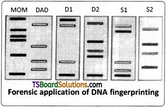
A given person can never have a VNTR which his parents do not have. Obtaining hybrid with radioactive probe and matching DNAs of different members of a family with biological children and adopted children, gives us an idea of how DNA Finger Prints help identification of paternity/maternity, by studying the ‘DNA Finger Prints’ of members of a Family – Biological and non-biological relationships.
The illustrations given below are the VNTR patterns for, Mrs. Rose (blue), Mr. Rao (yellow), and their four children : D1 (Mr. Rao’s biological daughter), D2 (Mr. Rao’s step -daughter, child of Mrs. Rose and her former husband (red), SI (Mr, Rao’s biological son), and S2 (Mr. Rao’s adopted son not biologically related, his parents’ DNA marked in light and dark green bands).
Applications of DNA Finger Printing :
- Conservation of wild life – protection of endangered species. By maintaining their DNA records for identification of tissues of the dead endangered organisms.
- Taxonomica! applications – study of phylogeny.
- Pedigree analysis – inheritance pattern of gene through generations.
- Anthropological studies-charting of origin and migration of human population.
- Medico – legal cases – establishing paternity and / or maternity more accurately.
- Forensic analysis – positive identification of a suspect in a crime.