Telangana TSBIE TS Inter 2nd Year Zoology Study Material Lesson 1(b) Breathing and Exchange of Gases Textbook Questions and Answers.
TS Inter 2nd Year Zoology Study Material Lesson 1(b) Breathing and Exchange of Gases
Very Short Answer Type Questions
Question 1.
Define vital capacity. What is its significance?
Answer:
Vital Capacity (VC) :
The maximum volume of air a person can breathe in after forced expiration. This includes ERV, TV and IRV or the maximum volume of air a person can breathe out after forced inspiration.
VC = TV + IRV + ERV
Question 2.
What is the volume of air remaining in lungs after a normal expiration?
Answer:
The volume of air that remains in the lungs after normal expiration is called Functional Residual Capacity (FRC).
FRC = ERV + RV
Question 3.
Diffusion of oxygen occurs in the alveolar region only and not in the other parts of respiratory system. How do you justify the statement?
Answer:
Alveoli are primary sites of exchange of gases in the lungs. The diffusion membrane of Alveoli is made up of 3 major layers like thin squamous epithelium of Alveolar wall, the endothelium of the alveolar capillaries and the basement material in between them. As it is a very thin border, it is favourable for diffusion of gases.
Question 4.
What is the effect of pCO2 on oxygen transport?
Answer:
The effect of pCO2 and H+ concentration on the oxygen affinity of haemoglobin is called Bohr effect. (A rise in pCO2 and fall in pH decreases the affinity of haemoglobin for oxygen. On other hand a fall in pCO2 and rise in pH increases affinity of haemoglobin for oxygen).
Question 5.
What happens to the respiratory process in a man going up a hill?
Answer:
At a height of about 6000 m the pO2 becomes almost half of what it is at the mean sea level, hence the mountain sickness in people ascending mountains.
![]()
Question 6.
What is Tidal volume? Find out the Tidal volume (approximate value) in a healthy human, in an hour?
Answer:
Volume of air inspired or expired during normal inspiration or expiration. It is approximately 500 ml. A healthy man can inhale or exhale approximately per hour is 3,60,000 -4,80,000 ml.
Question 7.
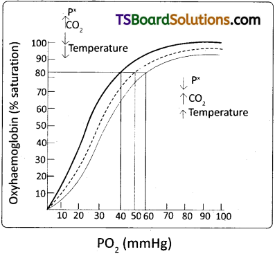
Define oxyhaemoglobin dissociation curve. Can you suggest any reason for its sigmoidal pattern?
Answer:
A sigmoid curve is obtained when percentage saturation of haemoglobin with O2 is plotted against the pO2. This curve is called oxyhaemoglobin dissociation curve. At normal condition that is on pCO2 of 40mm Hg concentration, this curve is sigmoid and normal. By increasing concentration of CO2 curve shifted towards rightside. By decreasing, concentration of CO2 curve shifted towards left side.
Question 8.
What are conchae?
Answer:
Nasal chamber of human being is divided into 3 parts namely vestibularpart, respiratory part and olfactory part. In respiratory part, it has three thin twisted bony plates called turbinals or conchae.
Question 9.
What is meant by chloride shift? [March 2014]
Answer:
The exchange of chloride and bicarbonate ions between RBC and plasma at the tissues is called chloride shift or Hamburger’s phenomenon or Hamburger’s shift.
Question 10.
Mention any two occupational respiratory disorders and their causes in human beings. [March 2018(A.P)]
Answer:
Two occupational respiratory disorders are
a) Asbestosis :
It occurs due to chronic exposure to asbestos dust in the people working in asbestos industry.
b) Black lung disease :
It is a lung disease that develops from inhalation of coal dust. It is common in long time coal mine workers.
![]()
Question 11.
Name the muscles that help in normal breathing movements.
Answer:
Normal breathing movements are aided by
- Phrenic muscles of diaphragm
- External and internal intercostal muscles of ribs.
Question 12.
Draw a diagram of oxyhaemoglobin dissociation curve.
Answer:
Short Answer Type Questions
Question 1.
Explain the process of inspiration and expiration under normal conditions? [March 2015 (T.S.)]
Answer:
Inspiration :
Intake of atmospheric air into the lungs is called inspiration. It is an active process, as it takes place by the contraction of the muscles of the diaphragm and the external inter-costal muscles, which extend in between the ribs. The contraction of the diaphragm (phrenic muscles) increases the volume of the thoracic chamber in the antero-posterior axis. The contraction of external intercostal muscles lifts up the ribs and sternum causing an increase in the volume of the thoracic chamber in the dorso – ventral axis. The overall increase in the thoracic volume causes a similar increase in the ‘pulmonary volume’. An increase in the pulmonary volume decreases the intra – pulmonary pressure to less than that of the atmosphere, which forces the air from the outside to move into the lungs, i.e., inspiration.
Expiration :
Release of alveolar air to the exterior is called expiration. It is a passive process. Relaxation of the diaphragm and the external inter – costal muscles returns the diaphragm and sternum to their normal positions, and reduces the thoracic volume and thereby the pulmonary volume. This leads to an increase in the intra – pulmonary pressure to slightly above that of the atmospheric pressure, causing the expulsion of air from the lungs, i.e., expiration.
Question 2.
What are the major transport mechanisms for CO2? Explain. [March 20191 May 2017 (A.P.)]
Answer:
Transport of Carbon Dioxide : CO2 is transported in three ways.
i) In dissolved state :
7 percent of CO2 is carried in a dissolved state (physical solution) through plasma.
CO2 + H2O → H2CO3
ii) As carbamino compounds :
About 20-25 percent of CO2 combines directly with free amino group of the haemoglobin and forms carbamino-haemoglobin in a reversible manner.
Hb – NH2 + CO2 → Hb – NHCOO– + H+
This binding of CO2 is related to the partial pressure of CO2. pO2 is a major factor which could affect this binding. When pCO2 is high and pO2 is low as in the tissues, binding of more carbon dioxide occurs. When pCO2 is low and pO2 is high as in the alveoli, dissociation of CO2 from carbamino – haemoglobin takes place, i.e., CO2 which is bound to haemoglobin from the tissues is delivered at the alveoli. Carbamino compounds are also formed by the union of CO2 with plasma proteins.
iii) As Bicarbonates: About 70 percent of CO2 is transported as bicarbonate. RBCs contain a very high concentration of the enzyme, carbonic anhydrase and a minute quantity of the same is present in the plasma too. This enzyme facilitates the following reaction in both the directions.
![]()
At the tissues where partial pressure of CO2 is high due to catabolism, CO2 diffuses into the blood (RBC and Plasma) and forms carbonic acid which dissociates into HCO–3 and H+. At the alveolar site where pCO2 is low, the reaction proceeds in the opposite direction leading to the formation of CO2and water. Thus CO2 is mostly trapped as bicarbonate at the tissues and transported to the alveoli where it is released out as CO2.
![]()
Question 3.
How is respiratory movements regulated in Man? [March 2018 (A.P); March 2014]
Answer:
Respiratory movements are regulated in Man by neural system
- A special centre present in the medulla region of brain called “Respiratory rhythm centre” is primarily responsible for this regulation.
- Another centre present in the pons of the brain stem called ‘Pneumotaxic Centre’ can moderate the functions of the ‘respiratory rhythm centre1. Neural signal from this centre can reduce the duration of inspiration and there by alter the respiratory rate.
- A chemo – sensitive area is situated adjacent to the respiratory rhythm centre which is highly sensitive to CO2 and hydrogen ions. Increase in these substances can activate this centre, which inturn can send signals to the respiratory rhythm centre to make necessary adjustments in the respiratory process by which these substances can be eliminated.
- Receptors associated with aortic arch and carotid artery also recognize changes in CO2 and H+ concentration and send necessary signals to the respiratory rhythm centre for necessary actions (increase in the rate and depth of breathing when their concentration is high). The role of oxygen in the regulation of the respiratory rhythm is quite insignificant.
4. Distinguish between
a) IRV and ERV
b) Inspiratory Capacity and Expiratory Capacity.
c) Vital capacity and Total lung capacity.
Answer:
a) IRV and EftV :
IRV is the maximum volume of air that can be inhaled during forced breathing in addition to the tidal volume. This is about 2500 ml 3000 ml. ERV is the maximum volume of air that can be exhaled during forced breathing in addition to the tidal volume. This is about 1000 ml to 1100 ml.
b) Inspiratory Capacity and Expiratory Capacity (1C):
Inspiratory Capacity :
The total volume of air a person can inhale after normal expiration. This includes the tidal volume and inspiratory reserve volume i.e., Ic = TV + IRV. It is about 3000 ml to 3500 ml.
Expiratory Capacity :
The total volume of air a person can exhale after normal inspiration. This includes tidal volume and expiratory reserve volume. TV + ERV. It is about 1500 ml to 1600 ml.
c) Vital capacity and Total lung capacity :
The maximum volume of air a person can breathe in after forced expiration is called vital capacity. This includes ERV, TV and IRV.
Total lung Capacity :
The total volume of air accommodated in the lungs at the end of forced inspiration. This includes RV, ERV, TV and IRV.
Question 5.
Describe disorders of respiratory system. [Mar. ’20, ’17, ’15 (A.P.); May ’14]
Answer:
Disorders of the Respiratory System :
i) Asthma is a difficulty in breathing caused due to inflammation of bronchi and bronchioles. It is characterized by the spasm of smooth muscles present in the walls of the bronchi and bronchioles. Symptoms include coughing, difficulty in breathing and wheezing. In the case of asthma, the allergen causes release of histamine and other inflammatory substances which cause constriction of the bronchi.
ii) Emphysema is a chronic disorder in which alveolar walls are damaged and their walls coalesce due to which respiratory surface area of exchange of gases is decreased. The lung shows larger but fewer alveoli and more fibrous and less elastic. One of the major causes of this is ‘smoking’ of tobacco.
iii) Bronchitis is the inflammation of the bronchi, resulting in the swelling of mucous lining of bronchi, increased mucus production and decrease in the diameter of bronchi. Symptoms include chronic cough with thick mucus/ sputum (phlegm).
iv) Pneumonia is infection of lungs caused by bacteria such as Streptococcus pneumoniae and also by certain viruses, fungi, protozoans and mycoplasmas. Symptoms include inflammation of lungs, accumulation of mucus in alveoli, and impaired exchange of gases, leading to death if untreated.
v) Emphysema, chronic bronchitis and asthma come under Chronic Obstructive Pulmonary Diseases (COPDs).
Occupational Respiratory disorders :
These are caused by exposure of the body to the harmful substances from certain industries, especially those involving grinding or stone breaking. Long term exposure of the body to such substances can give rise to inflammation of respiratory passage and lungs leading to several disorders.
i) Asbestosis :
It occurs due to chronic exposure to asbestos dust in the people working in asbestos industry.
ii) Silicosis :
It occurs because of long term exposure to ‘silica dust’ in the people working in mining industries, quarries etc.
iii) Siderosis :
It occurs due to deposition of iron particles in tissues. It can cause different types of siderosis such as pneumoconiosis due to inhalation of iron particles, hyperferremia and hemosiderosis (which causes recurrent alveolar hemorrhage).
iv) Black-lung disease :
It is a lung disease that develops from inhalation of coal dust. It is common in long time coal mine workers.
Long Answer Type Questions
Question 1.
Describe the respiratory system in man.
Answer:
Human Respiratory System : Respiratory system of man includes the following :
I) External nostrils (External Nares) :
A pair of external nostrils opens out above the upper lip. They lead into nasal chambers through the nasal passages.
II) Nasal Chambers :
They lie above the palate and are separated from each other by a nasal septum. Each nasal chamber can be differentiated into three parts namely, (i) vestibular part (which has hair and sebaceous glands to prevent the entry of dust particles), (ii) respiratory part (which is involved in the conditioning the temperature of inhaled air, it has three thin, twisted bony plates called turbinals / conchae) and (iii) olfactory part (which is lined by an olfactory epithelium).
III) Naso-pharynx :
Nasal chambers lead into nasopharynx through a pair of internal nostrils, located above the soft palate. Nasopharynx is a portion of the pharynx, the common chamber for the passage of food and air. Nasopharynx leads into oropharynx and opens through glottis of larynx into the trachea.
IV) Larynx :
Larynx is a cartilaginous box which helps in sound production, hence called the voice box. Wall of larynx is supported by nine cartilages. Thyroid, cricoid and epiglottis are the unpaired cartilages, whereas corniculate cartilages (cartilages of Santorini – two small conical nodules of elastic cartilage articulating with the arytenoid cartilages), arytenoids, and cuneiform cartilages are the paired cartilages. Epiglottis is a thin leaf like elastic cartilaginous flap attached to the thyroid cartilage to prevent the entry of food into the larynx through the glottis. The yellow elastic fibres which connect the thyroid and arytenoid cartilages are called vocal cords / vocal folds. The space between the true vocal cords and the arytenoids cartilages is called rima glottidis.
The mid ventral part of the thyroid cartilage forms the laryngeal prominence called Adam’s apple.
In males, the vocal cords are thicker, longer, and produce low pitch voice, where as in women and children the vocal cords are usually short and produce high pitch voice.
V) Trachea :
Trachea, the wind pipe is a straight tube extending up to the mid – thoracic cavity. The wall of the trachea is supported by ‘C’ shaped rings of hyaline cartilage. These rings are incomplete dorsally and keep the trachea always open preventing collapse. Internally the trachea is lined by pseudostratified ciliated epithelium.
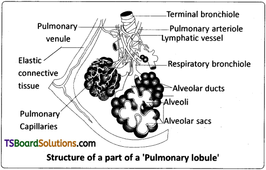
VI) Bronchi and Bronchioles :
On entering the mid thoracic cavity, trachea divides at the level of the fifth thoracic vertebra into right and left primary bronchi. Each primary bronchus enters the corresponding lung and divides into secondary bronchi that further divide into tertiary bronchi. Each tertiary bronchus divides and re – divides to form primary, secondary, tertiary terminal and respiratory bronchioles sequentially. Each respiratory bronchiole terminates in a cluster of alveolar ducts which end alveolar sacs. Bronchi and initial bronchioles re supported by incomplete cartilaginous rings. The branching network of trachea, bronchi and bronchioles constitute the’pulmonary tree’ (an upside down tree).
VII) Lungs :
Lungs occupy the greater part of the thoracic cavity. Lungs are covered by a double layered pleura, with pleural fluid between them. It reduces friction on the lung surface. The outer pleural membrane is in close contact with the thoracic lining whereas the inner pleural membrane is in contact with lung’s surface. The part starting with external nostrils up to the terminal bronchioles constitute the conducting part, whereas the alveoli and their ducts form the respiratory or exchange part of the respiratory system. The conducting part transports the atmospheric air to the alveoli, clears it from foreign particles, humidifies and also brings the inhaled air to the body temperature. Exchange part is the site of actual diffusion of and between blood and atmospheric air.
The lungs are situated in the thoracic chamber which is anatomically an air – tight chamber. It is formed dorsally by the vertebral column, ventrally the sternum, laterally by ribs and on the lower side by the dome – shaped diaphragm. The anatomical setup of lungs in the thorax is such that any change in the volume of thoracic cavity will be reflected in the lung cavity. Such an arrangement is essential for breathing, as the pulmonary volume cannot be directly altered.
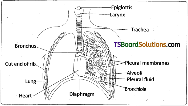
Diagrammatic view of human respiratory system
(Sectional view of the left lung is also shown)
![]()
Question 2.
Write an essay on the transport of oxygen and carbon dioxide by blood.
Answer:
Transport of gases :
Blood is the medium of transport for O2 and CO2.
I. Transport of Oxygen :
Oxygen is transported from the lungs to the tissues through the plasma and RBC of the blood. 100 ml of oxygenated blood can deliver 5 ml of o2 to the tissues under normal conditions.
i) Transport of oxygen through plasma :
About 3% of 02 is carried through the blood plasma in a dissolved state.
ii) Transport of oxygen by RBC :
About 97% of O2 is transported by the RBCs in the blood. Haemoglobin is a red coloured iron containing pigment present in the RBCs. Each haemoglobin molecule can carry a maximum of four molecules of oxygen. Binding of oxygen with haemoglobin is primarily related to the partial pressure of O2. At lungs, where the partial pressure of O2 (oxygen tension) is high, pO2 (mmHg) oxygen binds to haemoglobin (purplish – bluish-red in colour) in a reversible manner to form oxyhaemoglobin (bright red in colour). This is called oxygenation of haemoglobin.
Hb + 4O2 → Hb (O2)4
At the tissues, where the partial pressure of 02 is low, oxyhaemoglobin dissociates into haemoglobin and oxygen. The other factors that influence binding of oxygen with haemoglobin are the partial pressure of C02, the hydrogen ion concentration (pH) and the temperature.
iii) Oxygen – haemoglobin dissociation curve :
It explains the relation between percentage saturation of haemoglobin and partial pressure of oxygen. A sigmoid curve is obtained when percentage saturation of haemoglobin with O2 is plotted against the pO2. This curve is called ‘oxyhaemoglobin dissociation curve’ and is highly useful in studying the effect of factors such as pCO2, H+ concentration, temperature, etc., on the binding of O2 with haemoglobin. In the alveoli, where there is a high pO2, low pCO2, lesser H+ concentration (high pH) and lower temperature, the factors are all favourable for the formation of oxyhaemoglobin.
In the tissues where low pO2, high pCO2, high H+ concentration (low pH) and higher temperature exist, the conditions are favourable for dissociation of oxygen from oxyhaemoglobin. Under these conditions, oxygen dissociation curve shifts away from the Y – axis (to the right). The effect of pCO2 and H+ concentration on the oxygen affinity of haemoglobin is called Bohr Effect (increase of carbondioxide in the blood and decrease in pH results in the reduction of the affinity of hemoglobin for oxygen).
II. Transport of Carbon Dioxide :
CO2 is transported in three ways.
i) In dissolved state :
7 percent of CO2 is carried in a dissolved state (physical solution) through plasma.
CO2 + H2O → H2CO3
ii) As carbamino compounds :
About 20 – 25 percent of CO2 combines directly with free amino group of the haemoglobin and forms carbamino-haemoglobin in a reversible manner.
Hb – NH2 + CO2 → Hb – NHCOO– + H+
This binding of CO2 is related to the partial pressure of CO2. pO2 is a major factor which could affect this binding. When pCO2 is high and pO2 is low as in the tissues, binding of more carbon dioxide occurs. When pCO2 is low and pO2 is high as in the alveoli, dissociation of CO2 from carbamino – haemoglobin takes place, i.e., CO2 which is bound to haemoglobin from the tissues is delivered at the alveoli. Carbamino compounds are also formed by the union of CO2 with plasma proteins.
iii) As Bicarbonates :
About 70 percent of CO2 is transported as bicarbonate. RBCs contain a very high concentration of the enzyme, carbonic anhydrase and a minute quantity of the same is present in the plasma too. This enzyme facilitates the following reaction in both the directions.
![]()
At the tissues where partial pressure of CO2 is high due to catabolism, CO2 diffuses into the blood (RBC and Plasma) and forms carbonic acid which dissociates into HCO–3 and H+. At the alveolar site where pCO2 is low, the reaction proceeds in the opposite direction leading to the formation of CO2 and water. Thus CO2 is mostly trapped as bicarbonate at the tissues and transported to the alveoli where it is released out as CO2. Every 100 mL of deoxygenated blood delivers approximately 4mL of CO2 to the alveolar air.