Telangana TSBIE TS Inter 2nd Year Zoology Study Material Lesson 2(a) Body Fluids and Circulation Textbook Questions and Answers.
TS Inter 2nd Year Zoology Study Material Lesson 2(a) Body Fluids and Circulation
Very Short Answer Type Questions
Question 1.
Write the differences between ‘open’ and ‘closed’ systems of circulation.
Answer:
a) open type :
In this type, blood flows from the heart into the arteries. The arteries open into large spaces called sinuses. From sinuses, blood is carried by the veins to the heart. There are no inter connecting vessels, capillaries between arteries and veins. It is found in leeches, arthropods, molluscs, and echinoderms.
b) Closed type :
In this type blood flows through blood vessels. Blood flows from arteries to the veins through capillaries. Closed type of blood vascular system is found in annelids, cephalopods among nonchordates and all vertebrates.
Question 2.
Sino-atrial node is called the pacemaker of our heart. Why?
Answer:
Sino atrial node consists of specialized cardiomyocytes. It has the ability to generate action potentials without any external stimuli, hence called pace maker of our heart.
Question 3.
What is the significance of artrioventricular node and antrioventricular bundle in the functioning of the heart?
Answer:
Atrio ventricular node (AV node) is a relay point that relays the action potentials received from the SA node to the ventricular musculature. A bundle of nodal fibres called atrioventricular bundle (His bundle / AV bundle) continues from the AV node into the interventricular septum. The action potentials received by AVN are conducted through atrioventricular bundle causing simultaneous ventricular systole.
Question 4.
Name the valves that guard the left and right atrioventricular apertures in man. [March 2015 (T.S.)]
Answer:
The left atrio ventricular aperture is guarded by bicuspid or mitral valve. The right atrio ventricular aperture is guarded by tricuspid valve.
Question 5.
Where is the valve of Thebesius in the heart of man?
Answer:
The valve of the besius is situated where the coronary sinus opens into the right atrium of heart.
![]()
Question 6.
Name the aortic arches arising from the ventricles of the heart of man.
Answer:
The aortic arches arising from the ventricles of the heart of man are.
- Pulmonary arch whose opening is guarded by pulmonary valve.
- Systemic arch whose opening is guarded by aortic valve.
Both valves are made up of 3 semilunar flaps each.
Question 7.
Name the heart sounds. When are they produced?
Answer:
Heart sounds are named as LUB and DUP.
- The first heart sound LUB is produced when atrioventricular valves (AV valves) close preventing the back flow of blood.
- The second heart sound DUP is produced when the semilunar valves close preventing the back flow of blood.
Question 8.
Define cardiac cycle and cardiac output. [March 2020]
Answer:
- The cardiac events that occur from the beginning of one heart beat to the beginning of the next constitute a cardiac cycle.
- The volume of blood pumped out by the heart from each ventricle per minute is termed cardiac output.
Question 9.
What is meant by double circulation? What is its significance?
Answer:
In this type of circulation blood circulate twice (two times) through the heart to complete circuit. There are two circuits.
- Lesser circulation i.e., pulmonary circulation.
- Greater circulation i.e., systemic circulation. Oxygenated and deoxygenated bloods never mix.
Question 10.
Why the arteries are more elastic than the veins?
Answer:
Arteries are most elastic than the veins because arteries and arterioles have two elastic laminae one on either side of the muscle layer. Veins have one elastic lamina inner to the muscle layer. The muscle layer is much thicker in the arteries than in the veins.
Short Answer Type Questions
Question 1.
Describe the evolutionary change in the structural pattern of the heart among the vertebrates.
Answer:
In the vertebrates the principal differences in the blood vascular system involve the gradual differentiation of the heart into two separate pumps as they evolved from the gill breathing aquatic life to the lung breathing complete terrestrial life.
Fishes have a 2 – chambered heart with an atrium and a ventricle. It pumps out deoxygenated blood to gills for oxygenation, hence the name ‘branchial heart’. Blood passes through the heart only once in a complete circuit, hence called single circulation.
Amphibians have a 3 – chambered heart with two atria and one ventricle. Reptile have two atria and an incompletely divided ventricle (except in the crocodiles in which the ventricle is divided into two chambers). The left atrium receives oxygenated blood from the gills/ lungs /skin and the right atrium receives deoxygenated blood from the other parts of the body through the venae cavae. However, the two types of blood get mixed up in the single ventricle, which pumps out mixed type of blood. Thus these animals (amphibians and reptiles) show an incomplete double circulation.
Birds and mammals possess a 4 – chambered heart with two atria and two ventricles. In these animals the oxygenated and the deoxygenated types of blood received by the left and right atria passes on to the left and right ventricles, respectively. The ventricles pump the blood out without any mixing of the oxygenated and deoxygenated types of blood i.e., there are two completely separate circulatory pathways namely systemic and pulmonary circulations. Hence, these animals are said to be showing ‘double circulation’.
![]()
Question 2.
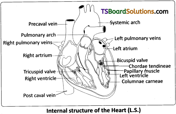
Describe atria of the heart of man.
Answer:
Atria of the heart of man :
Atria are thin walled ‘receiving chambers’ (upper chambers). The right one is larger than the left. The two atria are separated by thin inter -atrial septum. In the fetal heart, the atrial septum has a small pore called foramen ovale. Normally the foramen ovale closes at birth, when lungs become functional. It is represented by a depression in the septum between the right and left atria, called fossa ovalis (that marks the position of the foramen ovale in the fetus). If, the foramen ovale does not close properly, it is called a patent foramen ovale.
The right atrium receives deoxygenated blood from different parts of the body (except the lungs) through three caval veins viz. the two precavals (right and left) and a post caval vein. It also receives blood from the myocardium (wall of the heart) through the coronary sinus, whose opening into the right atrium is guarded by the valve of Thebesius. Opening of the postcaval vein is guarded by the valve of the inferior vena cava or Eustachian valve. It directs the blood to the left atrium through the foramen ovale, in the foetal stage, but in the adult it becomes rudimentary and non – functional. The openings of the precaval veins into the right atrium have no valves. The left atrium receives blood from each lung through two pulmonary veins, which open into the left atrium. The two left pulmonary veins open by a common aperture in some.
Atria and ventricles are separated by a membranous atrio – ventricular septum, which possesses left and right atrioventricular apertures. The left and right apertures are guarded by bicuspid (mitral valve) and tricuspid valves respectively.
Question 3.
Describe the ventricles of the heart of man.
Answer:
Ventricles :
These are the thick walled blood pumping chambers (lower chambers), separated by an interventricular septum. The wall of the left ventricle is thicker than that of the right ventricle. The inner surface of the ventricles is raised into muscular ridges or columns called columnae carneae / trabeculae carneae projecting from the inner walls of the ventricles. Some of these ridges are large and conical, and are called papillary muscles, whose apices are connected to the chordae tendineae, or ‘heart strings’. They are cord – like collagenous processes that connect the papillary muscles to the tricuspid valve and the mitral valve in the heart. They prevent the cusps of the atrioventricular valves from bulging too far into atria during ventricular systole.
Question 4.
Draw a labelled diagram of the L.S of the heart of man.
Answer:
Question 5.
Describe the events in a cardiac cycle; briefly.
Answer:
Cardiac cycle :
The cardiac cycle consists of three phases, namely atrial systole, ventricular systole and cardiac diastole.
To begin with, all the four chambers of the heart are in a relaxed state / joint diastole stage. Blood from the pulmonary veins and venae cavae flows into the respective atria. As the A – V valves are in open condition, blood flows into the left and right ventricles, through the left and right atrioventricular apertures. The semilunar valves of the pulmonary and aortic arches are closed at this stage.
Atrial systole :
The SAN now generates an action potential which stimulates both the atria to contract simultaneously causing the ‘atrial systole’. It lasts about 0. 1 sec. This increases the flow of blood into the ventricles by about 30%. It means atrial systole accounts for about 30% of the filling of the ventricles, the remaining blood flows into the ventricles before the atrial systole.
Ventricular systole :
The action potentials from the SAN reach the AVN from where they are conducted through the bundle of His, its branches and the Purkinje fibres to the entire ventricular musculature. This causes the simultaneous ventricular systole. It lasts for about 0.3 sec. The atria undergo relaxation coinciding with the ventricular systole. Ventricular systole increases the pressure causing the closure of the AV valves preventing the ‘backflow’ of blood. It results in the production of the first heart sound known as ‘Lub’. As the ventricular pressure increases further, the semilunar valves guarding the pulmonary artery and the aorta are forced open. This allows the blood in the ventricles to flow into the aortic arches and enter the circulatory pathway.
Cardiac diastole :
The ventricles now relax and the ventricular pressure falls causing the closure of the semilunar valves which prevents the back flow of blood. This results in the production of the second heart sound known as ‘Dup’. As the ventricular pressure declines further, the AV valves are pushed open by the pressure in the atria exerted by the blood, which flowed into them through the larger veins.
![]()
Question 6.
Explain the mechanism of clotting of blood.
Answer:
Mechanism of blood clotting:
Clotting takes place in three essential steps.
i) Step – 1 :
It involves the formation of a complex of activated substances collectively called, prothrombin activator. It is formed by a complex cascade of chemical reactions that occur in the blood by the involvement of clotting factors in two pathways.
a) Intrinsic pathway :
It occurs when the blood is exposed to collagen of injured wall of blood vessel. This activates Factor XII, and in turn it activates another clotting factor, which activates yet another reaction (cascade fashion) which results in the formation of the prothrombin activator.
b) Extrinsic pathway :
It occurs when the damaged vascular waH or extra vascular tissue comes into contact with blood. This activates the release of tissue thromboplastin, from the damaged tissue. It activates the Factor VII . As a result of these cascade reactions, the final product formed is the prothrombin activator.
ii) Step – 2 :
The prothrombin activator, in the presence of sufficient amounts of ionic Ca++, causes the conversion of inactive prothrombin to active thrombin (activation of prothrombin).
iii) Step – 3 :
Thrombin converts the soluble protein fibrinogen into soluble fibrin monomers, which are held together by weak hydrogen bonds. The fibrin stabilizing factor (Factor XIII, released from platelets) replaces hydrogen bonds with covalent bonds and cross links the fibres to form a ‘mesh work . The insoluble mesh work of fibrin fibers spreading in all directions adhere to the damaged surfaces and trap the blood cells and platelets.
Question 7.
Distinguish between SAN and AVN.
Answer:
A specialized cardiac musculature called the nodal tissue is also distributed in the heart. A patch of this tissue called the sinoatrial node (SAN) is present in the right upper corner of the right atrium near the openings of the superior venae cavae. Another mass of this tissue, called the atrioventricular node (AVN), is seen in the lower left corner of the right atrium close to the atrioventricular septum. A bundle of nodal fibres, called atrioventricular bundle (AV Bundle / ‘His’ bundle) continues from the AVN into the inter-ventricular septum. It divides into right and left bundle branches. These branches give rise to minute fibres called purkinje fibres that extend throughout the ventricular musculature / walls of the respective sides. SAN consists of specialized cardiomyocytes. It has the ability to generate action potentials without any external stimuli, hence called pacemaker. AV mode is a relay point.
Question 8.
Distinguish between arteries and veins.
Answer:
Differences between arteries and veins.
| arteries | Veins |
| 1) Arteries carry oxygenated blood, away from the heart (except the pulmonary artery). | 1) Veins carry deoxygenated blood, towards the heart (except the pulmonary veins). |
| 2) These are bright red in colour. | 2) These are dark red in colour. |
| 3) These are mostly deep seated in the body. | 3) Veins are generally superficial. |
| 4) Arteries are thick – walled as the tunica media is relatively thick, with elastin and smooth muscles. | 4) Veins are thin walled (tunica media is relatively thin with few elastin fibres) and slightly muscular. |
| 5) Lumen is narrow | 5) Lumen is wide |
| 6) Non – valvular. | 6) Valvular |
| 7) Blood in the arteries flows with more pressure and by jerks. | 7) Blood in the veins flows steadily with relatively low pressure |
| 8) Arteries end in capillaries. | 8) Veins start with capillaries. |
Long Answer Type Questions
Question 1.
Describe the structure of the heart of man with the help of neat labelled diagram. [March 2018 (A.P.); March 2014; May/June ’14]
Answer:
The heart is mesodermal in origin. It is a thick walled, muscular and pulsating organ, situated in the mediastinum (the region in the thorax between the two lungs), and with its apex slighty turned to the left. It is the size of a clinched fist.
The heart is covered by a double walled pericardium which consists of the outer fibrous pericardium and the inner serous pericardium is double – layered, formed of an outer parietal layer and an inner visceral layer. The parietal layer is fused with the fibrous pericardium, whereas the visceral layer adheres to the surface of the heart and forms its outer layer, the epicardium. The two layers are separated by a narrow pericardial space, which is filled with the pericardial fluid. This fluid reduces friction between the two membranes and allows free movement of the heart.
The wall of the heart consists of three layers. They are the outer epicardium, the middle myocardium (a thick layer of cardiac muscles), and the inner most endocardium (a thin layer of endothelium). The endothelium covers the heart valves also and is continuous with the endothelial lining of the large blood vessels connected to the heart.
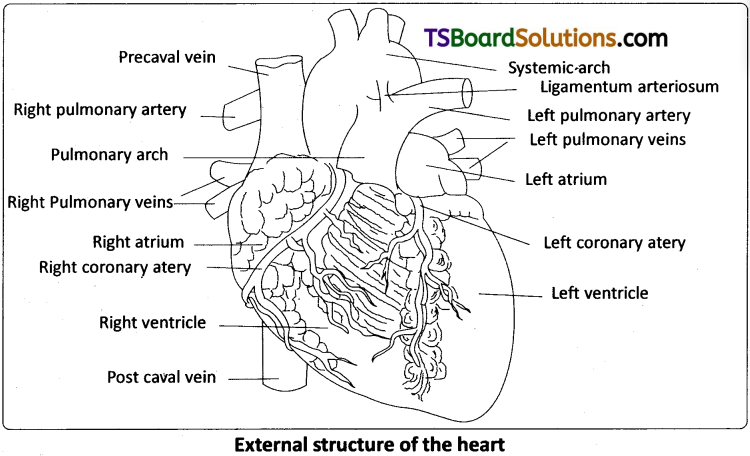
External structure:
Human heart has four chambers, with two relatively smaller upper chambers, called atria and two larger lower chambers called ventricles. Atria and ventricles are separated by a deep transverse groove called coronary sulcus (atrio – ventricular groove). The muscular pouch like projection from each atrium is called auricular appendix (auricular appendage). The ventricles are separated by two inter ventricular grooves (anterior and posterior), in which the coronary arteries and their branches are lodged.
Internal structure:
i) Atria :
Atria are thin walled ‘receiving chambers’ (upper chambers). The right one is larger than the left. The two atria are separated by thin inter-atrial septum. In the fetal heart, the atrial septum has a small pore called foramen ovale. Normally the foramen ovale closes at birth, when lungs become functional. It is represented by a depression in the septum between the right and left atria, called fossa ovalis (that marks the position of the foramen ovale in the fetus). If, the foramen ovale does not close properly, it is called a patent foramen ovale.
The right atrium receives deoxygenated blood from different parts of the body (except the lungs) through three caval veins viz. the two precavals (right and left) and a post caval vein. It also receives blood from the myocardium (wall of the heart) through the coronary sinus, whose opening into the right atrium is guarded by the valve of Thebesius. Opening of the postcaval vein is guarded by the valve of the inferior vena cava or Eustachian valve. It directs the blood to the left atrium through the foramen ovale, in the foetal stage, but in the adult it becomes rudimentary and non – functional. The openings of the precaval veins into the right atrium have no valves. The left atrium receives blood from each lung through two pulmonary veins, which open into the left atrium. The two left pulmonary veins open by a common aperture in some.
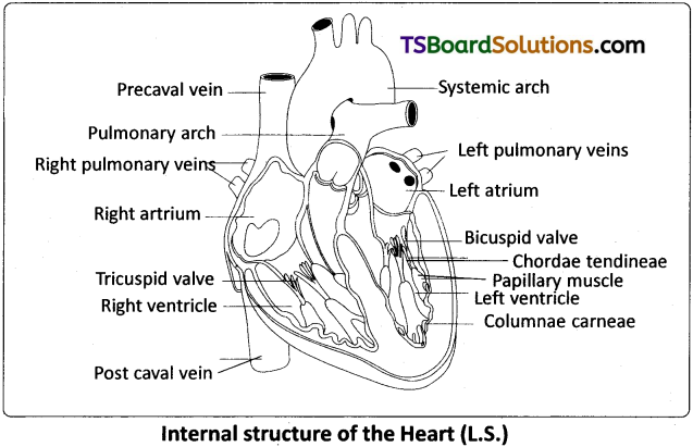
Atria and ventricles are separated by a membranous atrio – ventricular septum, which possesses left and right atrioventricular apertures. The left and right apertures are guarded by bicuspid (mitral valve) and tricuspid valves respectively.
ii) Ventricles :
These are the thick walled blood pumping chambers (lower chambers), separated by an interventricular septum. The wall of the left ventricle is thicker than that of the right ventricle. The inner surface of the ventricles is raised into muscular ridges or columns called columnae carneae / trabeculae carneae projecting from the inner walls of the ventricles. Some of these ridges are large and conical, and are called papillary muscles, whose apices are connected to the chordae tendineae, or ‘heart strings’. They are cord – like collagenous processes that connect the papillary muscles to the tricuspid valve and the mitral valve in the heart. They prevent the cusps of the atrioventricular valves from bulging too far into atria during ventricular systole.
![]()
Question 2.
Write notes on the working of the heart of man.
Answer:
The cardiac events that occur from the beginning of one heart beat to the beginning of the next constitute a cardiac cycle. This cardiac cycle consists of three phases, namely atrial systole, ventricular systole and cardiac diastole.
To begin with, all the fpur chambers of the heart are in a relaxed state / joint diastole stage. Blood from the pulmonary veins and venae cavae flows into the respective atria. As the A – V valves are in open condition, blood flows into the left and right ventricles, through the left and right atrioventricular apertures. The semilunar valves of the pulmonary and aortic arches are closed at this stage.
Atrial systole :
The SAN now generates an action potential which stimulates both the atria to contract simultaneously causing the ‘atrial systole’. It lasts about 0. 1 sec. This increases the flow of blood into the ventricles by about 30%. It means atrial systole accounts for about 30% of the filling of the ventricles, the remaining blood flows into the ventricles before the atrial systole.
Ventricular systole :
The action potentials from the SAN reach the AVN from where they are conducted through the bundle of His, its branches and the Purkinje fibres to the entire ventricular musculature. This causes the simultaneous ventricular systole. It lasts for about 0.3 sec. The atria undergo relaxation coinciding with the ventricular systole. Ventricular systole increases the pressure causing the closure of the AV valves preventing the ‘backflow’ of blood. It results in the production of the first heart sound known as ‘Lub’. As the ventricular pressure increases further, the semilunar valves guarding the pulmonary artery and the aorta are forced open. This allows the blood in the ventricles to flow into the aortic arches and enter the circulatory pathway.
Cardiac diastole :
The ventricles now relax and the ventricular pressure falls causing the closure of the semilunar valves which prevents the back flow of blood. This result in the production of the second heart sound known as ‘Dup’. As the ventricular pressure declines further, the AV valves are pushed open by the pressure in the atria exerted by the blood, which flowed into them through the larger veins. The blood now once again flows freely into the ventricles. All the heart chambers are now again in a relaxed state (joint diastolic phase). Soon, another cardiac cycle sets in.
Cardiac output :
The volume of blood pumped out by each ventricle, for each heart beat, is known as the stroke volume. The volume of blood pumped out by the heart from each ventricle per minute is termed cardiac output.
Cardiac output = Stroke volume × No. of beats per minute = 70 ml / beat × 72 beats / minute = 5040 ml/ min. or approximately 5 litres.