Telangana TSBIE TS Inter 2nd Year Zoology Study Material Lesson 2(b) Excretory Products and their Elimination Textbook Questions and Answers.
TS Inter 2nd Year Zoology Study Material Lesson 2(b) Excretory Products and their Elimination
Very Short Answer Type Questions
Question 1.
Name the blood vessels that enter and exit the kidney. [May 2017 (A.P.)]
Answer:
Renal artery enters the kidney and renal vein comes out of kidney.
Question 2.
What are renal pyramids and renal papillae?
Answer:
The medulla of kidney is divided into multiple cone shaped masses of tissue called renal pyramids. The base of each pyramid originates at the border between cortex and medulla and terminates in the renal papilla.
Question 3.
What are the columns of Bertin? [March 2019, 2015 (A.P.)]
Answer:
The renal pyramids are separated by the projections of the cortex called columns of Bertin (renal column).
Question 4.
Name the structural and functional unit of kidney. What are the two main types of structural units in it?
Answer:
Structural and functional unit of kidney is nephron. Each nephron has two types of structural units a) Bowman’s capsule and 2) the renal tubcile.
Question 5.
Distinguish between cortical and juxta medullary nephrons.
Answer:
- In a majority of nephrons, the loops of Henle is too short and extends only very little into the medulla. Such nephrons are called Cortical nephrons.
- In some of the nephrons, the loops of Henle are very long and run deep into the medulla. They are called Juxta medullary nephrons.
![]()
Question 6.
Define glomerular filtration. [Mar. ’14; May/June ’14; Mar. ’17 (A.P.)]
Answer:
Filtration of the blood from the glomerulus into the lumen of the Bowman’s capsule and this passive process is called glomerular filtration.
Question 7.
Define Glomerular Filtration Rate (GFR).
Answer:
The amount of filtrate formed by both the kidneys per minute is called Glomerular Filtration Rate (GFR).
Question 8.
What is meant by mandatory reabsorption? In which parts of nephron does it occur?
Answer:
About 85% of the filtrate formed is reabsorbed in a constant, unregulated fashion by the proximal convoluted tubule and descending limb of Henle’s loop. This is called obligatory or Mandatory reabsorption.
Question 9.
Distinguish between juxta glomerular cells and macula densa.
Answer:
- Juxta glomerular cells are present in Juxta glomerular apparatus where the afferent arteriole comes into contact with DCT. These are modified smooth muscle cells of the afferent arteriole.
- A group of modified epithelial cells of DCT which comes in contact with afferent arteriole are crowded in this region constitute macula densa.
Macula densa together with JG cells form the JGA.
Question 10.
What is juxta glomerular apparatus? [March 2018 (A.P.)]
Answer:
The juxta Glomerular Aoparatus plays a complex regulating role. The JGA is the region in each nephron where the afferent arteriole comes into contact with DCT. Macula densa together with JG cells from the JGA.
![]()
Question 11.
Distinguish between the enzymes renin and rennin.
Answer:
- A fall in glomerular blood flow / glomerular blood pressure / GFR can activate the JG cells to release an enzyme called renin into the blood. This enzyme catalyses the conversion of angiotensinogen.
- Rennin is a digestive enzyme produced by gastric glands that converts milk into curd in infants. ‘
Question 12.
What is meant by the term osmoregulation?
Answer:
The process of maintaining the quantity of water and dissolved solutes in balance is referred to as osmoregulation.
Question 13.
What is the role of atrial natriuretic peptide in the regulation of urine formation?
Answer:
An increase in the flow of blood to the right atrium of the heart stretches its wall. It causes the release of atrial natriuretic peptide (ANP). It causes vasodilation and there by decrease the blood pressure. ANP mechanism therefore, acts as a counter check on the RAAS.
Short Answer Type Questions
Question 1.
Terrestrial animals are generally either ureotelic or uricotelic and not ammonotelic. Why?
Answer:
Aquatic animals excrete ammonia (ammonotelic) through body surface, gill surface etc. by diffuse. They can send ammonia through dilute urine as they can afford to lose much water. But that is not the case with terrestrial animals which may be excreting urea (ureotelic) or uric acid (uricotelic) water is the most important constituent of protoplasm, the living substance. Dehydration kills a person much faster than lack of food. Terrestrial adaptation necessitated the production of lesser toxic nitrogenous wastes such as urea and uric acid, for the conservation of water. Hence terrestrial animals are generally either ureotelic or uricotelic and not ammonotelic.
![]()
Question 2.
Differentiate vertebrates on the basis of the nitrogenous waste products they excrete, giving examples.
Answer:
Based on the nitrogenous waste products vertebrates are differentiated into 3 categories.
1. Ammonotelic animals :
Excrete nitrogenous Wastes in the form of ammonia, e.g.: Hydra, some bony fishes.
2. Ureotelic animals :
Excrete nitrogenous wastes in the form of urea, e.g.: Cartilaginous fishes, amphibians and mammals.
3. Uricotelic animals :
Excrete nitrogenous wastes in the form of uric acid, e.g.: Insects, reptiles and birds.
Question 3.
Draw a labelled diagram of the V.S. of Kidney.
Answer:
Question 4.
Describe the internal structure of kidney of man.
Answer:
Internal structure of Kidney :
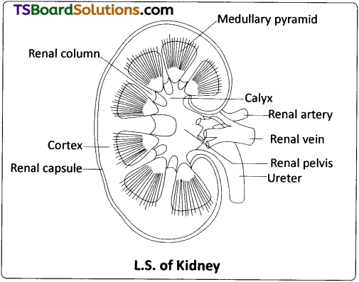
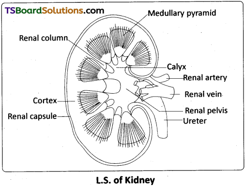
A longitudinal section of the human kidney shows two distinct regions, the outer cortex and the inner medulla. The medulla is divided into multiple cone shaped masses of tissue called renal pyramids. The renal pyramids are separated by the projections of the cortex called columns of Bertin (renal column). The base of each pyramid originates at the border between the cortex and the medulla and terminates in the renal papilla. Renal papillae project into cup like calyces, formed by the funnel shaped pelvis, which continues out as the ureter.
Question 5.
Explain micturition.
Answer:
Micturition :
Urine formed by the nephrons is ultimately carried to the urinary bladder where it is stored till a voluntary signal is given by the central nervous system (CNS). This signal is initiated by the stretching of the urinary bladder as it gets filled with urine. In response, the stretch receptors on the walls of the bladder send signals to the CNS. The CNS passes on motor messages to initiate the contraction of smooth muscles of the bladder and simultaneous relaxation of the urethral sphincter, causing the release of urine. The process of passing out urine is called micturition and the neural mechanism involved is called ‘micturition reflex’.
Question 6.
What is the significance ofJuxta Glomerular Apparatus (JGA) in Kidney function?
Answer:
The Juxta Glomerular Apparatus plays a complex regulating role. The JGA is the region in each nephron where the afferent arteriole comes into contact with the DCT. A group of modified epithelial cells of the DCT are crowded in this region, constituting the macula densa. The wall of the afferent renal arteriole has JG cells (they are modified smooth muscle cells of the afferent arteriole). Macula densa together with JG cells form the JGA.
A fall in glomerular blood flow / glomerular blood pressure / GFR can activate the JG cells to release an enzyme called renin into the blood. This enzyme catalyses the conversion of angiotensinogen (a protein produced by the liver) into angiotensin I, which is converted into angiotensin II, by angiotensin converting enzyme (ACE). This conversion occurs primarily as blood passes through the capillaries of the lungs, where most of the converting enzyme is present. Angiotensin II stimulates the adrenal cortex to secrete aldosterone. Aldosterone causes reabsorption of Na+ and water from the DCT and CD to reduce loss through urine, and also promotes secretion of K+ ions into the DCT and CD. It leads to an increase in the blood pressure and GFR. This complex mechanism is generally known as renin – angiotensin – aldosterone system (RAAS).
An increase in the flow of blood to the right atrium of the heart stretches its wall. It causes the release of atrial natriuretic factor (ANF) / atrial natriuretic peptide (ANP). ANP can cause vasodilation (dilation of blood vessels) and there by decrease the blood pressure. ‘ANP’ mechanism therefore, acts as a counter check on the ‘RAAS’.
Question 7.
Give a brief account of the counter current mechanism.
Answer:
Mammals have the ability to produce concentrated urine. The Henle’s loop and vasa recta play a significant role in this. The flow of the renal filtrate in the two limbs of Henle’s loop is in opposite directions and thus forms a counter current. The flow of blood through the two limbs of vasa recta is also in a counter current pattern. The proximity between the Henle’s loop and vasa recta, as well as the counter currents of renal fluid and blood in them help in maintaining an increasing osmolarity towards the inner medullary interstitium. i.e., from 300 mOsml/ L in the cortex to about 120 mOsml/L in the inner medulla.
This gradient is mainly caused by NaCI and urea. NaCI passes out of the ascending limb of Henle’s loop, and it enters the blood of the descending limb of vasa recta. NaCI is returned to the interstitium from the ascending portion of the vasa recta. Similarly, small amounts of urea enter the thin segment of the ascending limb of Henle’s loop which is transported back to the interstitium, from the collecting duct.
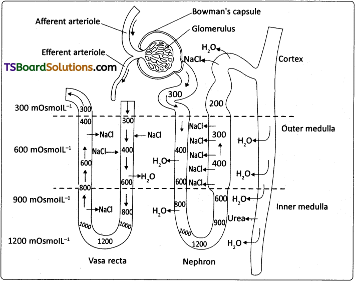
Diagrammatic representation of a nephron and vasa recta showing counter current mechanisms
The above described transport of substances facilitated by the special arrangement of Henle’s loop and vasa recta is called the countercurrent mechanism (the two limbs of the loop of Henle constitute a counter current multiplier system). This mechanism helps to maintain a concentration gradient in the medullary interstitium. Presence of such interstitial gradient helps easy passage of water from the collecting duet, thereby concentrating the filtrate (urine). Human kidneys can produce urine nearly four times concentrated than the initial filtrate formed.
Question 8.
Explain the auto regulatory mechanism of GFR.
Answer:
The tubular epithelial cells in different segments of a nephron reabsorb certain substances of the glomerular filtrate either by active or passive mechanisms. About 85% of the filtrate formed is reabsorbed in a constant, unregulated fashion by the PCT and descending limb of Henle’s loop (obligatory or mandatory reabsorption) and the reabsorption of the rest of the fluid is “regulated”. Based on the necessity of re- absorption, the substances of glomerular filtrate can be categorized into ‘high threshold substances’ (essential and are efficiently reabsorbed e.g. glucose, amino acids, vitamins, some salts etc.,) ‘low threshold substances’ (absorbed in very little amounts e.g. urea, uric acid etc.) or ‘athreshold substances’ (actual excretory products and are not reabsorbed at all e.g. creatinine).
During the formation of urine, the tubular cells secrete substances such as H+, K+ and NH3 into the filtrate. Tubular secretion is also an important step in the formation of urine as it helps in the maintenance of ionic and acid – base balance of the body fluids.
![]()
Question 9.
Describe the role of liver, lungs and skin in excretion.
Answer:
In addition to the kidneys, lungs, liver and skin also help in the elimination of excretory wastes.
a) Lungs :
Lungs regularly eliminat about 18L of C02 and also significant amount of water per day in the form of water vapour in normal resting condition. The quantity of water loss increases in dry climates. Various volatile materials are also eliminated through the lungs.
b) Liver :
Liver is the largest gland in our body. It changes the decomposed haemoglobin of the worn-out RBCs into bile pigments, namely, bilirubin and biliverdin. These pigments pass into the alimentary canal along with the bile for elimination. The liver also excretes cholesterol, degraded steroid hormones, certain vitamins and drugs via bile.
c) Skin :
Human skin possesses two types of glands for the elimination of certain substances through their secretion.
- Sweat glands secrete a watery fluid called sweat. Primary function of sweat is to facilitate a cooling effect on the body surface. It also helps in the removal of some of the wastes like NaCI, small amounts of urea, lactic acid etc.
- Sebaceous glands eliminate certain substances like sterols, hydrocarbons, waxes through sebum. This secretion provides a protective ‘oily covering’ to the skin.
Question 10.
Name the following:
a) A chordate animal having protonephridial type excretory structure.
b) Cortical portions projecting between the medullary pyramids in the human kidney.
c) Capillary network paralleling the loop of Henle.
d) A non-chordate animal having green lands as excretory structure.
Answer:
a) A chordate animal having protonephridial type excretory structures is Lancelet (with solenocytes) or Amphioxus.
b) Cortical portions projecting between the medullary pyramids in the human kidney are columns of Bertin.
c) Capillary network paralleling the loop of Henle is vaserecta.
d) A non-chordate animal having green glands as excretory structures is crustaceans like prawn & crab.
Long Answer Type Questions
Question 1.
Describe the excretory system of man, giving the structure of a nephron. [March 2015 (T.S.)]
Answer:
Human Excretory System :
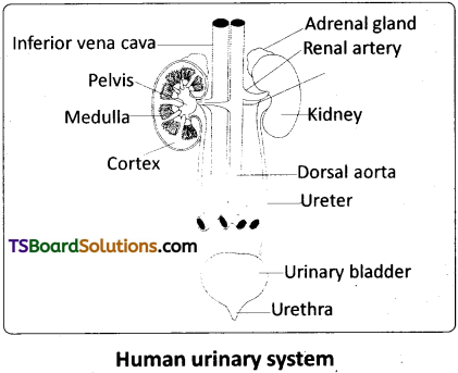
In humans, the excretory system consists of a pair of kidneys, a pair of ureters, a urinary bladder and urethra.
Kidneys :
Kidneys are reddish brown, bean shaped structures, situated on either side of the vertebral column between the levels of the last thoracic and third lumbar vertebrae, in a ‘retroperitoneal position’. The right kidney is slightly lower than the left one due to the presence of liver.
The outer surface of the kidney is convex and the inner surface has a deep notch called hilum, the point at which the renal artery and nerves enter and the renal vein and ureter leave. Each kidney is surrounded by a tough, fibrous capsule that protects its delicate inner surface.
Internal structure:
A longitudinal section of the human kidney shows two distinct regions, the outer cortex and the inner medulla. The medulla is divided into multiple cone shaped masses of tissue called renal pyramids. The renal pyramids are separated by the projections of the cortex called columns of Bertin (renal column). The base of each pyramid originates at the border between the cortex and the medulla and terminates in the renal papilla. Renal papillae project into cup like calyces, formed by the funnel shaped pelvis, which continues out as the ureter.
Ureters :
These are slender whitish tubes which emerge from the pelvis of the kidneys. Their walls are lined by ‘transitional epithelium’. The ureters run down wards and open into the urinary bladder.
Urinary bladder :
It is a median storage sac, situated in the lower abdominal cavity. It has thick, muscular, distensible wall lined by ‘transitional epithelium’. The neck of the bladder leads into the urethra, which has an internal urethral sphincter (made of smooth muscles) and external urethral sphincter (made of striped muscles). Urethra opens near the vaginal orifice in the female and through penis in the males.
Structure of a Nephron :
Each kidney has nearly one million nephrons which are the ‘structural’ and ‘functional’ units. Each nephron has two parts – the ‘Bowman’s capsule’ and the renal tubule. The Bowmans’ capsule encloses a tuft of capillaries called glomerulus, formed by the afferent renal arteriole – a fine branch of the renal artery. Blood from the glomerulus is carried away by an efferent renal arteriole of a lesser diameter. The blind end of the tubule forms a double walled cup called Bowman’s capsule, which surrounds the glomerulus.
The inner wall of the Bowman’s capsule has certain unique cells called podocytes which wrap around each capillary. The podocytes are arranged in an intricate manner so as to leave some minute spaces called ‘filtration slits’ or ‘slit pores’. The endothelial cells of the capillaries have numerous pores or ‘fenestrations’. The glomerulus along with the Bowman’s capsule constitutes the Malpighian body or renal corpuscle.
The tubule continues further and forms a highly coiled proximal convoluted tubule (PCT). A hairpin shaped Henle’s loop, which has descending and ascending limbs, is the next part of the tubule. The proximal part of the ascending limb is thin and the distal part is thick . The thick ascending limb continues into the distal convoluted tubule (DCT). The DCT continues as the ‘initial collecting duct’ in the cortex. Some initial collecting ducts unite to form a straight collecting duct, which passes through the medullary pyramid. In the medulla, the tubes of each pyramid join and form the duct of Bellini, which finally opens on the tip of the renal papilla. The contents of the duct of Bellini are discharged into the renal pelvis through the renal calyx.
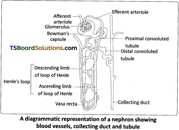
A diagrammatic representation of a nephron showing blood vessels, collecting duct and tubule
The Malpighian corpuscle, PCT and DCT of a nephron are situated in the cortical region of the kidney, whereas the loop of Henle is in the medulla. In a majority of nephrons, the loop of Henle is too short and extends only very little into the medulla. Such nephrons are called cortical nephrons. In some of the nephrons, the loops of Henle are very long and run deep into the medulla. These nephrons are called juxtamedullary nephrons.
The efferent arteriole emerging from the glomerulus forms a fine capillary network called the peritubular capillaries, around the renal tubule. The portion of the peritubular capillaries that surrounds the loop of Henle is called the vasa recta. The vasa recta is absent or highly reduced in the cortical nephrons. The juxtamedullary nephrons possess well developed vasa recta.
![]()
Question 2.
Explain the physiology of urine formation. [March 2020]
Answer:
Urine Formation :
The formation of urine involves three main processes namely, glomerular filtration, selective reabsorption and tubular secretion.
a) Glomerular filtration :
The first step in the formation of urine is the ‘filtration’ of the blood from the glomerulus into the lumen of the Bowman’s capsule and this ‘passive’ (non -energy consuming process) process is called glomerular filtration. The hydrostatic pressure of the blood while flowing in the glomerulus is 60 mm Hg. It is opposed by ‘glomerular colloidal osmotic pressure’ of 32 mm Hg (which is exerted by the non-filtered plasma proteins of the blood in the glomerular capillaries) and Bowman’s capsular hydrostatic pressure of 18mm Hg. The net filtration pressure is 10mm Hg (60 – 32 + 18 = 10). This causes the filtration of blood through the 3 layered filtrate membrane formed by the endothelial cells of glomerular capillary together with the basement membrane and podocytes of the Bowman’s cup.
Blood is filtered through the fine slit pores and fenestrations due to the NFP. Therefore, this process is called ‘ultrafiltration’. The filtrate contains almost all the constituents of the plasma, except the proteins. The filtrate thus formed is called ultra – filtrate or ‘glomerular filtrate’ or ‘primary urine’, which is hypotonic to the cortical fluid. It passes into the next part of .the renal tubule.
b) Selective reabsorption and secretion :
The tubular epithelial cells in different segments of a nephron reabsorb certain substances of the glomerular filtrate either by active or passive mechanisms. About 85% of the filtrate formed is reabsorbed in a constant, unregulated fashion by the PCT and descending limb of Henle’s loop (obligatory or mandatory reabsorption) and the reabsorption of the rest of the fluid is ‘regulated’. Based on the necessity of re-absorption, the substances of glomerular filtrate can be categorized into ‘high threshold substances’ (essential and are efficiently reabsorbed e.g. glucose, amino acids, vitamins, some salts etc.), ‘low threshold substances’ (absorbed in very little amounts e.g. urea, uric acid etc), or ‘athreshold substances’ (actual excretory products and are not reabsorbed at all e.g. creatinine).
During the formation of urine, the tubular cells secrete substances such as H+, K+ and NH3 into the filtrate. Tubular secretion is also an important step in the formation of urine as it helps in the maintenance of ionic and acid-base balance of the body fluids. Mechanism of selective reabsorption and secretion in different parts of a nephron takes place as follows.
i) In the proximal convoluted tubule :
PCT is lined by simple cuboidal epithelium with ‘brush border’, which increases the surface area of absorption. Nearly all the essential nutrients and 70-80% of electrolytes and water are reabsorbed by this segment. Na+ is actively transported into the cortical interstitial fluid. This transfer of positive charge drives the passive transport of Cl–. Glucose, amino acids, and other essential substances are also ‘actively’ transported. Movement of water occurs by ‘osmosis’.
PCT also helps to maintain the pH and ionic balance of the body fluids by selective secretion of hydrogen ions, and ammonia into the filtrate and by the absorption of HCO–3 from it.
ii) In the Henle’s loop :
Reabsorption in this segment is minimum. However, this region plays a significant role in the maintenance of high osmolarity of the medullary interstitial fluid.
The descending limb of loop of Henle is permeable to water and almost impermeable to electrolytes, hence reabsorption of water continues as the filtrate moves along the descending limb (passive transport). As a result, the filtrate concentration gradually increases as it moves towards the inner medulla. The ascending limb has two specialized regions, a proximal thin segment, in which NaCI diffuses out into the interstitial fluid passively, and a distal thick segment, in which NaCI is actively pumped out. The ascending limb is impermeable to water. Thus the filtrate becomes progressively more dilute as it moves up to the cortex (towards the DCT).
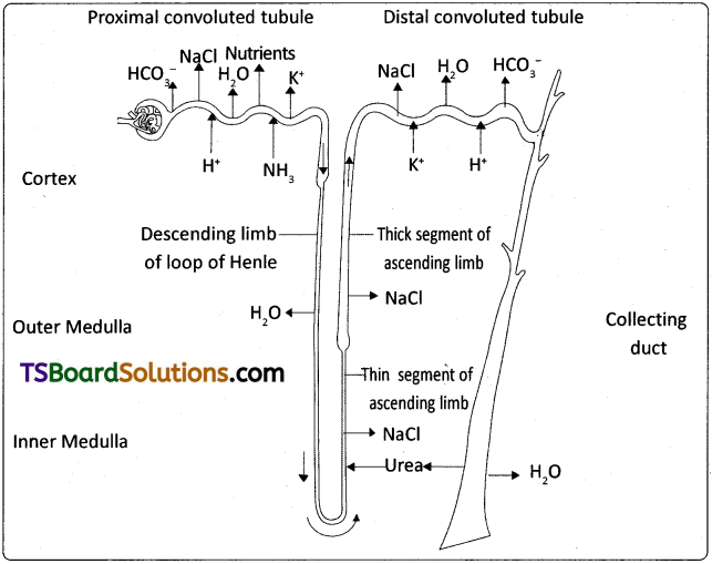
Reabsorption and secretion of major substances at different parts of the nephron (arrows indicate direction of movement of materials)
iii) In the distal convoluted tubule (DCT) :
The cells here are shorter than those in the proximal tubule and lack ‘microvilli’, indicating that they are not involved much in reabsorption ‘conditional reabsorption’ /’facultative reabsorption’ of Na+ and water takes place in this segment. The reabsorption of water is variable depending on several conditions and is regulated by ADH. DCT is also capable of reabsorption of HCO–3 and selective secretion of H+ and K+ ions and NH3 into the DCT from the peritubular network, to maintain the pH and sodium – potassium balance in the blood.
iv) In the collecting duct (CD) :
This long duct carries the filtrate through the medulla to the renal pelvis. Considerable amount of water could be reabsorbed from the region to produce concentrated urine. This segment allows passage of small amount of urea to the medullary interstitum to keep up its osmolarity. It also plays a role in the maintenance of pH and ionic balance of blood by the selective secretion of H+ and K+ ions. The renal fluid after the process of facultative reabsorption in the CD, influenced by ADH, constitutes the ‘urine’, that is sent out. Urine in the CD is hypertonic to the plasma of blood.