Telangana TSBIE TS Inter 2nd Year Zoology Study Material Lesson 3(a) Musculo-Skeletal System Textbook Questions and Answers.
TS Inter 2nd Year Zoology Study Material Lesson 3(a) Musculo-Skeletal System
Very Short Answer Type Questions
Question 1.
What is a ‘motor unit’ with reference to muscle and nerve?
Answer:
A motor neuron and the set of muscle fibres innervated by all the telodendrites constitute a motor unit.
Question 2.
What is triad system? [Mar. ’14; May/June ’14; Mar. ’15 (T.S.)]
Answer:
T- tubule and the two terminal cisternae at its sides form the triad system in a skeletal muscle fibre.
Question 3.
Write the difference between actin and myosin. [Mar. 2019, ’15, May ’17 (A.P.)]
Answer:
a) The light band in a myofibril contains actin and two regulatory proteins called troponin and tropomyosin. Actin filaments are thinner compared to myosin filaments.
b) The dark band in a myofibril contains (A band) myosin. Myosin filaments are thick and non-contractile.
Question 4.
Distinguish between red muscle fibers and white muscle fibers. [March 2018 (A.P.)]
Answer:
a) Red muscle fibres :
Myoglobin of these fibres is high which give a reddish appearance. Such muscle fibres are called red fibres. They also contain plenty of mitochondria.
b) White muscle fibres :
Some of the muscle fibres possess very less quantity of myoglobin in their muscle fibers and therefore appear pale or whitish. Hence called white muscle fibers.
Question 5.
Name two cranial sutures and their locations.
Answer:
1) Coronal suture :
Parietal bones that form major portion of sides and roof of cranial cavity are joined to the frontal bone by coronal suture.
2) lambdoid suture :
Parietal bones are posteriorly joined to the occiput by lambdoid suture.
![]()
Question 6.
Name the keystone bone of the cranium. Where is it located? [May 2017 (A.P.) March 2014]
Answer:
Sphenoid bone is called the keystone bone. It is present at the middle part of the base of the skull. It is named keystone bone because it articulates with all other cranial bones.
Question 7.
Human skull is described as dicondylic skull. Give the reason.
Answer:
In human skull two occipital condyles are present one on each side of the foramen magnum hence called dicondylic skull.
Question 8.
Name the ear ossicles and their evolutionary origin in human beings.
Answer:
Each middle ear contains 3 tiny bones – Malleus (modification of articular), Incus (modified quadrate) and Stapes (modified hyomandibula) collectively called as ear ossicles.
Question 9.
Name the type of joint between a) atlas/ axis b) carpal / metacarpal of the human thumb.
Answer:
1) Type of joint between atlas and axis is Pivot joint.
2) Type of joint between carpal / metacarpal is Condyloid joint.
Question 10.
Name the type of joint between a) atlanto – axial joint b) Femur-acetabulum joint.
Answer:
1) Type of joint between atlanto – axial joint is Pivot joint.
2) Type of joint between Femur – acetabulum is Ball and socket joint.
Question 11.
Name the type of joint between a) Cranial bones b) Inter – tarsal joint.
Answer:
- Joint between cranial bones is fibrous joint called Suture.
- Inter tarsal joint is Gliding joint.
Short Answer Type Questions
Question 1.
Write a short note on sliding filament theory of muscle contraction.
Answer:
Sliding Filament Hypothesis :
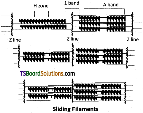
The sliding filament hypothesis of muscle contraction was put forth by Hugh Huxley and Hanson. According to their hypothesis, the contraction of sarcomere depends on the presence of two sets of filaments. During muscle contraction, thin filament slide over the thick filaments resulting in shortening of sarcomere. A series of events takes place during this process like, a) Stimulation of muscle b) Contraction of muscle c) Relaxation of muscle.
![]()
Question 2.
Describe the important steps in muscle contraction.
Answer:
Important steps in muscle contraction are
i) Excitation or stimulation of muscle :
Muscle contraction is initiated by a signal sent by the central nervous system (CNS) via a motor neuron. A neural signal reaching the neuromuscular junction releases a neurotransmitter (acetylcholine) which generates an ‘action potential’ in the sarcolemma. When the action potential spreads to the triad system through the T tubules, the cisternae of the sarcoplasmic reticulum release calcium ions into the sarcoplasm.
ii) Formation of Cross bridges :
Increase in the Ca2+ level leads to the binding of calcium ions to the subunit Tn – C of the troponin of the thin filaments. This makes troponin and tropomyosin complex to move away from the active sites of action molecules. Now, the active sites are exposed to the heads of the myosin. Utilizing the energy released from hydrolysis of ATP, the myosin head now binds to the exposed ‘active sites’ on the actin molecules to form a cross bridge and P1 is released.
iii) Power Stroke :
The cross bridge pulls the attached actin filaments towards the centre of the ‘A’ band. The ‘Z’ lines attached to these actin filaments are also pulled inwards from both the sides, thereby causing shortening of the sarcomere, i.e., contraction. During the shortening of the muscle, the T bands get reduced in size / length (Z membranes of the sarcomere are brought closer) whereas the’A’ bands retain their size / length. It is important to note that myofilaments do not actually shorten. As the thin filaments are pulled deep in to the A bands making the H bands narrow, the muscle shows the effect contraction.
Question 3.
Describe the structure of a skeletal muscle.
Answer:
Structure of a skeletal Muscle :
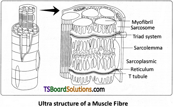
Let us examine a skeletal muscle in detail to understand the structure and mechanism of contraction. Each organised skeletal muscle in our body is made of a number of muscle bundles or fascicles. Each fascicle contains a number of cylindrical muscle fibers. The fascicles are held together by a common collagenous connective tissue layer called fascia. Each muscle fibre is lined by the plasma membrane called sarcolemma enclosing the sarcoplasm. Skeletal muscle fibre is a ‘syncytium’, as each fibre is formed by fusion of embryonic, mononucleate ‘myoblasts’.
Hence, the skeletal muscle cells are multinucleate, with characteristically peripheral nuclei (just below the sarcolemma). The endoplasmic reticulum, also called sarcoplasmic reticulum of the muscle fibers is the store house of calcium ions. A characteristic feature of the muscle fiber is the presence of a large number of parallel filaments called myofilaments or myofibrils, in the sarcoplasm.
Question 4.
Write short notes on contractile proteins.
Answer:
Structure of Contractile Proteins :
Each actin (thin) filament is made of two ‘F’ (filamentous) actin molecules helically wound around each other. Each ‘F’ actin is a polymer of monomeric ‘G’ (globular) actin molecules. Two filaments of another protein, called tropomyosin also run close to the ‘F’ actin molecules, throughout their length. A complex protein called ‘troponin’ is distributed at regular intervals on the tropomyosin.
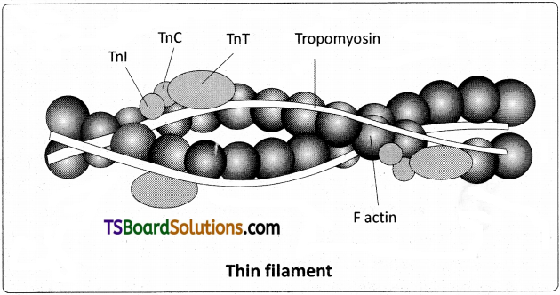
Troponin is made of three polypeptide units named Tn – T, Tn – I, and Tn – C. Tn – T binds to tropomyosin. Troponin – I (Tn – I), inhibits the myosin binding site on the actin. Tn – C can bind to Ca2+. When calcium ions are not bound to troponin (Tn – C), it stabilizes tropomyosin in its blocking position over the active sites of actin. When Calcium ions attach to the Tn – C of the troponin, the tropomyosin moves away/ is pulled away from the ‘active sites’ allowing the myosin heads to bind to the active sites of actin. Troponin and tropomyosin are often called ‘regulatory proteins’, because of their role in masking and unmasking the active sites.
Question 5.
Draw a neat labelled diagram of the Ultrastructure of muscle fibre.
Answer:
Question 6.
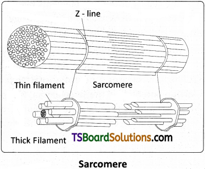
Draw the diagram of a Sarcomere of skeletal muscle showing different regions.
Answer:
Question 7.
What is Cori’s cycle – explain the process.
Answer:
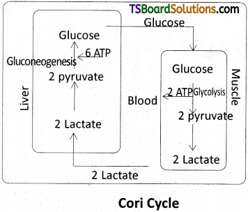
Cori cycle :
The lactic acid produced during rapid contractions of skeletal muscles under low availability of oxygen is partly oxidized and a major part of it is carried to the liver by the blood, where it is converted into pyruvic acid (pyruvate) and then to glucose through gluconeogenesis. The glucose can enter the blood and be carried to muscles and immediately used. If, by this time the muscles have stopped contraction, the glucose can be used to rebuild reserve of glycogen through glycogenesis. This two way traffic between skeletal muscle and liver is called the Cori cycle.
Question 8.
List out the bones of the human cranium. .
Answer:
Cranium, the brain box, is formed by eight flattened bones. They are a) frontal bone (1), b) Parietals (2), c) Temporal bones (2), d) occipital bone (1), e) spenoid bone (1) f) Ethmoid bone (1)
i) Frontal bone :
It forms the forehead, anterior part of the cranial floor, and the roof of the orbits.
ii) Parietal bones :
They form the major portion of the sides and roof of the cranial cavity. They are joined to the frontal bone by a coronal suture and posteriorly to the occiput by lambdoid suture.
iii) Temporal bones :
They form the lateral parts and the floor of the cranium.
iv) Occipital bone :
It forms the posterior part and most of the base of the cranium. It has a large opening, the foramen magnum. Medulla oblongata passes out through this foramen and joins the spinal cord. Two occipital condyles are present one on each side of the foramen magnum (dicondylic skull).
v) Sphenoid bone :
It is present at the middle part of the base of the skull. It is the keystone bone of the cranium because it articulates with all the other cranial bones.
vi) Ethmoid bone :
It is present on the midline of the anterior part of the cranial floor.
![]()
Question 9.
Write short notes on the ribs of human being. [March 2020]
Answer:
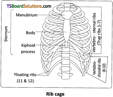
Ribs :
Twelve pairs of ribs are present in the human chest. Each rib is a thin flat bone connected dorsally to the vertebral column and ventrally to the sternum. It has two articulation surfaces on its dorsal end, hence called bicephalic. The first seven pairs of ribs are called true ribs (vertebro – sternal ribs). Dorsally they are attached to the thoracic vertebrae and ventrally connected to the sternum with the help of hyaline cartilages. The remaining ribs are called ‘false ribs’. The 8th, 9th and 10th pairs of ribs do not articulate directly with the sternum but join the cartilaginous (hyaline cartilage) parts of the seventh rib. These are called vertebro – chondral (false ribs’) ribs. Last 2 pairs (11th and 12th) of ribs are not connected ventrally either to the sternum or the anterior ribs, hence called floating ribs. The thoracic vertebrae, ribs and sternum together form the rib cage.
Question 10.
List the bones of human fore limb.
Answer:
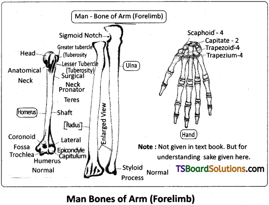
Bones of the Fore limb :
Each limb is made of 30 bones. The bones of the hand (forelimb) are humerus, radius and ulna, carpals (wrist bones – 8), metacarpals (palm bones – 5) and phalanges (digits -14).
Question 11.
List the bones of the human leg.
Answer:
Bones of the human leg : The bones of the leg are Femur (1) (thing bone – the longest bone), tibia (1) and fibula (1), tarsals (ankle bones – 7), metatarsals (5) and phalanges (digits – 14) are the bones of the legs (hind limbs). A cup shaped bone called patella (knee cap) (1) covers the knee joint ventrally.
Question 12.
Draw a neat labelled diagram of the skeleton of the fore limb of man.
Answer:
Question 13.
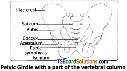
Draw a neat labeled diagram of pelvic girdle.
Answer:
Question 14.
Describe the structure of synovial joint with the help of a neat labelled diagram. [Mar. 2019, ’17 (A.P.); May/June; Mar.’14]
Answer:
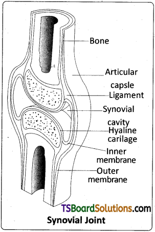
Synovial joint is covered by a double layered synovial capsule. The outer layer consists of dense fibrous irregular connective tissue with more collagen fibres. This layer is continuous with the periosteum and resists stretching and prevents the dislocation of joints. Some fibres of these membranes are arranged in bundles called ligaments. The inner layer of synovial capsule is formed of areolar tissue and elastic fibers. It secretes a viscous synovial fluid which contains hyaluronic acid, phagocytes etc., and acts as a ‘lubricant1 for the free movement of the joints. Synovial joints include Ball and socket joint. Hinge joint, Pivot joint, Gliding joint, Condyloid joint, Saddle joint.
Long Answer Type Questions
Question 1.
Explain the mechanism of Muscle contraction.
Answer:
Mechanism of Muscle contraction :
Mechanism of muscle contraction is best explained by the ‘Sliding Filament Theory’. It states that contraction of a muscle fibre takes piace by the sliding of the thin filaments over / in between the thick filaments. It was proposed by Jean Hanson and Hugh Huxley. The process of muscle contraction can be studied under the following heads.
i) Excitation of muscle :
Muscle contraction is initiated by a signal sent by the central nervous system (CNS) via a motor neuron. A neural signal reaching the neuromuscular junction releases a neurotransmitter (acetylcholine) which generates an ‘action potential’ in the sarcolemma. When the action potential spreads to the triad system through the T tubules, the cisternae of the sarcoplasmic reticulum release calcium ions into the sarcoplasm.
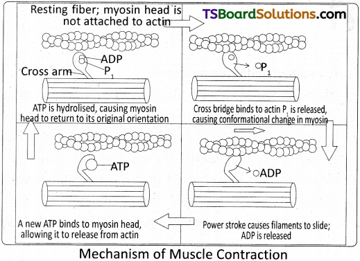
ii) Formation of Cross bridges :
Increase in the Ca2+ level leads to the binding of calcium ions to the subunit Tn – C of the troponin of the thin filaments. This makes troponin and tropomyosin complex to move away from the active sites of actin molecules. Now, the active sites are exposed to the heads of the myosin. Utilizing the energy released from hydrolysis of ATP, the myosin head now binds to the exposed ‘active sites’ on the actin molecules to form a cross bridge and P1 is released.
iii) Power Stroke :
The cross bridge pulls the attached actin filaments towards the centre of the ‘A’ band. The ‘Z’ lines attached to these actin, filaments are also pulls inwards from both the sides, thereby causing shortening of the sarcomere, i. e., contraction. During the shortening of the muscle, the T bands get reduced in size / length (Z membranes of the sarcomere are brought closer), whereas the ‘A’ bands retain their size / length. It is important to note that myofilaments do not actually shorten. As the thin filaments are pulled deep in to the A bands making the H bands narrow, the muscle shows the effect contraction.
iv) Recovery Stroke :
The myosin, goes back to its relaxed state and releases ADP. A new ATP moelcule binds to the head of myosin and the cross bridge is broken. The new ATP is again hydrolysed by the ATPase of the myosin head and the cycle of cross bridge formation, and breakage is repeated causing further sliding.
v) Relaxation of Muscle :
When motor impulses stop the Ca2+ ions are pumped back into the sarcoplasmic cisternae. It results in the masking of the active sites of the actin filaments. The myosin heads fail to bind with the active sites ofactin. These changes cause the return of ‘Z’ lines backtotheiroriginal position, i.e., relaxation.
![]()
Question 2.
List, in sequence, the events that take place during muscle contraction.
Answer:
[Refer the answer of above question and also add the following matter.]
For the contraction of muscle, continuous supply of energy is needed. ATP is the immediate source of energy for muscle contraction. As the ATP content is very low, it is actively replenished, continuously, by an energy rich muscle phosphagen.
ATP → ADP + Pi
The high energy phosphates of muscles that donate energy phosphate group to ADP are known as phosphagen. In vertebrate muscles creatine phsophate (CP) is the phsophagen, which is an immediate backup source. In invertebrate muscles, it is in the arginine phosphate. This reaction is catalysed by creatine kinase (CK). Creatine Phosphate is the immediate additional source of energy in the muscle.
Creatine phosphate + ADP → Creatine
When creatine phosphate gets exhausted, the next source of reserve energy is utilised, which includes the oxidation of glucose and fatty acids. The energy liberated in this process is transferred to ADP and creatine. Thus ATP and creatine phosphate are formed and which in turn supply energy for muscle contraction.
During rapid activity of a muscle, the respiratory system is unable to supply sufficient oxygen needed by it, which leads to oxygen debt. It is defined as the amount of extra oxygen required by a muscle during recovery from vigorous exercise. Thus the pyruvic acid produced by glycolysis is transformed into lactic acid in the absence of oxygen. Accumulation of lactic acid in the muscle leads to muscle fatigue.
Lactic acid, formed in the anaerobic degradation in the muscle, reaches the liver through blood circulation. In liver, during rest, 80% of lactic acid is utilised in the resynthesis of glycogen, which is transported back to muscle. This is known as Cori cycle. 20% of lactic acid is oxidised as CO2 and H2O.
![]()
Question 3.
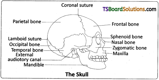
Describe the structure of human skull.
Answer:
The skull :
It is composed of two sets of bones – cranial and facial bones (22 bones in all). Cranium, the brain box, is formed by eight flattened bones. They are a) frontal bone (1), b) Parietals (2), c) Temporal bones (2), d) Occipital bone (1), e) Sphenoid bone (1) and f) Ethmoid bone (1).
i) Frontal bone :
It forms the forehead, anterior part of the cranial floor and the roof of the orbits.
ii) Parietal bones :
They form the major portion of the sides and roof of the cranial cavity. They are joined to the frontal bone by a coronal suture and posteriorly to the occiput by lambdoid suture.
iii) Temporal bones :
They form the lateral parts and the floor of the cranium.
iv) Occipital bone :
It forms the posterior part and most of the base of the cranium. It has a large opening, the foramen magnum. Medulla oblongata passes out through this foramen and joins the spinal cord. Two occipital condyles are present one on each side of the foramen magnum (dicondylic skull).
v) Sphenoid bone :
It is present at the middle part of the base of the skull. It is the keystone bone of the cranium because it articulates with all the other cranial bones.
vi) Ethmoid bone :
It is present on the midline of the anterior part of the cranial floor.
A) The facial region is made up of 14 skeletal elements which form the front part of the skull. The bones of the facial skeleton are the nasals (2), the maxillae (2), the zygomatic bones (2) the mondible (1) the lacrimal bones (2), the palatine bones (2), inferior nasal conchae (2), and the vomer (1)
i) Nasal bones :
These are paired bones that from the bridge of the nose.
ii) Maxilla :
Two maxillae join together and form the upper jaw. The maxilla bears sockets (alveoli) for lodging the maxillary teeth. The palatine process is involved in the formation of the hard palate.
iii) Zygomatic bones :
These are known as a cheek bones.
iv) Lacrimal bones :
These are the smallest bones of the face.
v) Palatine bones :
They form the posterior portion of the hard palate.
vi) Nasal conchae :
These are scroll like bones that form a part of lateral wall of the nasal cavity. Nasal conchae are 3 pairs, namely superior, middle and inferior.
vii) Vomers :
It is a triangular bone present on the floor of nasal cavity.
viii) Mandible (Lower jaw) :
It is ‘U’ shaped and is the longest and strongest of all the facial bones. It is the only movable skull bone (except the ear ossicles).
B) Skeletal structures associated with sense organs:
i) The nasal cavity is divided into left and right cavities by a vertical partition called the nasal septum.
ii) Orbits : Orbits are bony depressions which accommodate the eyeballs and associated structures.
iii) Ear Ossicles : Each middle ear contains three tiny bones – Malleus (modification of articular), Incus (modified quadrate) and Stapes (modified hyomandibula), collectively called ear ossicles.
C) Hyoid Bone :
It is a single U shaped bone present at the base of the buccal cavity between the larynx and the mandible. The hyoid bone keeps the larynx open.