Telangana TSBIE TS Inter 2nd Year Zoology Study Material Lesson 5(a) Human Reproductive System Textbook Questions and Answers.
TS Inter 2nd Year Zoology Study Material Lesson 5(a) Human Reproductive System
Very Short Answer Type Questions
Question 1.
Where are the testes located in man? Name the protective coverings of each testis.
Answer:
Testes are located outside the abdomen with in a pouch called scrotum. Each testis is enclosed in a fibrous envelope, the tunica albuginea.
Question 2.
Name the canals that connect the cavities of scrotal sac and abdominal cavity. Name the structures that keep the testes in their position.
Answer:
The cavity of scrotal sac is connected to the abdominal cavity through the inguinal canal. Structure that keeps testis in their position is guberhaculum.
Question 3.
What are the functions of Sertoli cells of the seminiferous tubules and the Leydig cells in man? [Mar. ’15 (A.P. & T.S.)]
Answer:
Sertoli cells :
Nourishes the growing sperms and also produce a hormone called inhibin.
Leidig cells :
Present in seminiferous tubules produce and rogens, the most important of which is testosterone.
Question 4.
Name the copulatory structure of man. What are the three columns of tissues in it?
Answer:
The copulatory structure of man is penis. Three columns of tissues are two upper corpora cavernosa and one lower (ventral) corpora spongiosum.
Question 5.
Define spermiogenesis and spermiation.
Answer:
Development of spermatozoa from sperm mother cells in male is called spermiogenesis. After spermiogenesis sperm heads become embedded in the Sertoli cells and are finally released from the seminiferous tubules by the process called spermiation.
![]()
Question 6.
Name the yellow mass of cells accumulated in the empty follicle after ovulation. Name the hormone secreted by it and what is its function? [March 2020]
Answer:
After ovulation, the granulosa cells in the follicle proliferate and are transformed into a yellowish glandular mass called corpus luteum. It secretes progesterone hormone. This hormone is essential for maintenance of pregnancy in first few months.
Question 7.
Define gestation period. What is the duration of gestation period in the human beings?
Answer:
Intra uterine development of the embryo or foetus is called gestation period. In human being gestation period is 266 days or 38 weeks.
Question 8.
What is implantation; with reference to embryo?
Answer:
Attachment of blastocyst to the uterine mucosa till the whole of it comes to lie with in the thickness of the endometrium. This is called interstitial implantation. In human the implantation beings on the 6th day after fertilization.
Question 9.
Distinguish between epiblast and hypoblast
Answer:
The mature oocyte lies eccentrically in the follicle surrounded by some surface facing the cavity. This cells layer develops into the hypoblast, which is the future extra embryonic endoderm. The remaining part of the embryonic disc is called epiblast.
Question 10.
Write two major functions, each of testis and ovary.
Answer:
Major functions of Produce
a) Testes :
Produce spermatozoa for fertilisation produce hormones which induce secondary sexual characters of males.
b) Ovary :
Produce mature ova for fertilisation. Produce before and after fertilisation estrogens and progesterone hormones.
![]()
Question 11.
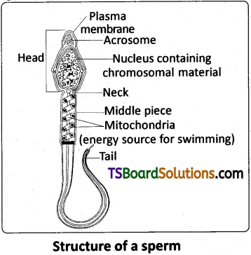
Draw a labelled diagram of a sperm.
Answer:
Question 12.
What are the major components of the seminal fluid?
Answer:
Seminal fluid is alkaline, viscous fluid. 60% of the volume of seminal fluid is constituted by secretion of the seminal vesicle. Seminal fluid contains fructose, proteins, citric acid, inorganic phosphorus, potassium and prostaglandins. Prostate secretion contributes 15 – 30 percent of semen.
Question 13.
What is menstrual cycle? Which hormones regulate menstrual cycle?
Answer:
The reproductive cycle in the female primates is called menstrual cycle. The cyclic changes that occur in the endometrium every month are together called menstrual cycle.
LH and FSH (Gonadotropic) from pituitary, estrogens from ovarian follicle, progesterone from corpus luteum regulate menstrual cycle.
Question 14.
What is parturition? Which homones are involved in inducing parturition?
Answer:
The process of delivery of the foetus (child birth) is called parturition. Parturition is induced by oxytocin.
Question 15.
How many eggs do you think were released by the ovary of a female dog which gave birth to six puppies?
Answer:
Only six (6) ova or eggs are released by the ovary of a femaleidog which gave birth to six puppies.
![]()
Question 16.
What is neurulation?
Answer:
The neural plate invaginates towards the notochord to form a neural groove, which deepens progressively to form a tube by fusion of the lateral neural folds. The process of formation of neural tube is referred to as NEURULATION.
Question 17.
What is capacitation of sperms?
Answer:
Spermatozoa acquire the ability to fertilize the ovum only after they undergo some changes in the female genital tract. These changes are called capacitation.
Question 18.
What is compaction in the human development? [March 2015 (A.P.)]
Answer:
In the morula due to unequal cleavage smaller and larger blastomeres are formed. The morula passes through a process called compaction. Now the embryo has a superficial flat cell layer and inner cell mass. Inner cell mass gives rise to the embryo proper. This is the first sign of cell differentiation in the human embryo.
Question 19.
Distinguish between involution and ingression in the human development.
Answer:
a) Involution :
The inward growth and curling inward of a group of cells (prospective mesodermal cells), as in the formation of a gastrula from a blastula is called involution.
b) Ingression :
The inward migration of future endodermal cells from the epiblast during gastrulation is called ingression.
![]()
Question 20.
What are the four extra embryonic membranes?
Answer:
The four extra embryonic membranes are Amnion, Chorion, Allantois and Yolk Sac.
Short Answer Type Questions
Question 1.
Describe microscopic structure of testis of man.
Answer:
Each testis is enclosed in a fibrous envelope the tunica albuginea which extends inward to form septa that partition the testis into lobules. There are about 250 testicular lobules in each testes. Each lobule contains 1-3 highly coiled seminiferous tubules. A pouch of serous membrane (peritoneal layer) called tunica vaginalis covers the testis.
Each seminiferous tubule is lined by the germinal epithelium which consists of undifferentiated male germ cells called spermatogonial mother cells and it also bears ‘nourishing cells’ called Sertoli cells. The spermatogonia produce the primary spermatocytes which undergo meiotic division, finally leading to the formation of spermatozoa or sperms (spermatogenesis). Sertoli cells provide nutrition to the spermatozoa and also produce a hormone called inhibin, which inhibits the secretion of FSH.
The regions outside the seminiferous tubules, called interstitial spaces, contain interstitial cells of Leydig or leydig cells. Leydig cells produce androgens, the most important of which is testosterone. Testosterone controls the development of secondary sexual characters and spermatogenesis. Other immunologically competent cells are also present. The seminiferous tubules open into the vasa efferentia through the rete testis (a network of tubules in of the testis carrying spermatozoaTrom the seminiferous tubules to the vasa efferentia).
Question 2.
Describe the microscopic structure of ovary of woman.
Answer:
The ovaries are covered on the outside by a layer of simple cuboidal epithelium called germinal (ovarian) epithelium. This is actually the visceral peritoneum that envelops the ovaries. Underneath this layer there is a dense connective tissue capsule, the tunica albuginea. The ovarian stroma is distinctly divided into an outer cortex and an inner medulla. The cortex appears more dense and granular due to the presence of numerous ovarian follicles in various stages of development. The medulla is a loose connective tissue with abundant blood vessels, lymphatic vessels, and nerve fibers.
![]()
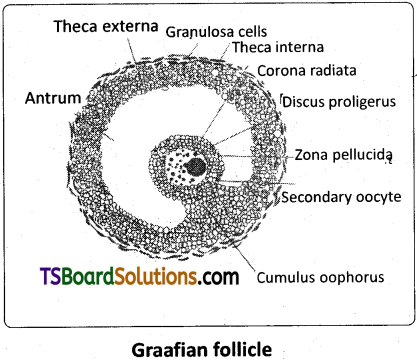
Question 3.
Describe the Graafian follicle in woman.
Answer:
A homogenous membrane, the zona pellucida, appears between the primary oocyte and granulosa cells. The innermost layer of granulosa cells are firmly attached to zona pellucida forming the corona radiata.
A cavity (antrum) appears within the membrana granulosa. The follicular cavity increases in size. As a result, the wall of the follicle becomes relatively thin. The oocyte now lies eccentrically in the follicle surrounded by some granulosa cells. It is called cumulus oophorus. As the follicle expands the stromal cells surrounding the membrana granulosa become condensed to form a covering called the theca interna. Outside the theca interna some fibrous tissue becomes condensed to form another covering called theca externa. Now these follicles are called secondary follicles.
The cells of theca interna later secrete a hormone called oestrogen. At this stage, the primary oocyte within the secondary follicle grows in size and completes Meiosis (.It is an unequal division resulting in the formation of a large haploid secondary oocyte and a tiny first polar body (haploid). The secondary oocyte retains bulk of the cytoplasm (nutrient rich) of the primary oocyte. Then the second meiotic division begins, but stops at metaphase. The secondary follicle further changes into the mature follicle called Graafian follicle.
Question 4.
Draw a labelled diagram of the female reproductive system.
Answer:
Question 5.
Diagrammatic sectional view of the female reproductive system
Answer:
Question 6.
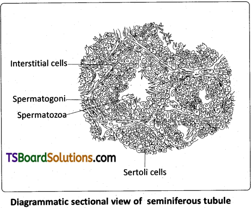
Describe the structure of seminiferous tubule.
Answer:
Each testis is enclosed in a fibrous envelope the tunica albuginea which extends inward to form septa that partition the testis into lobules. There are about 250 testicular lobules in each testes. Each lobule contains 1-3 highly coiled seminiferous tubules. A pouch of serous membrane (peritoneal layer) called tunica vaginalis covers the testis.
Each seminiferous tubule is lined by the germinal epithelium which consists of undifferentiated male germ cells called spermatogonial mother cells and it also bears ‘nourishing cells’ called Sertoli cells. The spermatogonia produce the primary spermatocytes which undergo meiotic division, finally leading to the formation of spermatozoa or sperms (spermatogenesis). Sertoli cells provide nutrition to the spermatozoa and also produce a hormone called inhibin, which inhibits the secretion of FSH.
The regions outside the seminiferous tubules, called interstitial spaces, contain interstitial cells of Leydig or leydig cells. Leydig cells produce androgens, the most important of which is testosterone. Testosterone controls the development of secondary sexual characters and spermatogenesis. Other immunologically competent cells are also present. The seminiferous tubules open into the vasa efferentia through the rete testis (a network of tubules in of the testis carrying spermatozoaTrom the seminiferous tubules to the vasa efferentia).
![]()
Question 7.
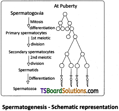
What is spermatogenesis? Briefly describe the process of spermatogenesis in man.
Answer:
Spermatogenesis :
In the testis, the immature male germ cells, spermatogonia produce sperms by spermatogenesis that begins at puberty. The spermatogonial stem cells (present in the seminiferous tubules) multiply by mitotic divisions and increase in numbers. Each spermatogonial stem cells is diploid and contains 46 chromosomes. Some of the spermatogonial stem cells develop into primary spermatocytes which undergo meiosis periodically. A primary spermatocyte completes the first meiotic division (Meiosis -1) leading to formation of two equal sized, haploid cells called secondary spermatocytes, Which have only 23 chromosomes each. The secondary spermatocytes undergo the second meiotic division (Meiosis – II) to produce four equal sized haploid spermatids.
The spermatids are transformed in to spermatozoa (sperms) by the process called spermiogenesis. After spermiogenesis, sperm heads become embedded in the Sertoli cells, and are finally released from the seminiferous tubules by the process called spermiation. Spermatogenesis starts at the age of puberty due to significant increase in the secretion of gonadotropin releasing hormone (GnRH). LH acts on the Leydig cells and stimulates secretion of androgens. Androgens, in turn, stimulate the process of spermatogenesis.
Question 8.
What is oogenesis? Give a brief account of oogenesis in a woman.
Answer:
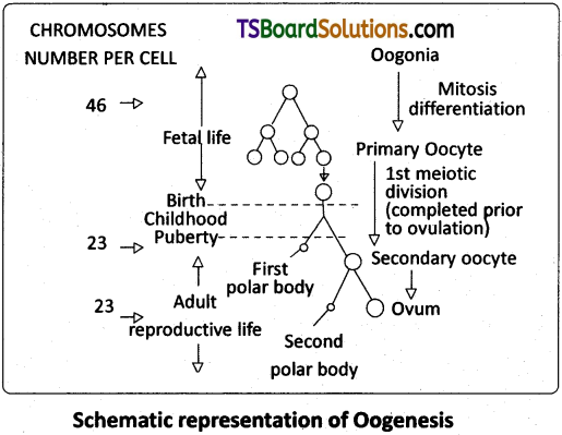
Oogenesis :
The process of formation of a mature female gamete is called oogenesis. Oogenesis is initiated during the embryonic development stage when a couple of million gamete mother cells (oogonia) are formed within each foetal ovary and do not multiply thereafter. These cells start division and stop the process at prophase -1 of the meiosis – I At this stage these are called primary oocytes.
Question 9.
Draw a labelled diagram of a Graafian follicle.
Answer:
Question 10.
In our society women are often blamed for giving birth to daughters. Can you explain why this is not correct?
Answer:
One has to remember that the sex of the baby has been decided at the time of fertilization itself. Let us see how? As we know the chromosome pattern in the human female is XX and that in the male is XY. Therefore, all the haploid gametes produced by the female (ova) have the sex chromosome X, whereas the male gametes (sperms) have either X- chromosome or Y-chromosome (50 percent of sperms carry the X-chromosome while the other 50 percent carry the Y chromosome).
After fusion of the male and female gametes the zygote would carry either XX or XY depending on what type of sperm fertilised the ovum. The zygote carrying ‘XX’ would develop into a female child and that with ‘XY’ would form a male child. So, the sex of a child depends on the male parent, (heterogametic parent). So it is not correct to blame women for giving birth to daughter very often.
Question 11.
Describe the accessory glands associated with male reproductive system of man.
Answer:
Male accessory genital glands :
The male accessory glands include paired seminal vesicles, a prostate and bulbourethal glands.
Seminal vesicles :
The seminal vesicles are a pair of simple tubular glands present postero inferior to the urinary bladder in the pelvis. Each seminal vesicle opens into the corresponding vas deferens, as the vas deferens enters the prostate gland. The secretion of the seminal vesicles constitutes about 60 percent of «.ne volume of seminal fluid. It is an alkaline, viscous fluid that contains fructose, proteins, citric acid, inorganic phosphorous, potassium, and prostaglandins. Once this fluid joins the sperm in the ejaculatory duct, fructose acts as the main energy source for the sperm outside the body. Prostaglandins are believed to aid fertilization by causing the mucous lining of the cervix to be more receptive to sperm as well as by aiding the movement of the sperm towards the ovum with peristaltic contractions of the uterus and fallopian tubes.
Prostate gland :
Prostate gland is located directly beneath the urinary bladder. The gland surrounds the prostatic urethra, and sends its secretions through several prostatic ducts. In man, the prostate contributes 15 – 30 percent of the semen. The fluid from the prostate is clear and slightly acidic. The prostatic secretion ‘activates’ the spermatozoa and provides nutrition.
Bulbourethral Glands :
Bulbourethral glands, also called Cowper’s glands, are located beneath the prostate gland at the beginning of the internal portion of the penis. They add an alkaline fluid to semen during the process of ejaculation. The fluid secreted by these glands lubricates the urethra. It is also throught to function as a ‘flushing agent’ that washes out the acidic urinary residues that remain in the urethra, before the semen is ejaculated.
![]()
Question 12.
Describe the placenta in a woman.
Answer:
Placenta :
After implantation, finger – like projections appear on the trophoblast called chorionic villi which are surrounded by the uterine tissue. The chorionic villi and uterine tissue become interdigitated with each other and jointly form a structural and functional unit called placenta between the developing embryo (foetus) and the mother. The maternal and foetal blood do not mix with each other. They are separated by the placental membranes.
The placenta consists of two essential portions :
a maternal part of the placenta derived from the endometrium of the uterus, and foetal membranes of the foetal part of the placenta. The maternal components of the placenta are : Uterine epithelium, Uterine connective tissue and Uterine capillary endothelium. The foetal components of the placenta are foetal capillary endothelium, foetal connective tissue and foetal chorionic epithelium.
The placenta of humans is called chorioallantoic placenta as allantois also fuses with he chorion in the nrocess of vascularisation. Placenta is discoidal as the villi are restricted to the dorsal surface of the blastodisc. Placenta is haemochorial as the maternal blood comes into direct contact with the foetal chorion. During parturition the placenta is cast of with loss of embryonic membranes and the encapsulating maternal tissues (decidua) causing extensive haemorrhage and thereby bleeding. So, it is also called as deciduate placenta.
Functions of Placenta :
The placenta facilitates the supply of oxygen and nutrients to the embryo and also removal of carbon dioxide and excretory / waste materials produced by the embryo. The placenta is connected to thl embryo through an umbilical cord which helps in the transport of substances to and from the embryo.
Long Answer Type Questions
Question 1.
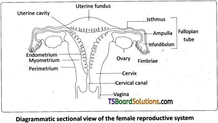
Describe female reproductive system of a woman with the help of a labelled diagram. [May 2017 (A.P.): Mar. ’15 (A.P. & T.S.)]
Answer:
The female reproductive system consists of a pair of ovaries along with a pair of oviducts, uterus, vagina and the extemaPgenitalia located in the pelvic region. These parts of the system along with a pair of the mammary glajids are integrated structurally and functionally to support the processes of ovulation, fertilization, pregnancy, birth and child care.
Ovaries :
Ovaries are the primary female sex organs that produce the female gametes (ova) and several steroid hormones (ovarian hormones). A pair of ovaries is located one on each side of the lower abdomen. The double layered fold of peritoneum connecting the ovary with the wall of the abdominal cavity is known as the mesovarium.
The ovaries are covered on the outside by a layer of simple cuboidal epithelium called germinal (ovarian) epithelium. This is actually the visceral peritoneum that envelops the ovaries. Underneath this layer there is a dense connective tissue capsule, the tunica albuginea. The ovarian stroma is distinctly divided into an outer cortex and an inner medulla. The cortex appears more dense and granular due to the presence of numerous ovarian follicle in various stages of development. The medulla is a loose connective tissue with abundant blood vessels, lymphatic vessels, and nerve fibers.
Fallopian tubes (Oviducts) :
Each fallopian tube extends from the periphery of each ovary to the uterus, and it bears a funnel shaped infundibulum. The edges of the infundibulum possess finger like projections called fimbriae, which help in collection of the ovum after ‘ovulation1. The infundibulum leads to a wider part of the oviduct called ampulla. The last part of the oviduct, isthmus has a narrow lumen and it joins the uterus. Fallopian tube is the site of fertilization. It conducts the ovum or zygote towards the uterus by peristalsis. The fallopian tube is attached to the abdominal wall by a peritoneal fold called mesosalpinx.
Uterus :
The uterus is single and it is also called womb. It is a large, muscular, highly vascular and inverted pear shaped structure present‘in the pelvis between the bladder and the rectum. The uterus is connected to the abdominal wall by the peritoneal fold called mesometrium. The lower, narrow part through which the uterus opens into the vagina is called the cervix. The cavity of the cervix is called cervical canal which along with vagina forms the birth canal.
The wall of the uterus has three layers of tissue. The external thin membranous perimetrium, the middle thick layer of smooth muscle called myometrium and inner glandular lining layer called endometrium. The endometrium undergoes cyclic changes during menstrual cycle while the myometrium exhibits strong contractions during parturition.
Vagina :
The vagina is a large, median, fibro – muscular tube that extends from the cervix to the vestibule (the space between the labia minora). It is lined by non – keratinised stratified squamous epithelium. It is highly vascular, and opens into the vestibule by the vaginal orifice.
Vulva :
The term vulva (vulva – to wrap around) or pudendum refers to the external genitals of the female. The vestibule has two apertures – the upper external urethral orifice of the urethra and the lower vaginal orifice of vagina. Vaginal orifice is often covered partially by a membrane called hymen which is a mucous membrane. Vestibule is bound by two pairs of fleshy folds of tissue called labia minora (inner) and larger pair called labia majora (outer). Clitoris is a sensitive , erectile structure, which lies at the upper junction of the two labia minora above the urethral opening. The clitoris is homologous to the penis of a male as both are supported by corpora cavernosa internally. There is a cushion of fatty tissue covered by skin and public hair present above the labia majora. It is known as mons pubis.
Accessory reproductive glands of female :
These glands include Bartholin’s glands, Skene’s glands and Mammary glands.
Bartholin’s glands :
The Bartholin’s glands (Greater vestibular glands) are two glands located slightly posterior and to the left and right of the opening of the vagina. They secrete mucus to lubricate the vagina and are homologous to the bulbourethral glands of the male reproductive system.
Skene’s glands :
The Skene’s glands (Lesser vestibular glands) are located on the anterior wall of the vagina, around the lower end of the urethra. They secrete a lubricating fluid when stimulated. The Skene’s glands are homologous to the prostate glands, of the male reproductive system.
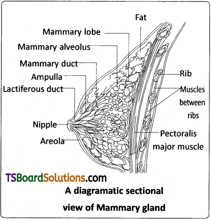
Mammary glands :
A functional mammary gland is characteristic of all female mammals. The mammary glands are paired structures ( breasts) that contain glandular tissue and variable amount of fat. The glandular tissue of each breast is divided into 15-20 mammary lobes containing clusters of cells called alveoli. The cells of the alveoli secrete milk, which is stored in the cavities (lumens) of the alveoli. The alveoli open into mammary tubules. The tubules of each lobe join to form a mammary duct. Several mammary ducts join to form a wider mammary ampulla which is connected to lactiferous duct through which milk is sucked out by the baby.
![]()
Question 2.
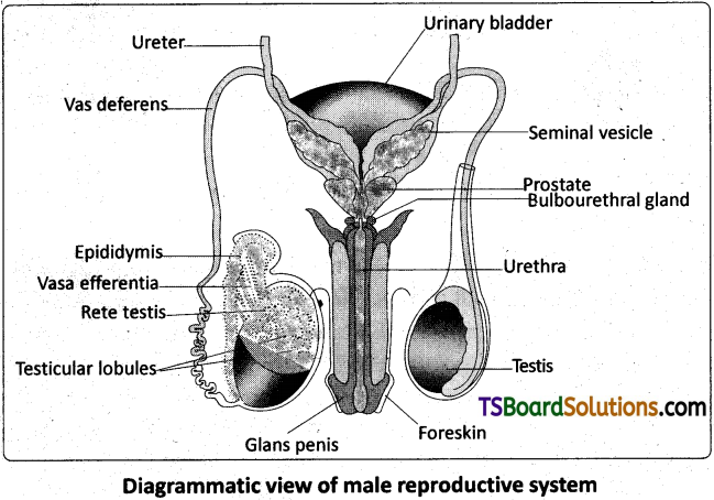
Describe the male reproductive system of a man. Draw a labelled diagram of it. [Mar. 2020, 2019, 18,’17 (A.P.); May/.June; Mar. 14]
Answer:
The Male Reproductive System :
The male reproductive system (male genital system) consists of a number of sex organs that are a part of the human reproductive process. The sex organs which are located in the pelvic region include a pair of testes (sing : testis) along with accessory ducts, glands and the external genitalia.
Testes :
The testes (testicles) are a pair of oval pinkish male primary sex organs suspended outside the abdominal cavity with in a pouch called scrotum. The scrotum helps in maintaining the low temperature of the testes (2 – 2.5°C lower than the normal internal body temperature) necessary for spermatogenesis. The cavity of the scrotal sac is connected to the abdominal cavity through the inguinal canal. Testis is held in position in the scrotum by the gubernaculum, a fibrous cord that connects the testis with the bottom of the scrotum and a spermatic cord, formed by the vas deferens, nerves, blood vessels and other tissues that run from the abdomen down to each testicle, through the inguinal canal.
Each testis is enclosed in a fibrous envelope, the tunica albuginea, which extends inward to form septa that partition the testis into lobules. There are about 250 testicular lobules in each testis. Each lobule contains 1 to 3 highly coiled seminiferous tubules. A pouch of serous membrane (peritoneal layer) called tunica vaginalis covers the testis.
Each seminiferous tubule is lined by the germinal epithelium which consists of undifferentiated male germ cells called spermatogonial mother cells and it also bears ‘nourishing cells’ called Sertoli cells. The spermatogonia produce the primary spermatocytes which undergo meiotic division, finally leading to the formation of spermatozoa or sperms (spermatogenesis). Sertoli cells provide nutrition to the spermatozoa and also produce a hormone called inhibin, which inhibits the secretion of FSH. The regions outside the seminiferous tubules, called interstitial spaces, contain interstitial cells of Leydig or Leydig cells.
Leydig cells produce androgens, the most important of which is testosterone. Testosterone controls the development of secondary sexual characters and spermatogenesis. Other immunologically competent cells are also present. The seminiferous tubules open into the vasa efferentia through the rete testis (a network of tubules in of the testis carrying spermatozoa from the seminiferous tubules to the vasa efferentia).
Epididymis :
The vasa efferentia leave the testis and open into a narrow, tightly coiled tube called epididymis located along the posterior surface of each testis. The epididymis provides a storage space for the sperms and gives the Sperms time to mature. It is differentiated into three regions – caput epididymis, corpus epididymis and cauda epididymis. The caput epididymis receives spermatozoa via the vasa efferentia t)f the mediastinum testis (a mass of connective tissue at the back of the testis that encloses the rate testis).
Vasa deferentia :
The vas deferens or ductus deferens is a long, narrow, muscular tube. The mucosa of the ductus deferens consists of pseudostratified columnar epithelium and lamina propria (areolar connective tissue). It starts from the tail of the epididymis, passes through the inguinal canal into the abdomen and loops over the urinary bladder. It receives a duct from the seminal vesicle. The vas deferens and the duct of the seminal vesicle unite to form a short ejaculatory duct / ductus ejaculatorius. The two ejaculatory ducts, carrying spermatozoa and the fluid secreted by the seminal vesicles, converge in the centre of the prostate and open into the urethra, which transports the sperms to outside.
Urethra :
In males, the urethra is the shared terminal duct of the reproductive and urinary systems. The urethra originates from the urinary bladder and extends through the penis to its external opening called urethral meatus. The urethra provides an exit for urine as well as semen during ejaculation.
Penis :
The penis and the scrotum constitute the male external genitalia. The penis serves as urinal duct and also intromittent organ that transfers spermatozoa to the vagina of a female. The human penis is made up of three columns of tissue; two upper corpora cavernosa on the dorsal aspect and one corpus spongiosum on the ventral side. Skin and a subcutaneous layer enclose all three columns, which consist of special tissue that helps in erection of the penis to facilitate insemination. The enlarged and bulbous end of penis called glans penis is covered by a loose fold of skin (foreskin) called prepuce. The urethra traverses the corpus spongiosum, and its opening lies at the tip of the glans penis (urethral meatus).
Male accessory genital glands :
The male accessory glands include paired seminal vesicles, a prostate and bulbourethral glands.
Seminal vesicles :
The seminal vesicles are a pair of simple tubular glands present postero- inferior to the urinary bladder in the pelvis. Each seminal vesicle opens into the corresponding vas deferens, as the vas deferens enters the prostate gland. The secretion of the seminal vesicles constitutes about 60 percent of the volume of seminal fluid. It is an alkaline, viscous fluid that contains fructose, proteins, citric acid, inorganic phosphorus, potassium, and prostaglandins. Once this fluid joins the sperm in the ejaculatory duct, fructose acts as the main energy source for the sperm outside the body. Prostaglandins are believed to aid fertilization by causing the mucous lining of the cervix to be more receptive to sperm as well as by aiding the movement of the sperm towards the ovum with peristaltic contractions of the uterus and fallopian tubes.
Prostate gland :
Prostate gland is located directly beneath the urinary bladder. The gland surrounds the prostatic urethra, and sends its secretions through several prostatic ducts. In man, the prostate contributes 15 – 30 percent of the semen. The fluid from the prostate is clear and slightly acidic. The prostatic secretion ‘activates’ the spermatozoa and provides nutrition.
Bulbourethral Glands :
Bulbourethral glands, also called Cowper’s glands, are located beneath the prostate gland at the beginning of the internal portion of the penis. They add an alkaline fluid to semen during the process of ejaculation. The fluid secreted by these glands lubricates the urethra. It is also thought to function as a ‘flushing agent’ that washes out the acidic urinary residues that remain in the urethra, before the semen is ejaculated.
![]()
Question 3.
Write an essay on different events that occur during development of a human.
Answer:
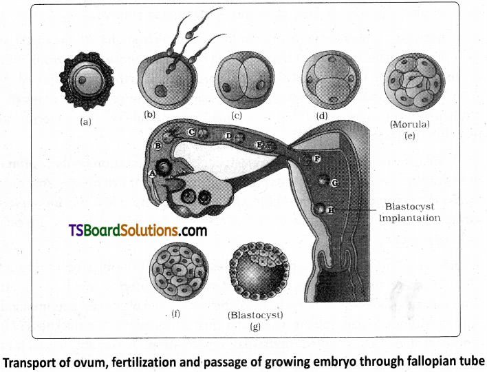
Cleavage :
The first phase of embryonic development, the cleavage is holoblastic (because of microlecithal condition of egg), radial, indeterminate and unequal. The mitotic division (cleavage) starts as the zygote moves through the isthmus of the oviduct towards the uterus. The daughter cells are called blastomeres.
Morula :
The embryo with 8 -16 blastomeres looks like a ‘mulberry’ and is called the morula. Morula is a solid mass of cells. The morula develops in the fallopian tube and reaches the uterus for further development. It is still surrounded by the zona pellucida. Due to unequal cleavage smaller and larger blastomeres are formed. The morula passes through a process called compaction. Now the embryo has a superficial flat cell layer and inner cell mass. The outer/supeficial layer becomes the trophoblast or trophectoderm. The trophoblast serves to attach the embryo to the uterine wall by forming trophoblastic villi. The inner cell mass constitutes formative cells. The inner cell mass gives rise to the embryo proper and is, therefore, also called the embryoblast. This is the first sign of cell differentiation (cells becoming different) in the human embryo.
Blastocyst :
Some fluid now passes into the morula from the uterine cavity, and partially separates the cells of the inner cell mass from those of the trophoblast. As the quantity of the fluid increases, the morula acquires the shape of a cyst. The cells of the trophoblast become flattened, and the inner cell mass comes to be attached to the inner side of the trophoblast on one side only. The morula now becomes a blastocyst.
The cavity of the blastocyst is the blastocoel or segmentation cavity or primary body cavity. The side of the blastocyst to which the inner cell mass is attached is called the embryonic or animal pole, while the opposite side is the bembryonic •pole. The cells of the trophoblast above the region of inner cell mass are called cells of Rauber.
Implantation :
The zona pellucida present around the blastocyst gradually disappears and the cells of the trophoblast stick to the uterine endometrium. The trophoblast invades the endometrium of the uterus. The blastocyst gets implanted into the uterine mucosa till the whole of it comes to lie within the thickness of the endometrium. This is called interstitial implantation. In humans, implantation begins on the 6th day after fertilisation. The process of implantation is aided by proteolitic enzymes produced by the trophoblast. The uterine mucosa also aids the process.
After the implantation of the embryo, the uterine endometrium is differentiated into what is called decidua. The portion of the decidua where the placenta is to be formed (i.e., deep to the developing blastocyst) is called the decidua basalis. The part of the decidua that separates the embryo from the uterine lumen is called the decidua capsularis. The part lining the rest of the uterine cavity is called the decidua parietalis. At the end of pregnancy the decidua is shed off, along with the placenta and membranes.
Formation of Bilaminar Embryonic Disc :
Implantation of the blastocyst is completed by the end of the second week. The inner cell mass forms into a disc called embryonic disc. Soon, the ‘cells of Rauber’ disappear exposing the embryonic disc. From the lower part of inner embryonic disc, some cells get separated by delamination and eventually form a layer of cells on the inner surface of the embryonic disc i.e., on the surface facing the cavity. This cell layer develops into the hypoblast, which is the future extra embryonic endoderm. The remaining part of the embryonic disc is called epiblast. Now the embryonic disc is called bilaminar embryonic disc.
The cells of the hypoblast increase in number and spread along the inner surface of the trophoblast. This hypoblast layer below the trophoblast finally encloses a cavity called yolk sac or umbilical vesicle. Meanwhile, the thickness of the embryonic disc increases towards the caudal end. Gradually the embryonic disc becomes oval.
Gastrulation :
Gastrulation involves proliferation, differentiation and movement of cells within the embryo. Along the longitudinal axis of the embryonic disc, a primitive streak is formed. A longitudinal furrow known as primitive groove forms along the middle of the primitive streak. On either side of it are the primitive folds. Anteriorly primitive streak has a shallow primitive pit. The anterior end of the primitive streak becomes thickened. This thickened part of the streak is called the primitive knot or primitive node or Hensen’s node.
Trilaminar Embryo – Formation of Primary Germ Layers :
Ingression, the future endodermal cells from the epiblast, replaces the hypoblast and forms the endoderm of the embryo. The future mesodermal cells converge towards the primitive folds, involute through the primitive groove and reach between epiblast and endoderm. The remaining epiblast is now known as the ectoderm. This invasion of the epiblast cells into the space between the epiblast and hypoblast is called gastrulation. Thus, the ectoderm, mesoderm and endoderm are all derived from the epiblast. The process of gastrulation converts the bilaminar embryonic disc into a trilaminar embryonic disc.
Extraembryonic Membranes :
During the development of the human embryo, as in all other amniotes four extraembryonic or foetal membranes are formed. They are chorion, amnion, allantois, and yolk sac. From the blastodisc, amniotic folds called head fold, tail fold and lateral fold are developed as somatopleure. As the folds fuse they are differentiated into outer chorion and inner amnion. Between the amnion and the embryo, there is an amniotic cavity filled with amniotic fluid. Amnion protects the embryo as the amniotic fluid acts as a shock absorber and also prevents the embryo form desiccation. The chorion develops a rich supply of blood vessels and forms an intimate association with the endometrium of the uterus.
Allantois and yolk sac are derived from the splanchnopleure. Allantois is formed from the hind gut as an evagination. It stores the waste materials. Allantois and chorion are fused to form chorio allantoic membrane which constitutes the placenta. Yolk sac encloses a fluid filled cavity. It is connected to the mid gut. It has no nutritive role.
After gastrulation is complete and any extra embryonic membranes are formed, the next stage of embryonic development begins with organ formation. Organogenesis: During organogenesis, regions of the three embryonic germ layers develop into the rudiments of organs. Whereas gastrulation involves mass movements of cells, organogenesis involves more localized changes.
Formation of the Notochord and Neural Tube :
The chorda mesodermal cells converge and involute through the Hensen’s node and extend forwards as notochordal process. This is later transformed into a solid rod – the notochord, the embryonic axial skeleton which is replaced by the vertebral column. The notochordal mesoderm induces the overlying ectodermal cells to form the neural plate. This is a good example of induction. At first the neural plate is oval but later elongates oves the underlying notochord along the longitudinal axis of the embryo. The plate invaginates towards the notochord to form a neural groove, which deepens progressively to form a tube by fusion of the lateral neural folds. The process of formation of neural tube is referred to as neurulation.
Differentiation of Mesoderm and Formation of Coelom :
The intra embryonic mesoderm spreads in all directions between the outer ectoderm and inner endoderm. The longitudinal column of mesoderm adjacent to the notochord and neural tube on either side is called epimere. The mesoderm around the gut is the hypomere. The mesoderm in between these two is the mesomere. The epimere becomes segmented into cubical blocks called somites or metameres. Each somite differentiates into myotome, sclerotome and dermatome.
The sclerotome forms the vertebral column. The dermatome forms the dermis of the skin and other connective tissues. The myotome forms the voluntary muscles of the body. The mesomere forms the urinogenital organs and their ducts. The hypomere splits into outer somatic and inner splanchnic mesodermal layers. Intra embryonic coelom is formed between these two layers. It gives rise to pericardial, pleural, peritoneal cavities etc.