TS Inter 1st Year Zoology Study Material Chapter 4 Animal Diversity-II: Phylum Chordata

Telangana TSBIE TS Inter 1st Year Zoology Study Material 4th Lesson Animal Diversity-II: Phylum Chordata Textbook Questions and Answers.

TS Inter 1st Year Zoology Study Material 4th Lesson Animal Diversity-II: Phylum Chordata

Very Short Answer Type Questions

Question 1.
List out the characters shared by chordates and echinoderms.
Answer:
Chordates share deuterostomeate condition, radial and indeterminate type of cleavage and enterocoelom with the echinoderms.

Question 2.
Write four salient features of cyclostomes.
Answer:
Salient features of cyclostomes

  1. These are jawless aquatic forms.
  2. Body is scaleless, long, slender and eel-like.
  3. Endoskeleton is cartilaginous
  4. Mouth is circular and suctorial. Tongue bears horny teeth.

Question 3.
What is the importance of endostyle in lancelets and ascidians?
Answer:
In ascidians and lancelets, ventral side of the pharynx possesses an endostyle which is believed to be the fore runner of the thyroid gland of a vertebrate. It helps in nutrition.

Question 4.
Name the type of caudal fin and scales that are present in a shark and Catla, respectively.
Answer:

  1. Shark → Heterocercal caudal fin and placoid scales are seen.
  2. Catla → Homocercal caudal fin, ctenoid or cycloid scales are seen.

Question 5.
What is the importance of air bladder in fishes?
Answer:
An air bladder is present with or without connection to the gut. It is either helpful in gas exchange or in maintaining buoyancy.

Question 6.
How do you justify the statement-“heart in fishes is a branchial heart”?
Answer:
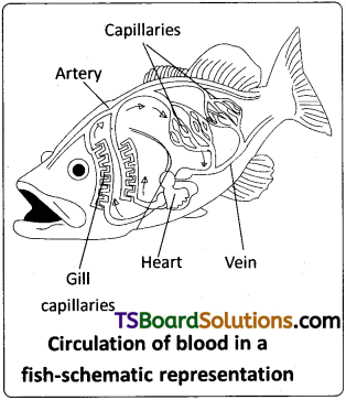
Fish heart is known as ‘branchial heart’ as it supplies blood only to the gills.

TS Inter 1st Year Zoology Study Material Chapter 4 Animal Diversity-II: Phylum Chordata

Question 7.
What are claspers? Which group of fishes possesses them?
Answer:
In the group chondrichthyes males possess claspers on pelvic fins to facilitate internal fertilization, (example : shark).

Question 8.
How does the heart of an amphibian differ from that of a reptile? [March 2015 – T.S.]
Answer:

  1. Heart in amphibians is 3 chambered with sinus venosus and conus arteriosus.
  2. Heart in reptiles is completely four chambered except in the crocodiles. Sinus venosus is present but conus arteriosus is absent.

Question 9.
Name the structures that appeared for the first time in amphibians, in the course of evolution.
Answer:
Tympanum, Lacrimal and Harderian glands (associated with eye) appeared for the first time in the amphibians in the course of evolution.

Question 10.
How do you distinguish a male frog from a female frog? [March 2018 – A.P.]
Answer:
Male frog can be distinguished by the presence of sound amplifying vocal sacs and also a copulatory pad on the first digit of each forelimb. They are absent in female frogs.

Question 11.
What is ‘force pump’ in frog? Why is it named so?
Answer:
In frog during the pulmonary respiration, the bucco – pharyngeal cavity acts like a “force pump”. Due to the elevation of bucco – pharyngeal cavity the air forces the glottis to open and enter the lungs, where exchange of gases takes place.

Question 12.
What are corporabigemina? Mention their chief function.
Answer:
In amphibians Mid brain is represented by a pair of optic lobes called corpora bigemina. The optic lobes are associated with the sense of sight.

TS Inter 1st Year Zoology Study Material Chapter 4 Animal Diversity-II: Phylum Chordata

Question 13.
Distinguish between mesorchium and mesovarium.
Answer:

  1. In male frog testes are attached to kidney and dorsal body wall by a double fold of peritoneum called mesorchium.
  2. In female frog ovaries are attached to the kidneys and dorsal body wall by a double fold of peritoneum called mesovarium.

Question 14.
Distinguish between milt and spawn. [Mar. 2017 – A.P ; May/June 2014,]
Answer:

  1. Mass of eggs released by a female frog into water is called Spawn.
  2. Mass of sperms released by a male frog into water is called Milt.

Question 15.
What are the “Golden ages” of the first jawed vertebrates and the first amniotes?
Answer:

  1. Golden age of first jawed vertebrates is “DEVONIAN PERIOD” (Fishes)
  2. Golden age of first amniotes is MESOZOIC ERA (Reptiles)

Question 16.
Name two poisonous and non-poisonous snakes found in South India. [March 2014]
Answer:

  1. Two poisonous snakes found in South India are a) Naja naja (cobra), b) Bungarus (Krait)
  2. Two non-poisonous snakes found in South India are a) Ptyas (rat snake) b) Tropidonotus (pond snake)

Question 17.
In which features does the skin of a reptile differ from that of a frog?
Answer:

  1. Skin of a frog is thin, scaleless and moist.
  2. Skin of a reptile is rough and dry. The exoskeleton occurs in the form of horny epidermal scales, shields and claws.

Question 18.
Describe a cat and a lizard on the basis of their chief nitrogenous wastes excreted.
Answer:

  1. Reptiles excrete uric acid as nitrogenous waste (uricotelic).
  2. Cat excrete urea as nitrogenous waste (ureotelic).

Question 19.
Name the four extra embryonic membranes. [March 2020]
Answer:
The four extra embryonic membranes are a) Amnion b) Allantois c) Chorion and d) Yolk sac.

TS Inter 1st Year Zoology Study Material Chapter 4 Animal Diversity-II: Phylum Chordata

Question 20.
What are Jacobson’s organs? What is their function?
Answer:
Jacobson’s organs, the specialized olfactory structures are highly developed in lizards and snakes.

Question 21.
What are pneumatic bones? How do they help birds?
Answer:
In birds bones are hollow or air filled and referred to as pneumatic bones. They help in reducing the weight, thus help in effective flying (aerial adaptation).

Question 22.
What is ‘wish bone’? What are the skeletal components that form it?
Answer:
In birds both clavicles are fused with the interclavicle to form a ‘V’- shaped bone, called furcula or ‘wish bone’ or merry thought bone.

Question 23.
What is continuous oxygenation of the blood? How is it made possible in birds?
Answer:
In birds the compact spongy lungs are associated with air sacs. Air sacs facilitate continuous oxygenation (oxygen supplied to lungs uninterruptedly and heavily) of blood and pneumacity of bones.

Question 24.
Distinguish between the crop and the gizzard in birds.
Answer:
a) In birds oesophagus is often dilated into crop for the storage of food.
b) Stomach is usually divided into glandular proventriculus and muscular gizzard (grinding mill).

Question 25.
Distinguish between altricial and precocial hatchlings.
Answer:

  1. Altricial hatchlings are seen in flying birds which are incapable of moving around on its own soon after hatching.
  2. PrecOcial hatchlings are seen in flightless birds which are capable of moving around on its own soon.

TS Inter 1st Year Zoology Study Material Chapter 4 Animal Diversity-II: Phylum Chordata

Question 26.
In which group of animals do we find three ear ossicles on each side and what are their names from the innermost to the outermost?
Answer:
In mammals, we find three ear ossicles on each side. From innermost to outer¬most they are stapes, incus and malleus.

Question 27.
How does a mature RBC of a mammal differ from that of other vertebrates?
Answer:
Mature RBC in mammals is unique because it is circular, biconcave and enucleate.

In other vertebrates RBC are oval, biconvex and nucleated.

Question 28.
Name the characteristic type of vertebrae found in reptiles, birds and mammals.
Answer:
Characteristic vertebrae

  1. In Reptiles → Procoelous.
  2. In Birds → Heterocoelous
  3. In Mammals → Amphiplatyan.

Question 29.
Name the three meninges. In which group of animals do you find all of them?
Answer:
The three meninges which are brain layers are
a) Dura Mater b) Arachnoid membrane c) Piamater.
The group mammals possess all the three layers.

TS Inter 1st Year Zoology Study Material Chapter 4 Animal Diversity-II: Phylum Chordata

Question 30.
Name the vertebrate groups in which ’renal portal system’ is absent.
Answer:
Renal portal system is absent in the group Mammals.

Short Answer Type Questions

Question 1.
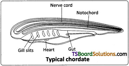
Give three major differences between chordstes and non-chordates and draw the sketch of a chordate’s body showing those features.
Answer:
Major differences between chordates and non-chordates:

ChordatesNon-chordates
1. Notochord is presentNotochord is absent
2. Pharynx is perforated by gill slitsGill slits are absent
3. Heart is ventralHeart is dorsal (if present).
4. A post-anal tail is present.Post-anal tail is absent

TS Inter 1st Year Zoology Study Material Chapter 4 Animal Diversity-II Phylum Chordata 1

Question 2.
Name the four ‘hallmarks’ of chordates and explain the principal function of each of them. [March 2020, 18]
Answer:
The four principal chordate characters and their functions are
a) Notochord :
Flexible rod like structure placed along the mid dorsal line between gut and nerve cord. It is a supporting structure.

b) Dorsal tubular nerve cord :
A single hollow tubular and fluid filled nerve cord is situated above notochord and below dorsal body wall. It is sensory in function. In higher chordates anteriorly it becomes brain and the rest becomes the spinal cord.

c) Pharyngeal gill slits :
These are a series of lateral perforations in the wall of pharynx which are ecto-endodermal in origin. They are helpful in the exchange of respiratory gases.

d) Post-anal tail :
Chordates have a tail extending posterior to the anus. It is lost in many species during embryonic development. It provides the propelling force in the locomotion of aquatic forms and act as a balancing organ in many terrestrial animals.

Question 3.
Describe the features of a tunicate that reveals its chordate identity.
Answer:
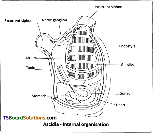
SUBPHYLUM – UROCHORDATA OR TUNICATA : All urochordates are marine and occur from the surface water to greater depths. They are either sessile (ascidians) or pelagic (Salpa, Doliolum) and solitary (Ascidia) or colonial (Pyrosoma).
TS Inter 1st Year Zoology Study Material Chapter 4 Animal Diversity-II Phylum Chordata 2

Body is un-segmented and covered by a ‘test’ or ‘tunic’ composed of cellulose1 which is uncommon in animals. Coelom is absent. However, an ectoderm – lined atrial cavity surrounds the pharynx, into which gill slits, anus and genital ducts open. Ventral side of the pharynx possesses an endostyle, (see Glossary) which is believed to be the ‘forerunner’ of the thyroid gland of a vertebrate. Atrium leads to the exterior by a dorsal or posterior atrial aperture. Digestive tract is ‘complete’.

Two to numerous gills slits are found in the pharyngeal wall. Circulatory system is of ‘open type’. There is a simple, tubular, ventral heart which alternately reverses the direction of the flow of blood. Nervous system is represented in the adult by a single dorsal ‘ganglion’. They are bisexual or hermaphrodites. Development generally includes a free-swimming tadpole larva with a tail, a dorsal hollow nerve cord, and a notochord confined to the tail, hence the name urochordata. e.g.: Ascidia, Salpa, Doliolum, Pyrosoma and Oikopleura.

Question 4.
Compare and contrast sea squirts and lancelets.
Answer:

Sea squirtsLancelets
1) Marine and occur from the surface water to greater depths. Sessile or pelagic and solitary or colonial.1) Marine forms lead a burrowing mode of life in shallow sea waters.
2) Body is unsegmented and covered by a test or tunic made up of cellulose.2) Body is fish like, translucent with median fins but without paired fins.
3) Larva stage exhibits a tail, dorsal hollow nerve cord and a notochord confined to the tail, hence the name urochordata.3) Often described as typical chordate because they possess principal chordate. Chordates such as notochord, tubular nerve cord and pharyngeal gill slits throughout their life.
4) Circulatory system is of open type. Simple, tubular, ventral heart which alternately reverses the direction of the flow of blood.
Example: Ascidia (sea squirt)
4) Circulatory system is of closed type and heart, blood corpuscles and respiratory pigment are absent.
Example: Branchiostoma (lancelet)

TS Inter 1st Year Zoology Study Material Chapter 4 Animal Diversity-II: Phylum Chordata

Question 5.
List out eight characteristics that help distinguish a fish from the other vertebrates. [March 2019]
Answer:
TS Inter 1st Year Zoology Study Material Chapter 4 Animal Diversity-II Phylum Chordata 3
Characteristics of group PISCES.

  1. Fishes are completely aquatic poikilo- thermic (cold blooded ) animals.
  2. Body of a fish is usually streamlined and differentiated into head, trunk and tail.
  3. Exoskeleton consists of mesodermal scales or bony plates. A few are scaleless.
  4. Endoskeleton may be cartilaginous or bony. Skull is monocondylic. Vertebrae are amphicoelous (centrum is concave at both anterior and posterior faces).
  5. Locomotion is assisted by unpaired (median and caudal) fins along with paired (pectoral and pelvic) fins.
  6. Mouth is ventral or terminal. Teeth are usually acrodont, homodont and polyphyodont.
  7. Exchange of respiratory gases is performed by the gills.
  8. Heart is ‘two chambered’ and is described as ‘branchial heart’ as it supplies blood only to the gills.
  9. Lateral line sensory system is well developed.

Question 6.
Compare and contrast cartilaginous and bony fishes. [May 2017 – A.P.; May/June. Mar. 2014 ; March 2013]
Answer:

Cartilaginous fishesBony fishes
1) Caudal fin is heterocercal1) Caudal fin is diphycercal or homocercal.
2) Scales if present placoid scales.2) Scales are ganoid, cycloid or ctenoid scales.
3) Endoskeleton is entirely cartilage.3) Endoskeleton is bony.
4) Mouth and nostrils are ventral.4) Mouth is usually terminal.
5) Digestive tract opens into cloaca, if present.5) Digestive tract opens out by anus.
6) Air bladder is absent.6) Air bladder is often present.
7) Fertilization is internal. Mostly viviparous.
eg: Scoliodon.
7) Fertilization is external. Mostly oviparous.
Eg: Catla catla

TS Inter 1st Year Zoology Study Material Chapter 4 Animal Diversity-II: Phylum Chordata

Question 7.
Describe the structure of the heart of frog.
Answer:
TS Inter 1st Year Zoology Study Material Chapter 4 Animal Diversity-II Phylum Chordata 4
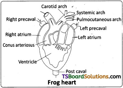
The heart is a muscular organ situated in the upper part of the body cavity. If has two separate atria and a single undivided ventricle. It is covered bye double layered membrane called pericardium. A triangular chamber called sinus venosus joins the right atrium on the dorsal side. It receives blood through three vena cavae (caval veins). The ventricle opens into the conus arteriosus on the ventral side. The conus arteriosus bifurcates into two branches and each of it divides into three aortic arches namely carotid, systemic and pulmocutaneous. Blood from the heart is distributed to all parts of the body by the branches of aortic arches. Three major veins collect blood from the different parts of the body and carry it to the sinus venosus.

Question 8.
Write eight salient features of the class Amphibia.
Answer:

  1. Body is divided into distinct’head’ and’trunk’. Tail may or may not be present.
  2. Skin is soft, scale-less (except the members of Apoda), moist and glandular.
  3. The body bears two pairs of equal or unequal pentadactyle limbs (caecilians are limbless).
  4. Skull is dicondylic as in mammals. Vertebrae are mostly procoelous (centrum is concave at its anterior face only) in the anurans, amphicoelous in the caecilians and usually opisthocoelous (centrum is concave at its posterior face) in the urodeles. Sternum appeared for the first time in the amphibians.
  5. Mouth is large; teeth are acrodont, homodont and polyphyodont.
  6. Respiratory gaseous exchange is mostly cutaneous; pulmonary and bucco pharyngeal respirations also occur. Branchial respiration is performed by larvae and some adult urodeles.
  7. Heart is three-chambered with sinus venosus and conus arteriosus. Three pairs of aortic arches and well-developed portal systems are present; erythrocytes are nucleate.
  8. Kidneys are mesonephric; ureotelic.
  9. Meninges are the inner piamater and outer duramater; cranial nerves are 10 pairs.
  10. Middle ear consists of a single ear ossicle, the columella auris which is the modified ‘hyomandibula’ of the fishes. Tympanum, lacrimal and harderian glands appeared for the first time in the amphibians.
  11. Sexes are separate and fertilization is mostly external. Development is mostly indirect, e.g.: Bufo (toad), Rana (frog), Hyla (tree frog), Salamandra (salamander), Ichthyophis (limbless amphibian), Rhacophorus (flying frog).

Question 9.
Describe the male reproductive system of frog with the help of a labelled diagram.
Answer:
Male Reproductive System of frog:
TS Inter 1st Year Zoology Study Material Chapter 4 Animal Diversity-II Phylum Chordata 5
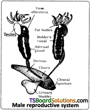
The male reproductive system consists of a pair of yellowish and ovoid testes, which are attached to the kidneys and dorsal body wall by a double fold of peritoneum called mesorchium. Each testis is composed of innumerable seminiferous tubules which are connected to form 10 to 12 narrow tubules, the vasa efferentia. They enter the kidneys and open into the Bidder’s canal which is connected to the ureter through transverse canals of the kidney. The urino-genital ducts of both the sides open into the cloaca.

Question 10.
Write short notes on organs of special senses in frog.
Answer:
Organs of special senses : Frog has sense organs such as the organs of touch, taste, smell, sight and hearing. The well-organised structures among them are eyes, internal ears and the rest are ‘cellular aggregations’ around nerve endings. The receptors of touch occur in the skin. Organs of taste are called taste buds that lie on small papillae of tongue. The organs of smell are a pair of nasal chambers.

The organs of sight are a pair of eyes located in the orbits of the skull. Eyes are protected by eyelids. The upper eyelid is immovable. The lower eyelid is folded into a transparent nictitating membrane, which can be drawn across the surface of the eye. The retina of the eye contains both rods and cones. Cones provide ‘colour vision’ and rods are helpful in ‘dimlight vision’.

Ear is useful for hearing and balance. It consists of a middle ear closed externally by a large tympanic membrane (ear drum) and a columella that transmits vibrations to the inner ear. The inner ear consists of a utriculus with three semicircular canals and a small sacculus.

TS Inter 1st Year Zoology Study Material Chapter 4 Animal Diversity-II: Phylum Chordata

Question 11.
List out the salient features of exo and endoskeleton in reptiles.
Answer:
Skin is rough and dry. The exoskeleton occurs in the form of horny epidermal scales, shields, and claws (which appeared for the first time in reptiles).

Dentition is acrodont, homodont and polyphyodont (thecodont in corcodiles as seen in the mammals). Chelonians are ‘edentate’.

Skull is monocondylic and many have temporal fossae. Each half of the lower jaw is formed by six bones. Vertebrae are mostly procoelous. The first two cervical vertebrae are specialized into atlas and axis to facilitate independent movement of the head from the rest of the body; sacral vertebrae are two in number.

Question 12.
List out the extant orders of the Class Reptilia. Give two examples for each order.
Answer:
The extant reptiles are grouped into four orders.
1. Chelonia –
Chelone (marine green turtle), Testudo (terrestrial form), Trionyx (fresh water form)

2. Rhynchocephalia –
Sphenodon (a ’living fossil’, endemic to New Zealand)

3. Crocodilia –
Crocodylus palustris (Indian crocodile or maggur), Alligator (alligator), Gavialis gangeticus (Indian gavial or gharial)

4. Squamata
a) Lizards – Hemidactylus (wall lizard), Chameleon, Draco (flying lizard)
b) Snakes
i) Poisonous snakes – Naja naja (cobra), Ophiophagushannah (King cobra), Bungarus(Krait), Daboia/Vipera russelli (chain viper).
ii) Non-poisonous snakes- Ptyas (rat snake), Tropidonotus (grass snake or pond snake)

Question 13.
What are the modifications that are observed in birds that help them in flight? [Mar. 2017 – A.P : Mar. 2015 – A.P & T.S]
Answer:
TS Inter 1st Year Zoology Study Material Chapter 4 Animal Diversity-II Phylum Chordata 6

  1. Body is streamlined
  2. The fore limbs are modified into ‘wings for flight.
  3. Skin is dry and devoid of glands, except the oil or preen gland or uropygial gland at the base of the tail to protect tail feathers.
  4. Exoskeleton consists of epidermal feathers (a unique feature), with interlocking system.
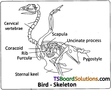
  5. Long bones are hollow with air cavities (pneumatic). Skull is monocondylic. Vertebrae are heterocoelous. The last thoracic, lumbar, sacral and anterior few caudal vertebrae are fused to form a synsacrum. it is fused with pelvic girdle to provide support to hind limbs. A few posterior most caudal vertebrae are fused to form the pygostyle that provides support to the tail feathers. Sternum has a keel/ carina for the attachment of flight muscles (except in the ratite birds). Both the clavicles are fused with the interclavicle to form a V – shaped bone, called furcula or ‘Wish bone1 or ‘Merry thought bone’.
  6. All modern flying birds are provided with powerful breast muscles (flight muscles), chiefly the pectoralis major and pectoralis minor.

Question 14.
What are the features peculiar to ratite birds? Give two examples of ratite birds.
Answer:
Ratitae / Palaeognathae :
They are modern flightless running birds. They are ‘discontinuous’ in their distribution like the lung fishes and marsupials. They are characterized by the presence of reduced wings, a raft like sternum without keel and males with penis. They do not possess syrinx, clavicles and usually preen gland, e.g. Struthio camelus (African ostrich), Kiwi (National bird of N6w Zealand), Rhea (American ostrich), Dromaeus (Emu), Casuarius.

TS Inter 1st Year Zoology Study Material Chapter 4 Animal Diversity-II: Phylum Chordata

Question 15.
Mention the most important features of nervous system and sense organs in mammals.
Answer:
Mammals have a relatively large brain when compared to that of other animals in relation to body size. The four optic lobes constitute corpora quadrigemina. The two halves of cerebrum are connected by corpus callosum. The CNS is enveloped by three meninges. The middle menix called, arachnoid membrane, is present in mammals only.

Cranial nerves are twelve pairs. Eyes have movable eye lids with ‘eye lashes’. External ear has a large fleshy and cartilaginous flap called pinna. Middle ear possesses three ear ossicles. They are malleus, incus and stapes. Cochlea of the internal ear is spirally coiled and bears the ‘organ of Corti’ which is the receptor of sound.

Question 16.
Write short notes on the following features of the eutherians.
a) Dentition
b) Endoskeleton.
Answer:
a) Dentition :
Teeth are thecodont, heterodont and diphyodont. Four pairs of salivary glands are present in association with the buccal cavity (3 pairs in man).

b) Endoskeleton :
Skull is dicondylic. Each halfofthe lowerjaw consists of a ‘single’ bone, the dentary. Most mammals have seven cervical vertebrae, six in Choloepus (two toed sloth) and Trichechus (manatee) and nine in Bradypus (three-toed sloth).Sacral vertebrae are two to five. Vertebrae are of the amphiplatyan type (centrum is flat at both faces). Ribs are double – headed. Buccal cavity is separated from the upper nasal cavity by a secondary palate.

Question 17.
Give an example for each of the following, a) A viviparous fish, b) A fish possessing electric organs, c) A fish possessing poison sting, d) An organ which regulates buoyancy in the body of fish, e) An oviparous animal with milk producing glands.
Answer:
a) Viviparous fish – Scoliodon (dog fish /shark)
b) Fish possessing electric organs – Torpedo (electric ray)
c) Fish possessing poison sting – Trygon (Sting ray)
d) Organ which regulates buoyancy in the body of fish – Air bladder.
e) Oviparous animal with milk producing glands – Ornithorhynchus (Duck-billed platypus)

Question 18.
Mention two similarities between a) Aves and mammals b) A frog and a crocodile c) A lizard and a snake.
Answer:
A) Aves and mammals :

  1. Ribs are double headed in both gorups.
  2. Heart is 4 chambered.
  3. Kidneys are metanephric.
  4. Cranial nerves are twelve pairs.

B) A frog and a crocodile :

  1. Both are amphibians and can lead life on land and in water.
  2. Cloaca is present.
  3. Middle ear consists of a single ear ossicle, the columella auris.

C) A lizard and a snake :

  1. Jacobson’s organs, the specialized olfactory structures are highly developed in lizards and snakes.
  2. In lizards and snakes also some are oviparous and some are viviparous.

TS Inter 1st Year Zoology Study Material Chapter 4 Animal Diversity-II: Phylum Chordata

Question 19.
Name the following animals, a) A limbless amphibian b) The largest of all living animals c) An animal possessing dry and cornified skin d) ‘National Animal’ of India.
Answer:
a) A limbless amphibian : Ichthyophis
b) The largest of all living animals : Balaenoptera (Blue whale)
c) An animal possessing dry and cornified skin : Reptiles (eg : Sphenodon)
d) National animal of India : Panthera tigris (tiger)
Ornithorhynchus (Duck-billed platypus)

Question 20.
Write the generic names of the following.
a) An oviparous mammal
b) Flying fox
c) Blue whale
d) Kangaroo
Answer:
a) An oviparous mammal – Ornithorhynchus (Duck-billed platypus)
b) Flying fox – Pteropus
c) Blue whale – Balaenoptera.
d) Kangaroo – Macropus.

TS Inter 1st Year Zoology Study Material Chapter 3 Animal Diversity-I: Invertebrate Phyla

Telangana TSBIE TS Inter 1st Year Zoology Study Material 3rd Lesson Animal Diversity-I: Invertebrate Phyla Textbook Questions and Answers.

TS Inter 1st Year Zoology Study Material 3rd Lesson Animal Diversity-I: Invertebrate Phyla

Very Short Answer Type Questions

Question 1.
What physical feature pertaining to the organism and its medium do you notice in a sponge body from which sponges can be/were identified as animals and not plants? What do you call the region in the sponge body in which you noticed that feature?
Answer:
Sponge body possesses two types of pores. Small pores are called ostia which are numerous and large one or two pores are called oscula. The osculum propels out water coming from body cavity called spongocoel. The presence of osculum confirm the sponge is an animal. It is situated at anterior free end.

Question 2.
What are the different structures that makeup the internal skeleton of a sponge? What are the chemicals involved in the formation of these structures?
Answer:
The body of sponge is supported by skeleton made up of calcareous or siliceous spicules or spongin fibres or both. Calcareous spicules are made up of CaCO3 Siliceous spicules are made up of glass (silicon dioxide). Spongin is fibre material.

Question 3.
What are the functions of canal system of sponges? [March 2018 – A.P.; March 2013]
Answer:
Sponges have a water transport system or canal system that constantly conducts water. It helps in gathering food (filter feeders), exchange of gases (respiration) and removal of wastes (excretion).

Question 4.
What are the two chief morphological ‘body forms’ of cnidarians? What are their chief functions? [March 2020]
Answer:
The two chief morphological body forms of cnidarians are
1. Polyp 2. Medusa.

Polyp is sessile cylindrical form and produce medusae asexually by budding. Medusa is umbrella-shaped and free swimming form and sexually produce polypoid forms.

Question 5.
What is metagenesis? Animals belonging to which phylum exhibit metagenesis?
Answer:
The cnidarians which exist in both forms namely polyp and medusa exhibit alternation of generations called metagenesis. Polyps by asexually method called budding produce medusae. Medusae by sexual method called syngamy give rise to polypoid forms.

TS Inter 1st Year Zoology Study Material Chapter 3 Animal Diversity-I: Invertebrate Phyla

Question 6.
What is the cnidarian group with quantitatively / relatively large mesoglea? What is the significance of such a well developed mesoglea pertaining to the aquatic life of that group?
Answer:
In cnidarians, the animals of class scyphozoa contain relatively large mesoglea containing amoebocytes. The mesoglea is important in buoyancy.

Question 7.
What is the chief difference between the hydrozoans and the rest of the cnidarians regarding the germinal layer(s) in which its ‘defencive structures or cells of defence’ occur?
Answer:
The chief difference between the hydrozoans and the rest of the cnidarians regarding the germinal layer in which its defencive structures or cells of defence called cnidocytes occur is only in ectoderm. In others, cnidocytes are present both in ectoderm and endoderm.

Question 8.
What are the excretory cells of flatworms called? What is the other important function of these specialized cells?
Answer:
The excretory cells of flatworms are called Flame cells (protonephridia). They help in osmoregulation and excretion.

Question 9.
Distinguish between amphids and phasmids [March 2019]
Answer:
a) Amphids are cuticular depressions around oral region performing chemoreceptor function.
b) Phasmids are posterior glandulo – sensory structures. Both are found in Nematodes.

Question 10.
What is the essential difference between a ‘flat worm’ and a ’round worm’ with reference to the perivisceral area of their bodies?
Answer:
Flat worms are the first bilaterally symmetrical triploblastic and acodomate animals. Round worms are bilaterally symmetrical, triploblastic and pseudocoelomate animals.

Question 11.
How do you account for the origin of the perivisceral space in the body of a nematode and an annelid?
Answer:

  1. In nematodes during embryonic development, mesoderm occupies only a part of the blastocoel adjoining the ectoderm. The unoccupied portion of the blastocoel persists as pseudocoelom.
  2. In annelids the perivisceral space is a true coelom formed by splitting of mesodermal blocks between ectoderm and endoderm which is a schizocoelom.

TS Inter 1st Year Zoology Study Material Chapter 3 Animal Diversity-I: Invertebrate Phyla

Question 12.
What is metamerism? What is the essential difference between the mode of formation of individual morphological body units of a tapeworm and those of an earthworm?
Answer:
Metamerism is division of body into segments divided by septa and are referred to as metameres. This is unique of annelids. Segments are formed at posterior tip.

Pseudometamerism is found in tape worm. There is no actual division of segments intervally. But gives false appearance because segments called proglottids are attached to each other. They are formed at anterior end.

Question 13.
How do you distinguish a ‘hirudinean’ from the rest of the annelids, based on the morphological features pertaining to metamerism? How does the coelom of a leech differ from the coelom of an earthworm with reference to its contents?
Answer:
In Hirudineans the body is with definite number of segments. The segments are externally subdivided into annuli. Internal segmentation is absent. Coelom of leech is filled with a characteristic tissue called botryoidal tissue. This is absent in earthworm.

Question 14.
What do you call the locomotor structures of Nereis? Why is Nereis called a polychaete?
Answer:
In polychaetes parapodia are the locomotory structures. Which hear many setae. Hence the name polychaeta. Nereis is a polychaete because of parapodia with many setae are seen.

Question 15.
What is botryoidal tissue?
Answer:
Botryoidal tissue is a characteristic tissue filled in the coelom of leeches (hirudineans). This tissue is believed to be excretory in funtion.

Question 16.
What is the difference between the epidermis of a nematode and that of an annelid? How does a nematode differ from an annelid with reference to the musculature of the body wall?
Answer:

  1. In nematodes the unsegmented body is covered by a transparent tough and protective collagenous cuticle. In some epidermis is synctial. Only longitudinal muscles are present (circular muscles are absent).
  2. In annelids the segmented body possesses dermo-muscular body wall containing cuticle, epidermis, dermis, longitudinal and circular muscles.

Question 17.
What do you call the first and second pairs of cephalic appendages of a scorpion?
Answer:
The first and second pairs of cephalic appendages of a scorpion are-called chelicerae and pedipalpi respectively.

TS Inter 1st Year Zoology Study Material Chapter 3 Animal Diversity-I: Invertebrate Phyla

Question 18.
What is the uniqueness about the first two pairs of cephalic appendages of a crustacean compared to those of the other extant arthropods?
Answer:
In Crustacea Head and Thorax are fused to form cephalothorax. The first two pairs of cephalic appendages are antennules and antennae which is unique feature.

Question 19.
What is the sub-phylum to which ‘ticks’ and ‘mites’ belong? How do you distinguish them from the insects with reference to their walking legs?
Answer:
Ticks and mites belong to sub-phylum chelicerata. They are differed from insects in having four pairs of walking legs. Insects possess three pairs of walking legs.

Question 20.
What are the respiratory structures of Limulus and Palamnaeus respectively?
Answer:

  1. Respiratory structures of Limulus (King crab) → Book – gills
  2. Respiratory structures of palaemnaeus (scorpion) → Book-lungs.

Question 21.
What are antennae? What is the arthropod group without antennae?
Answer:
Antennae are cephalic appendages of arthropods which are sensory in function (sense of touch). They are absent in the sub-phylum chelicerata.)

Question 22.
What do you call the perivisceral cavity of an arthropod? Where from is it derived during development?
Answer:
The perivisceral cavity of arthropods is called Haemocoel (blood cavity) derived from mostly the embryonic blastocoel.

Question 23.
Which arthropod, you have studied, is called a ‘living fossil’? Name its respiratory organs. [March 2015 – A.P.]
Answer:
Limulus (King crab) belonging to class Xiphosura of sub-phylum chelicerata is called living fossil. Its respiratory organs are book gills.

Question 24.
How do you identify a Chiton from its external appearance? How many pairs of gills help in the respiration of Chiton?
Answer:
Chiton is identified by a dorsal shell consisting of 8 transverse plates. 6 – 88 pairs of gills help in respiration of Chiton.

TS Inter 1st Year Zoology Study Material Chapter 3 Animal Diversity-I: Invertebrate Phyla

Question 25.
What is the function of radula? Give the name of the group of molluscs which do not possess a radula. [May/June, March 2014]
Answer:
The buccal cavity of molluscs contain a file like rasping organ called radula for feeding. It is absent in the class bivalve or pelecypoda.

Question 26.
What is the other name for the gill of a mollusc? What is the function of osphradium?
Answer:
Ctenedia is the other name of gill of a mollusc. Osphradium in molluscs tests the purity of water. It is present in the bivalves and gastropods.

Question 27.
What is Aristotle’s lantern? Give one example of an animal possessing it. [May 2017 ; Mar. ’17 – A.P ; Mar. 2015 – T.S]
Answer:
In the mouth of a sea urchin a complex five jawed masticatory apparatus called Aristotle’s lantern is present. Example : Echinus (sea urchin).

Question 28.
What is the essential difference between the juveniles and adults of echinoderms, symmetry wise?
Answer:

  1. Adult of echinoderms exhibit pentamerous radial symmetry.
  2. Juveniles of echinoderms exhibit bilateral symmetry.

Question 29.
What are blood glands in Pheretima?
Blood glands in pheretima are present in the 4th, 5th, and 6th segments. They produce blood cells and haemoglobin which is dissolved in plasma. Blood cells are phagocytic in nature.

TS Inter 1st Year Zoology Study Material Chapter 3 Animal Diversity-I: Invertebrate Phyla

Question 30.
What are spermathecae on the body of pheretima?
Answer:
4 pairs of spermathecae are located in the segments 6th to 9th (one pair in each segment). They receive and store sperms during copulation.

Short Answer Type Questions

Question 1.
Write short notes on the salient features of the anthozoans.
Answer:
TS Inter 1st Year Zoology Study Material Chapter 3 Animal Diversity-I Invertebrate Phyla 1
Class – Anthozoa or Actinozoa :

  1. They are commonly referred to as sea anemones.
  2. They are sedentary and only polypoid in form.
  3. Coelenteron is divided into several compartments by vertical septa called mesenteries.
  4. Mesoglea contains connective tissue.
  5. Cnidocytes occur both in the ectoderm and endoderm.
  6. Germ cells are derived from the endoderm.
    Examples : Adamsia (sea anemone), Corallium rubrum (precious red stone coral), Gorgonia (sea fan), Pennatula (sea pen).

Question 2.
What is the class to which the flukes belong? Write short notes on the chief characters of that group.
Answer:
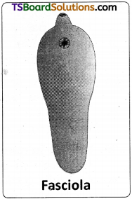
Flukes belong to class Trematoda of phylum platyhelminthes.
TS Inter 1st Year Zoology Study Material Chapter 3 Animal Diversity-I Invertebrate Phyla 2

Class – Trematoda:

  1. They are commonly called flukes.
  2. They are parasitic on other animals.
  3. Body is covered by a thick cuticle (tegument); bears two suckers, an oral and a ventral (acetabulum).
  4. Mouth is anterior and the intestine is bifurcated.
  5. They are bisexual (monoecious).
  6. Life history is complex with many hosts and different types of stages (miracidium, sporocyst, redia, cercaria etc.).

Examples : Fasciola (liver fluke), Schistosoma or Bilharzia (blood fluke).

Question 3.
What are the salient features exhibited by polychaetes?
Answer:
TS Inter 1st Year Zoology Study Material Chapter 3 Animal Diversity-I Invertebrate Phyla 3
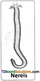
Class – Polychaeta salient features :

  1. Polychaeta (poly : many; chaetae : setae) includes marine annelids commonly called ‘bristle worms’.
  2. Some are free-moving and others burrowing or tubicolous (tube dwelling).
  3. Head is distinct with sense organs such as eyes, tentacles and palps.
  4. Parapodia which bear many setae (hence the name polychaeta) help in locomotion and respiration.
  5. Clitellum is absent.
  6. They are dioecious (unisexual); gonoducts are absent; gametes are shed into the coelom and passed out through the nephridiopores.
  7. Fertilization is external; development includes a trochophore larva.
    Examples : Nereis (sandworm or ragworm or clam worm), Aphrodite (sea mouse), Arenicola (lugwarm).

Question 4.
How do the hirudineans differ from the polychaetes and oligochaetes?
Answer:
Class: Hirudinea.

  1. Hirudinea (hirudo : leech) includes leeches.
  2. All are ectoparasites; majority live in fresh water; some are marine and others live on moist land (terrestrial).
  3. They have dorso-ventrally flattened body with a ‘definite number1 of segments; the segments are externally subdivided into ‘annuli’; internal segmentation is absent.
  4. Suckers help in locomotion.
  5. Clitellum is conspicuous during the breeding season only.
  6. Coelom is filled with a characteristic tissue called botryoidal tissue.
  7. They are monoecious (hermaphroditic); males possesses a copulatory organ, the cirrus.
  8. Fertilization is internal and development is direct.
    Examples: Hirudinaria (freshwater leech), Pontobdella (marine leech), Haemadipsa (land leech).

Question 5.
What are the chief characters of the crustaceans? [May – 2017 – A.P.; March 2015 T.S.]
Answer:
TS Inter 1st Year Zoology Study Material Chapter 3 Animal Diversity-I Invertebrate Phyla 4
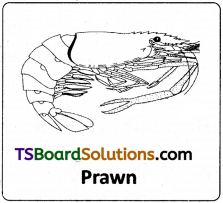
Class – Crustacea – Chief characters :

  1. They are aquatic.
  2. Head and thorax fuse forming the cephalothorax (covered by chitinous carapace). In some the exoskeleton is hardened by calcium carbonate (crabs and lobsters).
  3. Cephalic region bears two pairs of antennae (antennules and antennae – unique feature), one pair of mandibles and two pairs of maxillae.
  4. Thoracic and abdominal appendages are ‘biramous’
  5. Respiratory organs are gills (branchiae).
  6. Excretory organs are green glands or antennary glands.
  7. Sense organs include antennae compound eyes, statocysts, etc.
  8. Development is indirect and includes different larval forms.
    Examples : Palaemon (freshwater prawn), Cancer (crab), Balanus (rock barnacle),
    Sacculina (root-headed barnacle), Astacus (cray fish), Daphnia (water flea).

TS Inter 1st Year Zoology Study Material Chapter 3 Animal Diversity-I: Invertebrate Phyla

Question 6.
Mention the general characters of Arachnida. [May/June ’14]
Answer:
TS Inter 1st Year Zoology Study Material Chapter 3 Animal Diversity-I Invertebrate Phyla 5
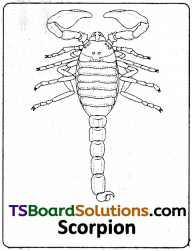
Class – Arachnida general characters :

  1. They are terrestrial.
  2. Prosoma bears a pair of chelicerae, a pair of pedipalpi and four pairs of walking legs.
  3. Mesosomal appendages are modified into book lungs.
  4. Four pairs of posterior abdominal appendages are modified into spinnerets in spiders.
  5. Respiratory organs are book – lungs (scorpions and some spiders), tracheae (some spiders) or both (some spiders).
  6. Respiratory pigments is ‘copper1 containing haemocyanin.
  7. Excretory organs are malpighian tubules and coxal glands.
  8. Development is direct; scorpions are viviparous.
    Example : Palamnaeus (scorpion), Aranea (spider), Sarcoptes (itch mite).

Question 7.
Compare briefly a centipede and a millipede.
Answer:

Centipede (chilopod)Millipede (Diplopod)
1) Commonly called hundred leggers.1) Commonly called thousand leggers.
2) Terrestrial, air breathing carnivorous animals.2) Terrestrial air breathing animals feeding on decaying plant material.
3) Body consists of head and trunk3) Body consists of head and trunk.
4) Each segment of trunk bears a pair of clawed appendages.4) Head bears paired antennae, mandibles and maxillae, (Maxillae modified into gnathochilarium)
5) First pair of trunk appendages bear poison claws.5) Each trunk segment bear two pairs of legs.
6) Respiratory structures are tracheae6) Respiratory structures are tracheae.
7) Excretory organs are Malpighian tubules.
Ex : Scolopendra, Scutigera
7) Excretory organs are Malpighian tubules
Ex: Spirostreptus, Julus.

TS Inter 1st Year Zoology Study Material Chapter 3 Animal Diversity-I: Invertebrate Phyla

Question 8.
Cephalopods show several unique or advanced features when compared to the other molluscs. Discuss briefly.
Answer:
Cephalopoda or Siphonopoda is an advanced group of Mollusca. This class includes cuttle fishes, squids, octopuses, nautili etc. Head is distinct with conspicuous eyes similar to those of vertebrates, a pair of horny beak like jaws and a radula occur in the buccal cavity. Shell may be external and multi – chambered (Nautilus) or internal (Sepia, Loligo) or absent (Octopus). The shell of Sepia is commonly called ‘cuttle bone’ and that of Loligo is commonly called ‘pen’. Foot is modified into eight (octopus) to ten arms (Sepia, Loligo) provided with suckers present around the mouth and a part of the foot is modified into a ‘siphon’ (useful in swift darting movements).

Some possess ink gland and eject a cloud of ink to escape from the predators (defensive adaptation). Ctenidia, atria and nephridia are two in dibranchiates (Sepia) and four in tetrabranchiates (Nautilus). Circulatory system is a closed type (unique feature of cephalopoda); heart’has two to four atria and a ventricle. Nervous system is well developed with a well developed brain enclosed in a cartilaginous cranium (braincase). They are dioecious (sexes are separate); development is direct.
examples : Sepia (cuttle fish), Architeuthis (giant squid – the largest living invertebrate), Nautilus, Octopus (devil fish).

Question 9.
Which class of Mollusca represents the primitive molluscs? What are their chief features?
Answer:
Class Aplacophora represents the primitive molluscs. They are primitive ‘worm like’ marine molluscs without mantle, shell, foot and nephridia. Head is poorly developed; a radula is present. Cuticle contains calcareous spicules. In some there is a mid-ventral groove which is homologous to the foot of the other molluscs. Examples: Neomenia, Chaetoderma.

Question 10.
What are the salient features of the echinoids? [March 2019, ’17 – A.P.]
Answer:

  1. The class Echinoidea includes sea urchins, heart urchins, sand dollars, sea biscuits, etc.
  2. The body is ovoid or discoid and covered with morable spines.
  3. Arms are absent; tube feet bear suckers.
  4. Calcarious ossicles of the body unite to form a rigid test or corona or case.
  5. Madreporite and anus are aboral in position.
  6. Ambulacral grooves are closed.
  7. Pedicellariae are three jawed.
  8. In the mouth of sea urchin a complex five jawed masticatory apparatus called Aristotle’s lantern is present (absent in the heart urchins).
  9. Development includes echinopluteus larava.
    Examples: Echinus (sea urchin), Echinocardium (heart urchin) Echinodiscus (sand dollar).

Question 11.
Mention the salient features of Holothuroidea. [March 2015 – A.P.]
Answer:
Holothuroidea-salient features.

  1. This class includes sea cucumbers.
  2. Body is elongated in the oro-aboral axis.
  3. Skin is leathery (coriaceous) and dermis contains loose spicules.
  4. Arms, spines and pedicellariae are absent. .
  5. Mouth is surrounded by retractile tentacles (modified tube feet useful for feeding).
  6. Ambulacral grooves are ‘closed1; tube feet bear suckers.
  7. Madreporite is internal (occurs in coelom).
  8. Respiratory organs are a pair of cloacal ‘respiratory trees’.
  9. Development is indirect and includes auricularia larva.
    Examples : Holothuria, Synapta, Thyone.

Question 12.
What is the function of Nephridia?
Answer:
The excretory organs occur as segmentally arranged called tubules, called nephridia. Earthworms are mostly ureotelic animals. The chief nitrogenous excretory waste is urea enteronephric nephridia have a role in asmoregulation/conservation/ homeostasis of water. Exonephric nephridia collect nitrogenous wastes and send them out through nephridiopores. They are ciliated coelomic ducts.

Question 13.
How many types of nephridia occur in pheretima and how do you distinguish them?
Answer:
There are 3 types of nephridia. 1) Septal nephridia present on both sides of the inter segmental septa of segments 15th/16th to the last. They open into the intestine. 2) Integumentary nephridia attached to inner body wall from 3rd segment to last. Open out by nephridiopores. 3) Pharyngeal nephridia present as three paired tufts in the segments 4th, 5th and 6th. They open into buccal cavity and pharynx. Septal nephridia with nephrostomes but without nephridiopore. Pharyngeal and integumentary without nephrostomes. Pharyngeal nephridia without Nephrostome and nephridiopore.

TS Inter 1st Year Zoology Study Material Chapter 3 Animal Diversity-I: Invertebrate Phyla

Question 14.
Give an account of the hearts on the circulatory system of pheretima.
Answer:
In pheretima there are 4 pairs of hearts which connect the dorsal and ventral blood vessels in 7, 9, 12,13 segments. They are muscular and valvular. They allow blood from dorsal blood vessel to ventral blood vessel only. The 12th, 13th segment lateral hearts are also called as lateral oesophageal hearts as they receive blood from supra oesophageal blood vessel and give to ventral blood vessel.

Essay Answer Type Questions

Question 1.
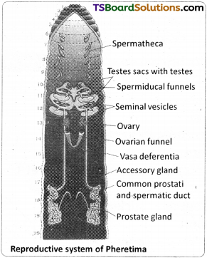
Draw a neat labelled diagram of the reproductive organs of pheretima.
Answer:
TS Inter 1st Year Zoology Study Material Chapter 3 Animal Diversity-I Invertebrate Phyla 6

Question 2.
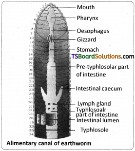
Describe the digestive system and process of digestion in pheretima.
Answer:
Digestive system :
The alimentary canal is a straight tube and runs from the first to the last segment of the body (Fig). The mouth opens into the buccal cavity (1-3 segments) which leads into the muscular pharynx (4th segment). A small narrow tube. Oesophagus (5-7 segments), continues into a muscular gizzard (8th segment). It helps in grinding the small particles of food the decaying leaves (grinding mill). The stomach extends fpm the segments 9 glands, present in the stomach, neutralise the humic acid present in the humus of the soil. The intestine starts from the 15th segment and continues till the last segment.

A pair of short and conical intestinal caecae project from the intestine in the 26th segment. An internal median fold of the dorsal wall of the intestine called typhlosole, helping in increasing the area of absorption, is poorly developed in Pheretima (between the 26th and the rectum, which occupies the last 23 to 28 segments). The alimentary canal opens to the exterior by a small rounded aperture called anus. The ingested soil rich in organic mater passes through the digestive tract where digestive enzymes breakdown complex food into smaller absorbable units. These simpler molecules are absorbed through intestinal membranes and are utilised for various metabolic activities.
TS Inter 1st Year Zoology Study Material Chapter 3 Animal Diversity-I Invertebrate Phyla 7

TS Inter 1st Year Zoology Study Material Chapter 2 Structural Organisation in Animals

Telangana TSBIE TS Inter 1st Year Zoology Study Material 2nd Lesson Structural Organisation in Animals Textbook Questions and Answers.

TS Inter 1st Year Zoology Study Material 2nd Lesson Structural Organisation in Animals

Very Short Answer Type Questions

Question 1.
The body of sponges does not possess tissue level of the organisation, though it is made up of thousands of cells. Comment on it.
Answer:
Sponges belonging to group Parazoa are example of cellular level of organisation. The cells are arranged as loose cells aggregates and do not form tissues. There is division of labour among the cells.

Question 2.
What is ’tissue’ level of organisation among animals? Which metazoans do exhibit this organisation?
Answer:
This is the lowest level of organisation among the eumetazoans, exhibited by diploblastic animals like the cnidarians. In these animals, the cells which perform the same function are arranged into tissues. There is a co-ordination between functioning of cells because of nerve cells and sensory cells.

Question 3.
Animals exhibiting which level of the organisation lead relatively more efficient way of life when compared to those of the other levels of organisation? Why?
Answer:
Animals exhibiting organ system level of organisation lead relatively more efficient way of life when compared to those of the other levels of organisation because highly specialized sensory and nerve cells bring about a higher level of co-ordination and integration among the various organ systems.

Question 4.
What is monaxial heteropolar symmetry? Name the group of animals in which it is the principal symmetry.
Answer:
When any plane passing through the central axis (oro-aboral axis) of the body divides an organism into two identical parts, it is called monaxial heteropolar symmetry. In cnidarians it is the principal symmetry.

Question 5.
Radial symmetry is an advantage to the sessile or slow moving organisms. Justify this statement.
Answer:
Animals showing radial symmetry live in water and they can respond equally to stimuli that arrive from all directions. Thus, radial symmetry is an advantage to sessile or slow moving animals.

TS Inter 1st Year Zoology Study Material Chapter 2 Structural Organisation in Animals

Question 6.
What is cephalization? How is it useful to its possessors? [May 2017 – A.P.; March 2015 – T.S.; March 2013]
Answer:
Cephalization is concentration of nerve and sensory cells at the anterior end. As a result of cephalization, bilaterally symmetrical animals can sense the new environment into which they enter and respond more efficiently and quickly.

Question 7.
Mention the animals that exhibited a ‘tube-within-a-tube’ organisation for the first time. Name their body cavity.
Answer:
A tube-with-in-a-tube organisation for the first time is seen in the group Nematoda. Their body cavity is named as Pseudocoelam.

Question 8.
Why is the true coelom considered a secondary body cavity? [March 2015 – T.S.]
A. During the embryonic development of the eucoelomates, the blastocoel is replaced by true coelom derived from the mesoderm. So, the true coelom is also called ‘Secondary body cavity’.

Question 9.
What are retroperitoneal organs? [March 2018 – A.P.]
Answer:
Certain organs such as the kidneys of the vertebrates are covered by the parietal peritoneum only on their ventral side. Such a peritoneum is called retroperitoneum and the organs lined by it are called ‘retroperitoneal organs’.

Question 10.
If the mesentoblast cell is removed in the early embryonic development of protostomes, what would be the fate of such animals?
Answer:
The 4d blastomere or mesentoblast cell Of protostomes divides to form mesodermal blocks between the ectoderm and endoderm. The split that appears in each mesodermal block leads to the formation of schizocoel (true body cavity). If mesentoblast cell is removed, the true coelom will not be formed.

Question 11.
What is enterocoelom? Name the enterocoelomate phyla in the animal kingdom. [March 2014]
Answer:
Animals in which the body cavity is formed from the mesodermal pouches of archenteron are called enterocoelomates. Echinoderms, hemichordates and chordates are the enterocoelomates.

Question 12.
Stratified epithelial cells have limited role in secretion. Justify their role in our skin.
Answer:
Stratified epithelium is made up of more than one layer of cells. Its main function is to provide protection against chemical and mechanical stress. It covers the dry surface of the skin. Hence it has limited role in secretion.

TS Inter 1st Year Zoology Study Material Chapter 2 Structural Organisation in Animals

Question 13.
Distinguish between exocrine and endocrine glands with examples. [March 2014]
Answer:

  1. Exocrine glands are provided with ducts : secrete mucus, saliva, earwax, oil, milk, digestive enzymes and other cell products.
  2. Endocrine glands are ductless and their products are hormones which are not sent out via ducts but are carried to the target organs by blood, eg : Thyroid glands secreting hormone Thyroxine.

Question 14.
Distinguish between holocrine and apocrine glands.
Answer:

  1. Apocrine glands (eg : mammary glands) in which the apical part of the cell is pinched off along with the secretory product.
  2. Holocrine glands (e.g : sebaceous glands) in which the entire cell disintegrates to discharge the contents.

Question 15.
Mention any two substances secreted by mast cells and their functions. [May/June, March 2014]
Answer:

  1. Heparin – an anticoagulant (prevents blood clotting)
  2. Histamine – vasodilators (cause inflammation in response to injury and infection.)

Question 16.
Distinguish between a tendon and a ligament. [May 2017-A.P.; March 2017, 15, March 2019]
Answer:

  1. Tendons attach the skeletal muscles to bones.
  2. Ligaments attach bones to other bones.

Question 17.
Distinguish between brown fat and white fat.
Answer:
1) White adipose tissue (WAT) :
It is mostly in adults. Adipocyte has a single large lipid droplet (monolocular). White fat is metabolically not active.

2) Brown adipose tissue (BAT) :
It is found in foetuses and infants. Adipocyte of BAT has several small lipid droplets and numerous mitochondria. Brown fat is metobolically active and generates heat to maintain body temperature required by infants.

TS Inter 1st Year Zoology Study Material Chapter 2 Structural Organisation in Animals

Question 18.
What is the strongest cartilage? In which regions of the human body, do you find it? [March 2020 ; Mar 15 – T.S.]
Answer:
Fibrous cartilage is the strongest of all types of cartilage. It occurs in the intervertebral discs and pubic symphysis of the pelvis.

Question 19.
Distinguish between osteoblasts, and osteoclasts. [March 2017 – A.P.]
Answer:
Osteoblasts (Immature bone cells) secrete the organic components (collagen fibres) of matrix and also play a major role in ‘mineralization of bone’.

Osteoclasts are phagocytic cells involved in resorption of bone.

Question 20.
Define osteon. [March 2015 – A.P.]
Answr:
In a dense bone a Haversian canal and the surrounding lamillae (rows of osteocytes) and lacunae are collectively called a Haversian system or Osteon.

Question 21.
What are Volkmann’s canals? What is their role?
Answer:
The Haversian canals communicate with one another, with the periosteum and also with the marrow cavity by transverse or oblique canals called Volkmann’s canals.

Question 22.
What is a Sesamoid bone? Give an example. [May/June 2014]
Answer:
Sesamoid bones are formed by ossification in tendons. Eg : Patella (Knee cap)

Question 23.
What is lymph? How does it differ from plasma?
Answer:

  1. Lymph is a colourless fluid. It lacks RBC, platelets and large plasma proteins, but has more number of Leucocytes.
  2. Plasma is the fluid matrix of blood. It consists of 92% of water and 8% of solutes.

Question 24.
What is the haematocrit value? [Mar. 2019, ’17, May ’17 – A.P ; May/June 2014]
Answer:
The percentage of total volume occupied by RBCs is called haematocrit value.

Question 25.
What are intercalated discs? What is their significance?
Answer:
The dark lines across cardiac muscle are called intercalated discs (IDs). These discs are highly characteristic of the cardiac muscle. They act as boosters of muscle contraction.

TS Inter 1st Year Zoology Study Material Chapter 2 Structural Organisation in Animals

Question 26.
“Cardiac muscle is highly resistant to fatigue.” Justify. [March 2020]
Answer:
The cardiac muscle js highly resistant to fatigue because it has numerous sarcosomes, many molecules of myoglobin (oxygen storing pigment) and copious supply of blood which facilitate “Continuous aerobic respiration”.

Question 27.
Distinguish between ‘nucleus’ and ‘ganglion’ with respect to the nervous system.
Answer:
A group of cell bodies in the central nervous system is called a ‘nucleus’ and in the peripheral nervous system, it is called a ‘ganglion’.

Question 28.
Distinguish between tracts and nerves with respect to the nervous system.
Answer:
Groups of axons (nerve fibres) in the central nervous system (CNS) are called tracts and in the peripheral nervous system (PNS) they are called nerves.

Question 29.
Name the glial cells that form myelin sheath around the axons of central nervous system and peripheral nervous system respectively.
Answer:
Myelin sheath around the axons of central nervous system is formed from ‘oligodendrocytes.‘ In peripheral nervous system myelin sheath around axon is formed from ‘Satellite cells’ a kind of glial cells.

Question 30.
Distinguish between white matter and grey matter of ‘CNS’
Answer:
Myelinated nerve fibres occur in the white matter of CNS. Non-myelinated nerve fibres occur in the grey matter of the CNS and autonomus nervous system.

TS Inter 1st Year Zoology Study Material Chapter 2 Structural Organisation in Animals

Question 31.
What are microglia and what is their origin and add a note on their function. [March 2018 – A.P.]
Answer:
Microglial cells which are phagocytic cells, of mesodermal origin. They become activated into ‘phagocytes’ when there is infection or injury in the nervous system.

Question 32.
What are pseudounipolar neurons? Where do you find them?
Answer:
Unipolar neurons are also called pseudounipolar neurons. They are found jn the ‘dorsal root ganglion’ of spinal nerve.

Short Answer Type Questions

Question 1.
Describe the four different levels of organization in metazoans.
Answer:
The levels of organisation in metazoans are as follows.
TS Inter 1st Year Zoology Study Material Chapter 2 Structural Organisation in Animals 1

Cellular level of organisation :
It is the lowest level of organisation among the metazoans and is exhibited by the sponges (parazoans). Different types of cells are functionally isolated due to the absence sensory and nerve cells. The cells are arranged as ‘loose cell aggregates’ and do not form tissues. There is division of labour among the cells.

Tissue level of organisation :
This is the lowest level of organization among the eumetazoans, exhibited by diploblastic animals like the cnidarians. In these animals, the cells which perform the same function are arranged into tissues. The cells of a tissue together perform their common function as a highly coordinated unit and this coordination is due to the presence of nerve cells and sensory cells.

Organ level of organisation :
An aggregation of different kinds of tissues which is specialized for a particular function is called an organ. Organ level of organisation appeared for the first time in the members of the Phylum Platyhelminthes.

Organ – system level of organisation :
It is the highest level of organisation among the animals and is exhibited by the triploblastic animals such as the flat worms, nematodes, annelids, arthropods, molluscs, echinoderms and chordates. In the triploblastic animals, the evolution of ‘mesoderm’ resulted in structural complexity. In these animals, the tissues are assembled to form organs and complex organ – systems. Highly specialized sensory and nerve cells bring about a higher level of coordination and integration among the various organ systems to lead an efficient way of life.

Question 2.
In which group of bilaterians do you find solid bauplan? Why it is called so?
Answer:
Solid bauplan is seen in acoelomate bilaterians eg : Platyhelminthes.
TS Inter 1st Year Zoology Study Material Chapter 2 Structural Organisation in Animals 3

The bilaterian animals in which the body cavity is absent are called acoelomates. In these animals, the mesenchyme derived from the third germinal layer, called mesoderm, occupies the entire blastocoel, between the ectoderm and the endoderm, so that the adults have neither the primary cavity (blastocoelom) nor the secondary cavity (coelom). As there is no body cavity, the acoelomates exhibit solid body plan. Problems faced by the acoelomates due to absence of perivisceral cavity are – their internal organs cannot move freely, as they are embedded in the mesenchyme, diffusion of material from the gut to the body wall is made slow and less efficient.

TS Inter 1st Year Zoology Study Material Chapter 2 Structural Organisation in Animals

Question 3.
Mention the advantages of coelom over pseudocoelom.
Answer:
Advantages of coelom over pseudocoelom :

  1. Visceral organs of eucoelomates are muscular (because of their association with mesoderm) and so they can contract and relax freely independent of the muscular movements of the body wall in the coelomic space, e.g. peristaltic movements of alimentary canal.
  2. Gametes are released into the coelom in some invertebrates (which do not have gonoducts) and in the female vertebrates.
  3. Coelomic fluid receives excretory products and stores them temporarily before their elimination.
  4. In the eucoelomates, the mesoderm comes into contact with the endoderm of the alimentary canal, and it causes ‘regional specialization of the gut, such as the development of gizzard, stomach etc… This is referred to as ‘primary induction’. In the case of the pseudocoelomates, due to the absence of such a contact between the gut and the mesoderm, the wall of the gut does not show complex and highly specialized organs.

Question 4.
Describe the formation of schizocoelom and enterocoelom.
Answer:
Formation of Schizocoelom: Animals in which the body cavity is formed by ‘splitting of mesoderm’ are called schizocoelomates. Annelids, arthropods and molluscs are schizocoelomates in the animal kingdom. All the schizocoelomates are protostomians and they show ‘holoblastic’, ‘spiral’ and ‘determinate’ cleavage. The 4d blastomere or mesentoblast cell of the early embryo divides to form mesodermal blocks between the ectoderm and the endoderm and replaces the blastocoel. The split that appears in each mesodermal block leads to the formation of Schizocoelom. Eg : Annelida, Arthropoda, Mollusca.
TS Inter 1st Year Zoology Study Material Chapter 2 Structural Organisation in Animals 4

Formation of enterocoelom :
Animals in which the body cavity is formed from the mesodermal pouches of archenteron are called enterocoelomates. Echinoderms, hemichordates and chordates are the enterocoelomates. In these animals, mesodermal pouches that evaginate from the wall of the archenteron into the blastocoel are fused with one another to form the enterocoelom. All the enterocoelomates are deuterostomes and they show radial and indeterminate cleavage.
TS Inter 1st Year Zoology Study Material Chapter 2 Structural Organisation in Animals 5

Question 5.
Describe briefly about the three types of intercellular junctions of epithelial tissues.
Answer:
There are three types of intercellular junctions of epithelial tissues.

A. Tight junctions :
These junctions between epithelial cells prevent ‘leakages’ of body fluids. For example, they prevent leakage of water into the surrounding cells in our sweat glands (making our skin water-tight).

B. Desmosomes :
Muscle cells are provided with ‘desmosomes (anchoring junctions) which act as ‘rivets’ binding the cells together into strong sheets. Intermediate filaments made of the protein ‘keratin’, anchor desmosomes in the cytoplasm.
TS Inter 1st Year Zoology Study Material Chapter 2 Structural Organisation in Animals 6

C. Gap junctions :
They provide continuous ‘cytoplasmic channels’ between adjacent cells (comparable to the ‘plasmodesmata’ between adjacent plant cells). Various types of ions, sugar molecules, amino acids etc., can pass from a cell to an adjacent cell through ‘gap junctions’. They occur in many types of tissues including the ‘cardiac muscles’, where they allow rapid conduction of impulses or depolarisation.

Question 6.
Give an account of glandular epithelium. [March 2015 – A.P.]
Answer:
Some of the columnar or cuboidal cells that get specialised for the production of certain secretions, form glandular epithelium. The glands are of two types – unicellular glands consisting of isolated glandular cells such as goblet cells of the gut, and multicellular glands consisting of clusters of cells such as salivary glands.
TS Inter 1st Year Zoology Study Material Chapter 2 Structural Organisation in Animals 7

On the basis of the mode of pouring of their secretions, glands are divided into two types namely exocrine and endocrine glands. Exocrine glands are provided with ducts; secrete mucus, saliva, earwax (cerumen), oil, milk, digestive enzymes and other cell products. In contrast, endocrine glands are ductless and their products are ‘hormones’, which are not sent out via ducts, but are carried to the target organs by blood. Based on the mode of secretion, exocrine glands are further divided into i. Merocrine glands (e.g. pancreas) which release the secretory granules without the loss of other cellular material ii. Apocrine glands (e.g. mammary glands) in which the apical part of the cell is pinched off along with the secretory product and iii. Holocrine glands (e.g. sebaceous glands), in which the entire cell disintegrates to discharge the contents.

TS Inter 1st Year Zoology Study Material Chapter 2 Structural Organisation in Animals

Question 7.
Give a brief account of the cells of areolar tissue.
Answer:
TS Inter 1st Year Zoology Study Material Chapter 2 Structural Organisation in Animals 8
Cells of the areolar tissue are fibroblasts, mast cells, macrophages, adipocytes and plasma cells.

  1. Fibroblasts are the most common cells which secrete fibres. The inactive cells are called fibrocytes.
  2. Mast cells secrete heparian (an anticoagulant), histamine, bradykinin (vasodilators), and serotonin (vasoconstrictor). Vasodilators cause inflammation in response to injury and infection.
  3. Macrophages are amoeboid cells, phagocytic in function and act as internal scavengers. They are derived from the monocytes of blood. ‘Tissue fixed macrophages’ are called histiocytes and others are ‘wandering macrophages’.
  4. Plasma cells are derived from the B-lymphocytes and produce antibodies.
  5. Adipocytes are specialized cells for the storage of fats.

Question 8.
Describe the three types of cartilage. [March 2020, ’17]
Answer:
The three types of cartilage are 1. Hyaline cartilage 2. Elastic cartilage 3. Fibrous cartilage.

1. Hyaline cartilage :
It is bluish-white, translucent and glass – like cartilage. Matrix is homogeneous and shows delicate collagen fibres. It is the weakest and the most common type of all the cartilages. Perichondrium is present except in articular cartilages. It forms the embryonic endoskeleton of bony vertebrates, endoskeleton of cyclostomes and cartilaginous fishes. It forms the articular cartilages (free surfaces of long bones that form joints), costal cartilages (sternal parts of ribs), and the epiphyseal plates. It also forms the nasal septal cartilage, cartilaginous rings of trachea, bronchi and cartilages of larynx.
TS Inter 1st Year Zoology Study Material Chapter 2 Structural Organisation in Animals 9

2) Elastic cartilage :
It is yellowish due to elastic fibres. Matrix has abundance of yellow elastic fibres in addition to collagen fibres. It provides strength and elasticity. Perichondrium is present. It is found in the pinnae of the external ears, Eustachian tubes and epiglottis.

3) Fibrous cartilage :
Matrix has bundles of collagen fibres. Perichondrium is absent. It is the strongest of all types of cartilages. It occurs in the intervertebral discs and pubic symphysis of the pelvis.

Question 9.
Explain Haversian system. [May 2017 – A.P; March 2014]
Answer:
In a bone between the outer and inner circumferential lamellae, there are many Haversian systems. The spaces between the Haversian systems are filled with interstitial lamellae. Haversian system consists of a Haversian canal that runs parallel to the marrow cavity. It contains an artery, a vein and a lymphatic vessel. Haversian canal is surrounded by concentric lamellae, small fluid filled spaces called ‘lacunae’ provided with minute canaliculi lie in between the lamellae. Canaliculi connect the lacunae with one another and with Haversian canal. Each lacuna encloses one osteocyte (inactive form of osteoblast).

The cytoplasmic processes of osteocytes extend through canaliculi. A Haversian canal and the surrounding lamellae and lacunae are collectively called a Haversian system or osteon. The Haversian canals communicate with one another, with the periosteum and also with the marrow cavity by transverse or oblique canals called Volkmanns canals. Nutrients and gases diffuse from the vascular supply of Haversian canals.
TS Inter 1st Year Zoology Study Material Chapter 2 Structural Organisation in Animals 10

Question 10.
Write short notes on Lymph.
Answer:
Lymph :
Lymph is a colourless fluid. It lacks RBC, platelets and large plasma proteins, but has more number of leucocytes. It is chiefly composed of plasma and lymphocytes. When compared to the tissue fluid, it contains very small amounts of nutrients and oxygen but has abundant C02 and other metabolites. The most important site of formation of lymph is interstitial space. As blood passes through the blood capillaries, some portion of blood that includes water, solutes and proteins of low molecular weight passes through the walls of capillaries, into the interstitial spaces due to hydrostatic pressure at the arteriolar ends.

This fluid forms the interstitial fluid (tissue fluid). Most of the interstitial fluid is returned directly to the capillaries due to osmotic pressure at the venular ends. Little amount of this tissue fluid passes through a system of lymphatic capillaries (lymph capillaries of the intestinal villi are called ’lacteals’), vessels, ducts and finally reach the blood through the subclavian veins. The extracecellular ’tissue fluid’ that passes into the lymph capillaries and lymph vessels is called ‘lymph’. Lymphatic system represents an ‘accessory route’ by which interstitial fluid flows from tissue spaces into blood.

Question 11.
Describe the structure of a skeletal muscle. [March 2018 – A.P.; March 2015 – T.S.]
Answer:
TS Inter 1st Year Zoology Study Material Chapter 2 Structural Organisation in Animals 11
Skeletal (striped and voluntary) muscle: It is usually attached to skeletal structures by ‘tendons’. In a typical muscle such as the ‘biceps’ muscle, skeletal muscle fibre is surrounded by a thin connective tissue sheath, the endomysium. A bundle of muscle fibres is called a fascicle. It is surrounded by a connective tissue sheath called perimysium. A group of fascicles form a ‘muscle’ which is surrounded by an epimysium (outermost connective tissue sheath). These connective tissue layers may extend beyond the muscle to form a chord-like tendon or sheet-like aponeurosis.

A skeletal muscle fibre is a long, cylindrical and unbranched cell. It is a multinucleated cell with many oval nuclei characteristically in the “peripheral” cytoplasm (a syncytium formed by fusion of cells). Sarcoplasm has many myofibrils which show alternate dark and light bands. So it is called striped or striated muscle. Skeletal muscle usually works under the conscious control of an organism (a voluntary muscle). Skeletal muscle contracts quickly and undergoes fatigue quickly. They are innervated by the ‘somatic nervous system’. Satellite cells are quiescent (quiet and inactive), mononucleate and myogenic cells and help in regeneration, which is ‘limited’.

TS Inter 1st Year Zoology Study Material Chapter 2 Structural Organisation in Animals

Question 12.
Describe the structure of a cardiac muscle. [March 2013]
Answer:
TS Inter 1st Year Zoology Study Material Chapter 2 Structural Organisation in Animals 12
Cardiac (striped and involuntary) muscle :
The cardiac muscle is striated like the skeletal muscle (shows sarcomeres). Cardiac muscle is found in the ‘myocardium’ of the heart of vertebrates. The cardiac muscle cells or the ‘myocardial cells’ are short, cylindrical, mononucleate or binucleate cells whose ends branch and form junctions with other cardiac muscle cells. Each myocardial cell is joined to adjacent myocardial cells by ‘electrical synapses’ or ‘gap junctions’. They permit ‘electrical impulses’ to be conducted along the long axis of the cardiac muscle fibre. The dark lines across cardiac muscle are called intercalated discs (IDs). These discs are highly characteristic of the cardiac muscle.

The cardiac muscle is highly resistant to fatigue, because it has numerous sarcosomes, many molecules of myoglobin (oxygen storing pigment) and copious supply of blood which facilitate ‘continuous aerobic respiration’.

Question 13.
Give an account of the supporting cells of Nervous tissue.
Answer:
Neuroglia (supporting cells):
These are the supporting and non-conducting cells that provide a microenvironment suitable for neuronal activity. Unlike neurons, they continue to divide throughout the life. Neuroglial cells of the CNS include oligodendrocytes; astrocytes (star shaped cells) that form interconnected network and bind neurons and capillaries (helping in providing blood-brain barrier); ependymal cells, which are ciliated cells that line the cavities of brain and spinal cord to bring movements in the cerebrospinal fluid; microglial cells, which are phagocytic cells, of mesodermal origin. Neuroglial cells of the peripheral nervous system include the satellite cells and Schwann cells. Satellite cells surround the cell bodies in ganglia, and Schwann cells form neurilemma around axons.

Question 14.
Describe the structure of a multipolar neuron.
Answer:
A neuron usually consists of a “cell body” with one to many dendrites and a single axon.
TS Inter 1st Year Zoology Study Material Chapter 2 Structural Organisation in Animals 13

Cell body :
It is also called perikaryon, cyton or soma. It contains abundant granular cytoplasm and a large spherical nucleus. The cytoplasm has Nissl bodies (they represent RER, the sites of protein synthesis), neurofibrils and lipofuscin granules (the products of cellular wear and tear, accumulating in lysosomes with age). A group of cell bodies in the central nervous system is called a ‘nucleus’, and in the peripheral nervous system, it is called a ‘ganglion’.

Dendrites :
Several short, branched processes which arise from the cyton are called dendrites. They also contain Nissl bodies and neurofibrils. They conduct nerve impulses towards the cell body
(afferent processes).

Axon :
An axon is a single, long, cylindrical process that originates from a region of the cyton called axon hillock. Plasmalemma of an axon is called axolemma, and the cytoplasm is called axoplasm, which contains neurofibrils. However, Nissl bodies are absent. An axon may give rise to collateral branches. Distally it branches into many fine filaments called telodendria, (axon terminals), which end in bulb like structures called synaptic knobs or terminal boutons. Synaptic knobs possess ‘synaptic vesicles’ containing chemicals called neurotransmitters. Axon transmits nerve impulse away from the cyton (efferent process) to an interneuronal or neuromuscular junction called synapse.

TS Inter 1st Year Zoology Study Material Chapter 2 Structural Organisation in Animals

Question 15.
Write short notes on (A) Platelets (B) Synapse.
Answer:
TS Inter 1st Year Zoology Study Material Chapter 2 Structural Organisation in Animals 14
A) Blood platelets (Thrombocytes) :
These are colourless non-nucleated, round or oval biconvex discs. Number of platelets per cubic mm of blood is about 2,50,000 – 4,50,000. They are formed from giant megakaryocytes produced in the red bone marrow by fragmentation. The average life-span of blood platelets is about 5 to 9 days. They secrete thromboplastin and play an important role in blood clotting. They adhere to the damaged endothelial lining of capillaries and seal minor vascular openings.

B) Synapse :
An axon distally branches into many fine filaments called telodendria (Axon terminals) which end in bulb like synaptic knobs or terminal boutons. Synaptic knobs possess synaptic vesicles containing chemicals called neurotransmitters. Axon transmits nerve impulse away from the cyton into an interneuronal or neuromuscular junction called synapse. Synapse is the smallest gap between telodendria of a neuron and dendrites of next neuron.

Essay Answer Type Questions

Question 1.
What is a coelom? Explain the different types of coelom with suitable examples and neat labelled diagrams.
Answer:
Coelom :
The term ‘coelom’ was coined by Haeckel. The body cavity, which is lined by mesoderm, is called coelom. More elaborately, coelom is a fluid-filled space between the body wall and visceral organs and lined by mesodermal epithelium, the peritoneum. Animals possessing coelom are called coelomates/ eucoelomates. Evolution of efficient organ systems was not possible until the evolution of coelom for supporting the organs and distributing material.

Acoelomate bilaterians:
TS Inter 1st Year Zoology Study Material Chapter 2 Structural Organisation in Animals 15
The bilaterian animals in which the body cavity is absent are called acoelomates, e.g. Platyhelminthes (lowest bilaterians). In these animals, the mesenchyme derived from the third germinal layer, called mesoderm, occupies the entire blastocoel, between the ectoderm and the endoderm, so that the adults have neither the primary cavity (blastocoelom). nor the secondary cavity (coelom) As there is no body cavity, the acoelomates exhibit solid body plan. Problems faced by the acoelomates due to absence of perivisceral cavity are – their internal organs cannot move freely, as they are embedded in the mesenchyme, diffusion of material from the gut to the body wall is made slow and less efficient.

Pseudocoelomate bilaterians :
TS Inter 1st Year Zoology Study Material Chapter 2 Structural Organisation in Animals 16
In some animals, the body cavity is not lined by mesodermal epithelia. Such animals are called Pseudocoelomates. They include the members of phylum Aschelminthes (Nematoda, Rotifera and some minor phyla). During the embryonic development mesoderm (mesenchyme) occupies only a part of the blastocoel adjoining the ectoderm. The unoccupied portion of the blastocoel persists as pseudocoelom, which is filled with pseudocoelomic fluid. Pseudocoelomates are the first animals to exhibit a ‘tube-within-a-tube’ organisation.

As the gut wall is made of only endodermal epithelium, diffusion of digested food from the lumen of the gut into the surrounding pseudocoelomic fluid becomes easier and the absence of circulatory system is thus compensated. Though it is called pseudocoelom (false coelom), it performs almost all the functions of a regular coelom. Pseudocoelomic fluid of pseudocoelomates serves as a hydrostatic skeleton and a ‘shock absorber’. It allows the free movements of visceral organs, helps in the circulation of nutrients, and storage of nitrogenous wastes.

Eucoelomate bilaterians:
TS Inter 1st Year Zoology Study Material Chapter 2 Structural Organisation in Animals 17
Coelom or ‘true coelom’ is a fluid-filled cavity, that lies between the body wall and the visceral organs and is lined by mesodermal epithelium, the peritoneum. The portion of the peritoneum that underlines the body wall is the parietal peritoneum or somatic peritoneum. The portion of the peritonieum that covers the visceral organs is the splanchnic peritoneum or visceral peritoneum. In coelomates, the visceral organs are suspended in the coelom by the peritoneum. A double layered peritoneum that connects some visceral organs to the body wall is called mesentery.

In some eucoelomates such as the annelids, the dorsal and ventral mesenteries divide the coelom into paired compartments. Certain organs such as the kidneys of the vertebrates are covered by the parietal peritoneum only on their ventral side. Such a peritoneum is called the ‘retroperitoneum’ and the organs lined by it are called ‘retroperitoneal organs’.

During the embryonic development of the eucoelomates, the blastocoel is replaced by true coelom derived from the mesoderm. So, the true coelom is also called ‘secondary body cavity’. In the eucoelomates, mesodermal epithelium (peritoneum) lines both the body wall and the walls of the visceral organs. So, visceral organs become ‘muscular’ and exhibit free movements ‘independent’ of the movement of the body wall in the coelomic fluid. As the wall of the gut becomes thick and muscular, digested food and other nutrients cannot diffuse from the lumen of the gut into the coelom. Circulatory system (blood vascular system) is developed in the eucoelomates to overcome this problem. Based on the mode of formation of coelom, the eucoelomates are classified into two types :

I. Schizocoelomates:
Animals in which the body cavity is formed by spilitting of mesoderm’ are called schizocoelomates. Annelids, arthropods and molluscs are schizocoelomates in the animal kingdom. All the schizocoelomates are protostomians and they show ‘holoblastic’, ‘spiral’ and ‘determinate’ cleavage. The 4d blastomere or mesentoblast cell of the early embryo divides to form mesodermal blocks between the ectoderm and the endoderm and replaces the blastocoel. The split that appears in each mesodermal block leads to the formation of’schizocoelom’ (split coelom).
TS Inter 1st Year Zoology Study Material Chapter 2 Structural Organisation in Animals 18

In annelids, the functional body cavity (perivisceral cavity) is schizocoelom. It is in the form of a series of paired coelomic cavities, but in arthropods and molluscs, the functional body cavity that lies around visceral organs is filled with blood (haemolymph) and is called haemocoel. It is formed by the fusion of the embryonic blastocoel with some coelomic spaces and the tissues are directly bathed in the blood (haemolymph).

II. Enterocoelomates:
Animals in which the body cavity is formed from the mesodermal pouches of archenteron are called enterocoelomates. Echinoderms, hemichordates and chordates are the enterocoelomates. In these animals, mesodermal pouches that evaginate from the wall of the archenteron into the blastocoel are fused with one another to form the enterocoelom. All the enterocoelomates are deuterostomes and they show radial and indeterminate cleavage.
TS Inter 1st Year Zoology Study Material Chapter 2 Structural Organisation in Animals 19

TS Inter 1st Year Zoology Study Material Chapter 2 Structural Organisation in Animals

Question 2.
What is symmetry? Describe the different types of symmetry in the animal kingdom with suitable examples.
Answer:
Importance of Symmetry :
The concept of symmetry is fundamental in understanding the organisation of an animal. Symmetry in animals is balanced distribution of paired body parts. The body plan of a vast majority of metazoans exhibits some kind of symmetry. However, most of the sponges and snails show asymmetry (lack of symmetry). The symmetry of an animal and its mode of life are correlated.

Asymmetry :
TS Inter 1st Year Zoology Study Material Chapter 2 Structural Organisation in Animals 20
The animals, which cannot be cut into two equal parts (antimeres) in any plane passing through the centre of the body are called asymmetrical, e.g. most sponges and adult gastropods. In the asymmetrical animals, the body lacks a definite form. Asymmetry cannot be said to be an adaptation or advantage to an organism. Most of the asymmetrical organisms do not develop complex sensory and locomotor functions.

Symmetry :
The regular arrangement of body parts in a geometrical design relative to the axis of the body is called symmetry. The animals, which can be cut into two equal parts, or antimeters in one or more planes passing through the ‘principal axis’ of the body are called symmetrical animals. In a symmetrical animal, paired body parts are arranged on either side of the plane passing through the principal axis, such that they are equidistant from the plane. The unpaired body parts are located mostly on the plane, passing through the principal axis. Basically, the symmetry in animals is of two kinds.
i) Radial symmetry and
ii) Bilateral symmetry

i) Radial Symmetry or Monaxial heteropolar (axis is single; poles are different) Symmetry :
TS Inter 1st Year Zoology Study Material Chapter 2 Structural Organisation in Animals 21 TS Inter 1st Year Zoology Study Material Chapter 2 Structural Organisation in Animals 22
When any plane passing through the central axis (oro-aboral axis/ principal axis) of the body divides an organism into two identical parts, it is called radial symmetry. The animals with radial symmetry are either sessile or planktonic or sluggish forms. It is the principal symmetry of the diploblastic animals such as the cnidarians and ctenophores (considered as biradial animals by some authors).

Animals showing radial symmetry live in water and they can respond equally to stimuli that arrive from all directions. Thus, radial symmetry is an advantage to sessile or slow moving animals. However, triploblastic animals such as echinoderms are ‘secondarily radially symmetrical’ (as it is five angled, it is also called pentamerous radial symmetry). Radially symmetrical animals have many planes of symmetry, whereas pentamerous radially symmetrical animals have five planes of symmetry.

ii) Bilateral symmetry :
When only one plane (median sagittal plane) that passes through the central axis (anterior – posterior axis) divides an organism into two identical parts, it is called bilateral symmetry. It is the ‘principal type of symmetry’ in the triploblastic animals. Among the triploblastic animals, some gastropods become secondarily asymmetrical, though they have primarily bilaterally symmetrical larvae.
TS Inter 1st Year Zoology Study Material Chapter 2 Structural Organisation in Animals 23

Question 3.
Classify and describe the epithelial tissues on the basis of structural modification of cells with examples.
Answer:
There are two types of epithelial tissues namely ‘simple epithelia’ and ‘compound epithelia’ based on the number of layers or strata. Various glands in the body involved in secretions are made up of epithelial tissue (glandular epithelium).

A) Simple epithelium :
Simple epithelium is composed of a single layer of cells and forms the lining of body cavities, ducts and vessels. It helps in diffusion, absorption, filtration and secretion of substances. On the basis of the shape of the cells, it is further divided into three types :

i) Simple squamous epithelium (Pavement epithelium) :
TS Inter 1st Year Zoology Study Material Chapter 2 Structural Organisation in Animals 24
It is composed of a single layer of flat and tile-like cells, each with a centrally located ‘ovoid nucleus’. It is found in endothelium of blood vessels, mesothelium of body cavities (pleura, peritoneum, and pericardium), wall of Bowman’s capsule of nephron, lining of alveoli of lungs, etc.

ii) Simple cuboidal epithelium :
TS Inter 1st Year Zoology Study Material Chapter 2 Structural Organisation in Animals 25
It is composed of a single layer of cube-like cells with centrally located spherical nuclei. It is found in germinal epithelium, proximal and distal convoluted tubules of nephron. Cuboidal epithelium of proximal convoluted tubule of nephron has ‘microvilli’.

iii) Simple columnar epithelium :
It is composed of a single layer of tall and slender cells with oval nuclei located near the base. It has mucus – secreting’ goblet cells’ in some places. It is of two types.

a) Ciliated columnar epithelium :
TS Inter 1st Year Zoology Study Material Chapter 2 Structural Organisation in Animals 26
Columnar epithelial cells have cilia on their free surface. It is mainly present in the inner surface of hollow organs like fallopian tubes, ventricles of brain, central canal of spinal cord, bronchioles etc.

b) Non-ciliated columnar epithelium :
TS Inter 1st Year Zoology Study Material Chapter 2 Structural Organisation in Animals 27
Columnar cells are without cilia. It is found in the lining of Stomach and intestine. Microvilli are present in the columnar epithelium of intestine to increase the surface area of absorption.
TS Inter 1st Year Zoology Study Material Chapter 2 Structural Organisation in Animals 28

B) Compound epithelium (stratified epithelium):
It is made up of more than one layer of cells. Its main function is to provide protection against chemical and mechanical stress. It covers the dry surface of the skin as stratified, keratinized, squamous epithelium. It covers the moist surface of buccal cavity, pharynx, oesophagus and vagina as stratified non – keratinized squamous epithelium. It forms the inner lining of the larger ducts of salivary glands, sweat glands and pancreatic ducts as stratified cuboidal epithelium. It forms the wall of the urinary bladder as transitional epithelium.

c) Glandular epithelium :
Some of the columnar or cuboidal cells that get specialised for the production of certain secretions, form glandular epithelium. The glands are of two types – unicellular glands consisting of-isolated glandular cells such as goblet cells of the gut, and multicellular glands, consisting of clusters of cells such as salivary glands. On the basis of the mode of pouring of their secretions, glands are divided into two types namely exocrine and endocrine glands. Exocrine glands are provided with ducts; secrete mucus, saliva, earwax (cerumen), oil, milk, digestive enzymes and other cell products. In contrast, endocrine
TS Inter 1st Year Zoology Study Material Chapter 2 Structural Organisation in Animals 29
glands are ductless and their products are ‘hormones’, which are not sent out via ducts, but are carried to the target organs by blood. Based on the mode of secretion, exocrine glands are further divided into i. Merocrine glands (e.g. pancreas) which release the secretory granules without the loss of other cellular material ii. Apocrine glands (e.g. mammary glands) in which the apical part of the cell is pinched off along with the secretory product and iii. Holocrine glands (e.g. sebaceous glands), in which the entire cell disintegrates to discharge the contents.
TS Inter 1st Year Zoology Study Material Chapter 2 Structural Organisation in Animals 30

TS Inter 1st Year Zoology Study Material Chapter 2 Structural Organisation in Animals

Question 4.
Describe the various types of connective tissue proper with suitable examples.
Answer:
Connective tissue proper:
It is of two types.

A) Loose connective tissue :
Cells and fibres are loosely arranged in a semi fluid ground substance. There are three types of loose connective tissues – areolar tissue, adipose tissue and reticular tissue.

i. Areolar tissue :
It is one of the most widely distributed connective tissues in the body. It forms the packing tissue in almost all the organs. Areolar tissue forms subcutaneous layer of the skin. It has cells and fibres. Cells of the areolartissue are fibroblasts, mast cells, macrophages, adipocytes and plasma cells.

  1. Fibroblasts are the most common cells which secrete fibres. The inactive cells are called fibrocytes.
  2. Mast cells secrete heparin (in anticoagulant), histamine, bradykinin (vasodilators), and serotonin (vasoconstrictor). Vasodilators cause inflammation in response to injury and infection.
  3. Macrophages are amoeboid cells, phagocytic in function and act as internal scavengers. They are derived from the monocytes of blood. ‘Tissue fixed macrophages’ are called histiocytes and others are ‘wandering macrophages’.
  4. Plasma cells are derived from the B- lymphocytes and produce antibodies.
  5. Adipocytes are specialized cells for the storage of fats.

TS Inter 1st Year Zoology Study Material Chapter 2 Structural Organisation in Animals 31

Fibres of areolar tissue are of three types. They are collagen, reticular and elastic fibres. Collagen and reticular fibres are composed of the protein collagen, elastic fibres are made of the protein elastin. Collagen (white) fibres occur in bundles, and they are strong and stretch resistant. Reticular fibres are thin and form a network and they provide strength and support to certain tissues such as bone marrow. Elastic (yellow) fibres are branched and form a network. They are also found in elastic cartilages, elastic ligaments, etc.

ii. Adipose tissue :
It is specialized for fat storage. It consists of a large number of adipocytes and few fibres. Adipose tissue which is found beneath the skin provides thermal insulation. It forms blubber of aquatic mammals such as whales and sea cows and the hump of camel. It acts as shock absorber in palms and soles. Adipose tissue is of two types; white adipose tissue, brown adipose tissue. Excess nutrients which are not used immediately are converted into fats and stored in this tissue.

White adipose tissue (WAT) :
It is the predominant type in the adults, and the adipocyte has a single large lipid droplet (monolocular). White fat is metabolically not active.

Brown adipose tissue (BAT) :
It is found in foetuses and infants. Adipocyte of BAT has several small ‘lipid droplets’ (multilocular) and numerous mitochondria. Brown fat is metabolically active and generates ‘heat’ to maintain body temperature required by infants.

iii. Reticular tissue :
It has specialized fibroblasts called reticular cells. They secrete ‘reticular fibres’ that form an inter connecting network. It forms the ‘supporting frame work’ of lymphoid organs such as bone marrow, spleen and lymph nodes and forms the reticular lamina of the ‘basement membrane’.

B) Dense connective tissue :
This tissue consists of more fibres, but fewer cells. It has very little ground substance. Based on the arrangement of fibres, dense connective tissue is of three types.

TS Inter 1st Year Zoology Study Material Chapter 2 Structural Organisation in Animals 32

i. Dense regular connective tissue :
In this tissue, collagen fibres are arranged parallel to one another in bundles. Tendons which attach the skeletal muscles to bones and ligaments which attach bones to other bones are examples of this type of connective tissue,

ii. Dense irregular connective tissue :
TS Inter 1st Year Zoology Study Material Chapter 2 Structural Organisation in Animals 33
In this type of connective tissue, bundles of collagen fibres are irregularly arranged. Periosteum, endosteum, pericardium, heart valves, joint capsule and deeper region of dermis of skin contain/ are made up of this type of connective tissue.

iii. Elastic connective tissue :
It is mainly made of yellow elastic fibres, capable of considerable extension and recoil. This tissue can recoil to its original shape, when the forces of stretch are released. It occurs in the wall of arteries, vocal cords, trachea, bronchi and ‘elastic ligaments’ present between vertebrate.

In addition to the above mentioned connective tissues, mucous connective tissue occurs as foetal or embryonic connective tissue. It is present in the umbilical cord as Wharton’s jelly.

Question 5.
What is a skeletal tissue? Describe the various types of skeletal tissue.
Answer:
Skeletal tissue (supporting tissue): It forms the endoskeletonof the vertebrates. It supports the body, protects various organs, provides surface for the attachment of muscles and helps in locomotion. It is of two types.

A) Cartilage (Gristle) :
Cartilage is a solid, but semi-rigid (flexible) connective tissue. It resists compression. Matrix is firm, but somewhat pliable. It has collagen fibres, elastic fibres (only in the elastic cartilage) and matrix-secreting cells called chondroblasts. These cells are enclosed in fluid filled spaces called lacunae, chondrocytes are the inactive cells of a cartilage. Cartilage is surrounded by a fibrous connective tissue sheath called perichondrium. Cartilage is ‘avascular’ and it is nourished by ‘diffusion of nutrients’ from the blood capillaries of the perichondrium. Growth, regeneration and repair of cartilage take place by the activity of perichondrial cells. Cartilage is of three types, which differ from each other chiefly in the composition of the matrix.

1. Hyaline cartilage :
It is bluish-white, translucent and glass-like cartilage. Matrix is homogeneous and shows delicate collagen fibres. It is the weakest and the most common type of all the cartilages. Perichondrium is present except in articular cartilages. It forms the embryonic endoskeleton of bony vertebrates, endoskeleton of cyclostomes and cartilaginous fishes. It forms the articular cartilages (free surfaces of long bones that form joints), costal cartilages (sternal parts of. lbs), and the epiphyseal plates. It also forms the nasal septal cartilage, cartilaginous rings of trachea, bronchi and cartilages of larynx.
TS Inter 1st Year Zoology Study Material Chapter 2 Structural Organisation in Animals 34

2) Elastic cartilage :
It is yellowish due to elastic fibres. Matrix has abundance of yellow elastic fibres in addition to collagen fibres. It provides strength and elasticity. Perichondrium is present. It is found in the pinnae of the external ears, Eustachian tubes and epiglottis.

3) Fibrous cartilage :
Matrix has bundles of collagen fibres. Perichondrium is absent. It is the strongest of all types of cartilages. It occurs in the intervertebral discs and pubic symphysis of the pelvis.

B. Bone (osseous) tissue :
Bone is highly calcified (mineralized), solid, hard and rigid connective tissue. It is the major component of the endoskeleton of most adult vertebrates. It is the main tissue that provides structural frame work to the body. It supports the soft tissues, protects the delicate organs. Limb bones of animals serve the weight bearing functions. Bones also interact with muscles attached to them to bring about movements. Bones have a hard and non-pliable matrix, rich in calcium salts and collagen fibres which give the bone its strength. During ageing, the proportion of inorganic materials increases in a bone, making it more brittle. Bone forms homeostatic reservoir of calcium, magnesium, phosphorus, etc. Bone is highly vascular.

Bone has an outer fibrous connective tissue sheath called periosteum, the inner connective tissue sheath that lines the marrow cavity called endosteum, non-living extra cellular matrix, living cells and bone marrow. Bone cells include osteoblasts, osteocytes and osteoclasts. Osteoblasts (immature bone cells) secrete the organic components (collagen fibres) of matrix and also play an important role in ‘mineralization of bone’ and become osteocytes (mature bone cells). Osteocytes are enclosed in fluid filled lacunae. Osteoclasts are phagocytic cells involved in resorption of bone.

Types of bones based on the method of formation :

  1. Cartilage bones (replacing bones or endochondral bones) are formed by ossification within the cartilage e.g. bones of limbs, girdles and vertebrae.
  2. Investing bones (membrane bones or dermal bones) are formed by the ossification in the embryonic mesenchyme e.g. most of the bones of cranium.
  3. Sesamoid bones are formed by ossification in tendons e.g. patella (knee cap) and pisiform bone of the wrist of a mammal.
  4. Visceral bones are formed by ossification in the soft tissues, e.g. Oscordis (Inside the heart of ruminants), Os penis (inside the glans-penis of many mammals such as the rodents, bats and carnivores).

Types of bones based on the structure :
1) Spongy bone (Cancellous bone or trabecular bone) :
It occurs in the epiphyses and metaphyses of long bones. It looks spongy and contains columns of bone called ‘trabeculae’ with irregular interspaces filled with red bone marrow.

2) Compact bone :
The diaphysis of a long bone is made up of ‘compact bone’. It has dense continuous lamellar matrix between periosteum and endosteum.

Structure of a compact bone :
Diaphysis (shaft) is a part of a long bone that lies in between expanded ends (epiphyses). In a growing bone there is a region called metaphysis between the diaphysis and epiphysis. It consists of an epiphyseal plate (formed by hyaline cartilage). It helps in the elongation of the bone. In adults it is represented by a bony epiphyseal line. Diaphysis is covered by a dense connective fibrous tissue called periosteum.

Diaphysis of a long bone has a hollow cavity called marrow cavity which is lined or surrounded by the endosteum. In between periosteum and endosteum, the matrix of the bone is laid down in the form of ‘lamellae’. Outer circumferential lamellae are located immediately beneath the periosteum; inner circumferential lamellae are located around the endosteum. Between the outer and inner circumferential lamellae, there are many Haversian systems (osteons – units of bone).

The spaces between the Haversian systems are filled with interstitial lamellae. Haversian system consists of a Haversian canal that runs parallel to the marrow cavity. It contains an artery, a vein and a lymphatic vessel. Haversian canal is surrounded by concentric lamellae. Small fluid filled spaces called ‘lacunae’ provided with minute canaliculi lie in between the lamellae. Canaliculi connect the lacunae with one another and with Haversian canal. Each lacuna encloses one osteocyte (inactive form of osteoblast).

The cytoplasmic processes of osteocytes extend through canaliculi. A Haversian canal and the surrounding lamellae and lacunae are collectively called a Haversian system or osteon. The Haversian canals communicate with one another, with the periosteum and also with the marrow cavity by transverse or oblique canals called Volkmann’s canals. Nutrients and gases diffuse from the vascular supply of Haversian canals.
TS Inter 1st Year Zoology Study Material Chapter 2 Structural Organisation in Animals 35

TS Inter 1st Year Zoology Study Material Chapter 2 Structural Organisation in Animals

Question 6.
Give an account of the “formed elements” of Blood.
Answer:
Formed elements :
They include erythrocytes (red blood corpuscles), leucocytes (white blood corpuscles) and platelets. The process of formation of blood cells is called haemopoiesis or haematopoiesis. In the earliest stages of embryogenesis, blood cells are formed from the yolk sac mesoderm. Later on, the liver and the spleen serve as temporary haemopoietic tissues”. In the final stage of embryonic development and after birth, the red bone marrow is the primary site of haemopoiesis.

Red blood corpuscles (Erythrocytes) :
TS Inter 1st Year Zoology Study Material Chapter 2 Structural Organisation in Animals 36
Erythrocytes of mammals are circular (elliptical in camels and Llamas), biconcave and enucleate. The biconcave shape provides a large surface area to – volume ratio, thus providing more area for the exchange of gases. These are 7.8 mm in diameter. The number of RBC per cubic millimeter of blood is about 5 million in a man, and 4.5 million in a woman. Decrease in the number of erythrocytes is called erythrocytopenia and it leads to anaemia. An abnormal rise in RBC count is called polycythemia.Shortage of oxygen stimulates the kidneys to secrete a hormone called erythropoietin into the blood. Erythropoietin stimulates the bone marrow to increase the production of RBC. Vitamin B12 and folic acid are required for maturation of RBC.

Mammalian RBC is surrounded by plasma membrane. Nucleus and other cell organelles are lost in the reticulocyte stage of its development. Cytoplasm of RBC contains a chromo protein, the ‘haemoglobin’. Each Haemoglobin molecule consists of 4 polypeptide (2α & 2β) chains and 4 haeme molecules. In the centre of each haeme group is one Fe2+, which can combine with one molecule of O2. Life span of RBC in humans is about 120 days. The worn out RBC are destroyed in the ‘spleen’ and ‘liver’.

White blood corpuscles (Leucocytes) :
These are.nucleate, colourless, complete cells. They are spherical or irregular in shape, and are capable of exhibiting amoeboid movement into the extravascular areas by diapedesis. They are larger than RBC in size, and less than RBC in number. The total leucocyte count is 6,000 – 10,000 per cubic millimeter of blood under normal conditions. The process of formation of WBC is called Leucopoiesis. Slight increase in the WBC count is called Leucocytosis (during infection and allergy).

An abnormal increase in the number of WBC is indicated in a type of cancer called Leukemia. Fall in WBC count is called Leucocytopenia. WBC are of two main types : Granulocytes and Agranulocytes. Granulocytes : They possess cytoplasmic granules that may take three different types of stains, neutral or acidic or basic. Nucleus of the granulocytes is divided into lobes and assumes different shapes, hence, these are also called Polymorph – nuclear leucocytes. Based on the staining properties these are of three types.

TS Inter 1st Year Zoology Study Material Chapter 2 Structural Organisation in Animals 37
Eosinophils (acidophils) :
They constitute about 2.3% of the total leucocytes. Nucleus is distinctly bilobed. Cytoplasm has large granules which stain with acidic dyes such as ‘eosin’. They play a role in allergic reactions. Their number increases during ‘allergic reactions’ and ‘helminth infections’. They remove ‘antigen – antibody complexes’.

Neutrophils :
They constitute about 62% of the total leucocytes. Nucleus is many lobed (2-5). Specific cytoplasmic granules are small and abundant. They stain with ‘neutral dyes’. These are active phagocytic cells commonly described as ‘microscopic policemen’. Certain neutrophils of female mammals have sex chromatin body or Drumstick body (an extra ‘X’ chromosome) attached to the nucleus.

Agranulocytes :
TS Inter 1st Year Zoology Study Material Chapter 2 Structural Organisation in Animals 38
Cytoplasmic granules are absent in agranulocytes. Nucleus of these cells is not divided into lobes. These are of two types; a) Lymphocytes: They constitute about 30% of the total leucocytes. They are small, spherical cells with large spherical nucleus and scanty peripheral cytoplasm. There are functionally two types of lymphocytes – ‘B’ lymphocytes, which produce ‘antibodies’ and ‘T’ lymphocytes which play the key role in the immunological reactions of the body.

Some lymphocytes live only a few days while others survive for many years.

Monocytes :
TS Inter 1st Year Zoology Study Material Chapter 2 Structural Organisation in Animals 39
They constitute about 5.3% of the leucocytes. The nucleus is kidney shaped (reniform). These are the largest, motile phagocytes. They engulf bacteria and cellular debris. They differentiate into macrophages, when they enter the connective tissues.

Blood platelets (Thrombocytes) :
TS Inter 1st Year Zoology Study Material Chapter 2 Structural Organisation in Animals 40
These are colourless non- nucleated, round or oval biconvex discs. Number of platelets per cubic mm of blood is about 2,50,000 – 4,50,000. They are formed from giant megakaryocytes produced in the red bone marrow by fragmentation. The average life-span of blood platelets is about 5 to 9 days. They secrete thromboplastin and play an important role in blood clotting. They adhere to the damaged endothelial lining of capillaries and seal minor vascular openings.

TS Inter 1st Year Zoology Study Material Chapter 2 Structural Organisation in Animals

Question 7.
Compare and contrast the three types of muscular tissues.
Answer:
Muscular tissue is mesodermal in origin except iris and ciliary body muscles which are ectodermal in origin. Muscles are 3 types- ‘Skeletal, smooth and cardiac muscles.

Skeletal muscleSmooth muscleCardiac muscle
1. Otherwise called as volunatry or striated or striped muscle.Otherwise called as unstriped or involuntary or visceral muscle.Otherwise called as striped involuntary muscle.
2. Long cylindrical unbranched multinucleated cell.Arranged in layers /sheets. Spindle shaped uninucleate cell.Short, cylindrical mononucleate or binucleate cells whose ends branch and form junctions with other cardiac muscle cells.
3. Skeletal muscle usually works under the conscious control of an organism.Smooth muscles do not work under the conscious control and so they are called involuntary muscles.Cardiac muscles are also involuntary in function. However the rate of beat is regulated by an autonomic innervation and hormones like epinephrine/ adrenaline
4. Contracts quickly and undergoes fatigue quickly. They are innervated by somatic nervous system.Smooth muscles exhibit slow and prolonged contractions. They may remain contracted for long periods without fatigue. The contractions are under the control of autonomons nervous system.Contractions are very fast. Cardiac muscle is highly resistant to fatigue because of numerous sarcosomes and many molecules of myoglobin. Intercalated discs are characteristic.
5. They are always attached to bones to bring movements of the body.They help in internal movements to regulate daily metabolic activities like digestion, micturition etc.They help in pumping of blood by contraction (systole) and expansion (diastole) of heart and facilitate continuous “aerobic respiration”.

TS Inter 1st Year Zoology Study Material Chapter 1 Diversity of Living World

Telangana TSBIE TS Inter 1st Year Zoology Study Material 1st Lesson Diversity of Living World Textbook Questions and Answers.

TS Inter 1st Year Zoology Study Material 1st Lesson Diversity of Living World

Very Short Answer Type Questions

Question 1.
Define the term metabolism. Give any one example.
Answer:
The sum total of all the chemical reactions occurring in the bodies of organisms constitute metabolism and it is a defining feature of all living organisms without exception. Eg : Photosynthesis and respiration.

Question 2.
How do you differentiate between growth in a living organism and non living object?
Answer:
Growth in living beings is ‘growth from inside’ whereas growth in the non-living things is by accumulation of material on the surface.

Question 3.
What is biogenesis?
Answer:
‘Life comes only from life and not from non-living substances’ is known as biogenesis.

Question 4.
Define the term histology. What is it otherwise called? [March 2019]
Answer:
Histology is the study of microscopic structure of different tissues. This branch is also referred to as “Microanatomy”.

Question 5.
Distinguish between embryology and ethology.
Answer:

  1. Embryology deals with the study of events that lead to fertilization, cleavages, early growth and differentiation of zygote into an embryo.
  2. The study of the animal behaviour based on the systematic observation, recording, analysis of functions of animals, with special attention to ecological, physiological and evolutionary aspects is called Ethology.

TS Inter 1st Year Zoology Study Material Chapter 1 Diversity of Living World

Question 6.
‘In a given area, remains of an animal that lived in the remote past are excavated for study. Which branch of science is it called?
Answer:
Palaeontology, (specifically palaeozoology – study of fossils of animals)

Question 7.
“Zoos are tools for classification” Explain.
Answer:
Zoos are the places where wild animals, taken out of their natural habitat, are placed in protected environment under human care. This enables us to study the various aspects of animal living. Thus it enables us to systematise the organism and position it in the animal world.

Question 8.
Where and how do we preserve skeletons of animals, dry specimens etc?
Answer:
In Museums animal specimens may also be preserved as dry specimens. Museur often have collections of skeletons of animals too.

Question 9.
What is trinominal nomenclature ? Give an example. [March 2018 – A.P.; March 2015 – T.S.]
Answer:
Trinominal nomenclature is the extension of the binominal system of nomenclature. This system permits the designation of subspecies with a three-worded name called ‘trinomen’. Subspecies is a category below the level of species. Eg : Corvus splendens splendens.,

Question 10.
What is meant by tautonymy. Give two examples.[ May 2017 – A.P.; May/June, Mar. 2014]
Answer:
The practice of naming the animals, in which the generic name and species name are the same, is called tautonymy. So the name is called tautonym. Eg : Naja naja (the Indian cobra), Axis axis (spotted deer).

Question 11.
Differentiate between Protostomia and Deuterostomia.
Answer:
a) Protostomia :
The eumetazoans in which blastopore develops into mouth are referred to as the protostomians. (eg : Annelida)

b) Deuterostomia :
These are eucoelomates in which anus is formed from or near the blastopore, (eg: Echinodermata)

Question 12.
‘Echinoderms are enterocoelomates’. Comment.
Answer:
Enterocoel is a true coelom formed from the archenteron. In phylum Echinodermata, true coelom is formed from the primitive gut called archenteron. Hence Echinoderms are enterocoelomates.

TS Inter 1st Year Zoology Study Material Chapter 1 Diversity of Living World

Question 13.
What does ICZN stand for? [March 2015 – A.P.]
Answer:
ICZN stands for International Code of Zoological Nomenclature.

Question 14.
Give the names of any four protostomian phyla.
Answer:
1) Platyhelminthes 2) Nematoda 3) Annelida 4) Arthropoda.

Question 15.
Nematode is a protostomian but not a eucoelomate. Justify the statement.
Answer:
In Nematodes the blastopore becomes mouth and the group belongs to protostomia. However the body cavity is not a true coelom as it is not lined by mesodermal epithelial layers. Their body cavity is a pseudocoel.

Question 16.
What is ecological diversity? Mention the different types of ecological diversities.
Answer:
Diversity at the ecosystem level is called “Ecological diversity”. The different types of ecological diversities are Alpha, Beta and Gamma diversities.

Question 17.
Define species richness. [March 2017 – A.P.]
Answer:
Species richness in simple terms, it is the number of species per unit area. The more the number of species in an area the more is the species richness.

Question 18.
Mention any two products of medicinal importance obtained from Nature.
Answer:

  1. Quinine (drug of Malaria) obtained from the bark of cinchona officinalis.
  2. Vin blastin (anti cancer drug) from Vinca rosea.
  3. Digitalin from ‘fox glove’ plant (Digitalis purpurea).

Question 19.
Invasion of an Alien species leads to extinction of native species. Justify this with two examples.
Answer:

  1. Nile perch introduced into Lake Victoria, in East Africa lead to the extinction of 200 species of cichlid fish in the lake.
  2. Illegal introduction of exotic African cat fish, Clarias gariepinus, for aquaculture purpose is posing a threat to the indigenous cat fishes.

TS Inter 1st Year Zoology Study Material Chapter 1 Diversity of Living World

Question 20.
List out any four sacred groves in India.
Answer:

NameState
1) Khasi and Jaintia Hills ………….Meghalaya.
2) Aravalli Hills ………….Rajasthan and Gujarat.
3) Sarguja, Bastar ………….Chhattisgarh.
4) Western Ghat region ………….Karnataka and Maharastra.

Question 21.
Write the full form of IUCN. in which book threatened species are enlisted. [March 2020]
Answer:

  1. International Union for the Conservation of Nature and Natural Resources (IUCN)
  2. All the threatened species are listed in the Red Data Books published by the IUCN.

Short Answer Type Questions

Question 1.
Explain the Phylogenetic system of biological classification.
Answer:
Carolus Linnaeus introduced the system of hierarchical classification. Phylogenetic system of biological classification (cladistic classification).

It is an evolutionary classification based on how a common ancestry was shared. Cladistic classification summarizes the ‘genetic distance’ between all species in the ‘phylogenetic tree’. In cladistic classification characters such as analogous characters (Characters shared by a pair of organisms due to convergent evolution e.g. wings in sparrows and patagia (wing like structures in flying squirrels) and homologous characters (characters shared by a pair of organisms, inherited from a common ancestor e.g., wings of sparrows and finches) are followed/ taken into consideration. Ernst Haeckel introduced the method of representing phylogeny by ‘trees’ or branching diagrams.

Question 2.
Explain the hierarchy of classification.
Answer:
Linnaeus was the first taxonomist to establish a definite hierarchy of taxonomic categories called taxa (singular: taxon) like kingdom, class, order, genus and species. Haeckel introduced the taxon phylum. A species sometimes may have more subspecies, which shows some morphological variations (intra – specific variations).

Taxonomic Categories :
Nowadays the three Domain classification is followed. CARL WOESE and co – workers observed that many prokaryotes previously classified under ‘Prokaryota/ Monera1 are more closely related to the ‘eukaryotes’ and classified them under a separate Domain the ARCHAEA. This type of study is called ‘MOLECULAR SYSTEMATICS’.

Now there is a general agreement on the THREE DOMAIN CLASSIFICATION of the living organisms namely DOMAIN -1: BACTERIA, DOMAIN – II: ARCHAEA and ; DOMAIN – III: EUKARYA. (Note : DOMAIN is a taxon higher than ‘Kingdom’.)

TS Inter 1st Year Zoology Study Material Chapter 1 Diversity of Living World

Question 3.
What is meant by classification? Explain the need for classification.
Answer:
NEED FOR CLASSIFICATION :
It is impossible to study all living organisms. So, it is | necessary to devise some means to make this possible. This process is called ‘classification’. Classification is defined as the process by which anything is grouped into convenient categories based on some easily observable characters. The scientific term used for these categories is ‘TAXA’ (singular: taxon). Taxa can indicate categories at different levels e.g. Animalia (which includes multicellular animals), chordata, mammalia, etc., represent taxa at different levels.

Hence, based on characteristics, all the living organisms can be classified into different taxa. This process of classification is called taxonomy.

Question 4.
Define species. Explain the various aspects of ‘species’. [March 2020, ’13, ’14]
Answer:
Species :
Species is the ‘basic unit’ of classification. Species is a Latin word meaning ‘kind’ or ‘appearance’. John Ray in his book ‘Historia Generalis Plantarum’, used the term ‘species’ and described it on the basis of common descent (origin from common ancestors) as a group of morphologically similar organisms. Linnaeus considered species, in his book ‘Systema Naturae’, as the basic unit of classification.

Buffon’s biological concept of species explains that species is an interbreeding group of similar individuals sharing the common ‘gene pool’, and producing fertile offspring. Species is considered as a group of individuals which are :

  1. Reproductively isolated from the individuals of other species – a breeding unit.
  2. Sharing the same ecological niche – An ecological unit.
  3. Showing similarity in the karyotype – a genetic unit.
  4. Having similar structure and functional characteristics – an evolutionary unit.
  5. Species is dynamic.

Question 5.
What is genetic diversity and what are the different types of genetic diversity?
Answer:
Genetic diversity :
It is the diversity of genes with in a species. A single species may show high diversity at the genetic level over its distributional range. For e.g. Rauwolfia vomitoria, a medicinal plant growing in the Himalayan ranges shows great genetic variation, which might be in terms of potency and concentration of the active chemical (reserpine extracted from it is used in treating high blood pressure) that the plant produces. India has more than 50,000 different strains of rice, and 1,000 varieties of mangoes. Genetic diversity increases with environmental variability and is advantageous for its survival.

TS Inter 1st Year Zoology Study Material Chapter 1 Diversity of Living World

Question 6.
What are the reasons for greater biodiversity in the tropics?
Answer:
Reasons for greater biodiversity in the tropics:

Reason 1 :
Tropical latitudes have remained relatively undisturbed for millions of years and thus had a long ‘evolutionary time’. As long duration was available in this region for speciation, it led to the species diversification.

Reason 2 :
Tropical climates are relatively more constant and predictable than that of the temperate regions. Constant environment promotes niche specialization (how an organism responds, behaves with environment and other organisms of its biotic community), and this leads to greater species diversity.

Reason 3 :
Solar energy, resources like water etc., are available in abundance in this region. This contributed to higher productivity in terms of food production, leading to greater diversity.

Question 7.
What is the “evil quartet”? [March 2018 – A.P.; March 2015 – A.P. & T.S.]
Answer:
The following are the four major causes for accelerated rates of species extinction in the world. These causes are referred to as evil quartet,
a) Habitat loss and fragmentation :
These are most important reasons for the loss of biodiversity.

b) Over exploitation :
When need turns to greed, it leads to over exploitation.

c) Invasion of Allen species :
When Alien species are introduced into a habitat, they turn invasive and establish themselves at the cost of indigenous species.

d) Co – extinctions :
In an obligate association between a plant and an animal, if a plant becomes extinct, the animal also becomes extinct as seen in a parasitic and host association.

Question 8.
Explain in brief “Biodiversity Hot Spots”. [March 2019]
Answer:
Conservationists identified certain regions by name ‘Biodiversity hot spots’ for maximum protection as they are characterized by very high levels of species richness & high degree of endemism. By definition ‘Biodiversity hot spot’ is a ‘Biogeographic Region’ with a significant reservoir of biodiversity that is under threat of extinction from humans. They are Earth’s biologically ‘richest’ and ‘most threatened’ terrestrial Ecoregions.

Biodiversity hot spots :
The concept of biodiversity originated by Norman Myers. There are about 34 biodiversity hot spots in the world. As these regions are threatened by destruction, habitat loss is accelerated e.g. I) Western Ghats and Srilanka; II) Indo Burma; III) Himalayas in India. Ecologically unique and biodiversity rich regions are legally protected as in
1. Biosphere Reserves -14, 2. National Parks – 90, 3. Sanctuaries -448.

Question 9.
Explain ‘Rivet Popper” hypothesis. [May ’17; Mar. ’17 – A.P; May/June ’14]
Answer:
This hypothesis is mainly for a reason, what happens if we lose a few species? Will it affect man’s life?

Paul Ehrlich’s experiments ‘The RIVET POPPER’ hypothesis, taking an aeroplane as an ecosystem, explains how removal of one by one ‘rivets’ (species of an ecosystem) of various parts can slowly damage the plane (ecosystem) – shows how important a ‘species’ is in the overall functioning of an ecosystem. Removing a rivet from a seat or some other relatively minor important parts may not damage the plane, but removal of a rivet from a part supporting the wing can result in a crash. Likewise, removal of a critical species’ may affect the entire community and thus the entire ecosystem.

TS Inter 1st Year Zoology Study Material Chapter 1 Diversity of Living World

Question 10.
Write short notes on In-situ conservation.

Answer:
In-situ conservation is the process of protecting an animal species in its natural habitat. The following are the types.

Biosphere Reserves
An area which is set aside, minimally disturbed for the conservation of the resources of the biosphere is ‘Biosphere reserve’. Latest biosphere reserve (17th biosphere reserve in India) is Seshachalam hills.

National Parks
A National Park is a natural habitat strictly reserved for protection of natural life. National Parks, across the country, offer a fascinating diversity of terrain, flora, and fauna. Some important National Parks in India are – Jim Corbett National Park (the first National Park in India located in Uttarakhand), Kaziranga National Park (Assam), Kasu Brahmananda Reddy National Park, Mahavir Harina Vanasthali National Park (Telangana), Keoladeo Ghana National Park (Rajasthan), etc.

Sanctuaries:
Specific endangered faunal species are well protected in wildlife sanctuaries which permits eco-tourism (as long as animal life is undisturbed). Some important sanctuaries in India (AP) include – Koringa Sanctuary, Eturnagaram Sanctuary, Papikondalu Sanctuary.

Sacred Groves:

  1. A smaller group of trees than a forest is called grove.
  2. A grove of trees of special religious importance to a particular culture is called sacred grove.
  3. In these regions all the trees of wild life were venerated (respected) and given total protection.

The following is a list of Sacred Groves in INDIA.

NameState
Khasi and Jaintia HillsMeghalaya
Aravalli HillsRajasthan and Gujarat
Western Ghat regionKarnataka and Maharashtra
Sarguja, BastarChhattisgarh
ChandaMadhya Pradesh

In Meghalaya, Sacred Groves are the last refuge for a large number of rare and threatened species.

TS Inter 1st Year Botany Study Material Chapter 13 Ecological Adaptations, Succession and Ecological Services

Telangana TSBIE TS Inter 1st Year Botany Study Material 13th Lesson Ecological Adaptations, Succession and Ecological Services Textbook Questions and Answers.

TS Inter 1st Year Botany Study Material 13th Lesson Ecological Adaptations, Succession and Ecological Services

Very Short Answer Type Questions

Question 1.
Climax stage is achieved quickly in secondary succession as compared to primary succession. Why? [May ’14]

Answer:

  1. Secondary succession begins in areas where natural communities have destroyed.
  2. It is achieved quickly than primary succession, it occurs in abandoned farm lands burned or cut forests with some soil or sediment.

Question 2.
Among bryophytes, lichens and ferns which one is a pioneer species in a xeric succession?
Answer:

  1. Lichens are pioneer species that growth on rocks in rocks in xeric succession.

  2. They lead to soil formation through weathering of rocks by secreating acids.

Question 3.
Give any two examples of xerarch succession.

Answer:

  1. Xerach succession takes place is dry areas and series progress from xeric to mesic conditions.

  2. Formation of plant communities on dry rocks, on cooled and hardened lava from volcanoes.

Question 4.
Name the type of land plants that can tolerate the salinities of the sea. [Mar. ’14]
Answer:

  1. Halophytes
  2. Ex : Rhizophora

Question 5.
Define heliophytes and sciophytes. Name a plant from your locality that is either heliophyte or sciophyte.
Answer:

  1. Plants which grow in direct sunlight are called heliophytes Ex : Tridax, grass plants
  2. Plants which grow in shady places are called sciophytes Ex : Ferns, Mosses

TS Inter 1st Year Botany Study Material Chapter 13 Ecological Adaptations, Succession and Ecological Services

Question 6.
Define population and community. [Mar. – 2019, May ’17, Mar. ’15 – T.S. ; Mar. ’13]
Answer:
Population :
It is a group of similar individuals belonging to the same species found in an area.

Community :
It is an assemblage of all the populations (belonging to different species) occurring in an area.

Question 7.
Define communities. Who classified plant communities into hydrophytes, mesophytes and xerophytes?
Answer:

  1. Community : An assemblage of all populations (belonging to different species) occurring in an area is called community.
  2. Warming classified plant communities into hydrophytes, mesopytes and xerophytes.

Question 8.
Hydrophytes show reduced Xylem. Why? [Mar. ’20, ’18, ’17, ’15]
Answer:
In hydrophytes, the absorption of water takes place through all over the surface of the plant body. So xylem is reduced.

Short Answer Type Questions

Question 1.
What are hydrophytes ? Briefly discuss the different kinds of hydrophytes with examples. [Mar. ’15 – T.S.]
Answer:
Plants growing in water or in very wet conditions are called hydrophytes. Hydrophytes are 5 types based on the way of developing in water.

1. Free floating hydrophytes :
They float freely on water. They have no contact with soil. Ex : Pistia, Eichhornia.

2. Rooted hydrophytes with floating leaves :
Roots of these plants are fixed in mud. Leaves have long petioles and float on water surface. Ex : Nelumbo, Nymphaea.
TS Inter 1st Year Botany Study Material Chapter 13 Ecological Adaptations, Succession and Ecological Services 1
TS Inter 1st Year Botany Study Material Chapter 13 Ecological Adaptations, Succession and Ecological Services 2

3. Submerged suspended hydrophytes :
These are not rooted. But they are completely submerged and suspended in water.
Ex : Hydrilla, Ceratophyllum,

4. Submerged rooted hydrophytes :
They are rooted at the bottom of the pond and remain submerged in water.
Ex : Vallisneria, Potamogeton

5. Amphibious plants :
They live partly in air and partly in water.
Ex : Ranunculus, Limnophila

Some plants growing around the water bodies are occasionally touched by water currents. They also survive in dry periods.
Ex : Cyperus, Typha.

Question 2.
Enumerate the morphological adaptations of hydrophytes. [Mar. – 2018]
Answer:
Morphological adaptations in hydrophytes :

  1. Roots may be absent or poorty developed. In some plants submerged leaves compensate for roots.
  2. Root caps are usually absent. However in some amphibious plants which grow in mud, roots are well developed with distinct root caps. In some plants, root caps are replaced by root pockets.
  3. Roots, if present, are generally fibrous, adventitious, reduced in length, unbranched or poorly branched.
  4. Stem is long, slender and flexible.
  5. Leaves are thin, and either long and ribbon-shaped or long and linear or finely dissected. Floating leaves are large and flat with their upper surfaces coated with wax.

TS Inter 1st Year Botany Study Material Chapter 13 Ecological Adaptations, Succession and Ecological Services

Question 3.
List out the anatomical adaptations of hydrophytes. [Mar. ’14, ’13]
Answer:
Anatomical Adaptations:

  1. Cuticle is totally absent in the submerged parts of the plants. It may be present as very fine fiim on the surface of parts that are exposed to atmosphere.
  2. Thin walled epidermal cells help in absorption and assimilation by having chloroplasts.
  3. Stomata are totally absent in submerged forms or non-functional as in Potamogeton. The floating leaves of Nelumbo possess stomata only on the upper surface (epistomatous).
  4. Mechanical tissues like collenchyma and sclerenchyma are absent, because the plants are not exposed to stress and strain.
  5. Water is absorbed through all over the surface of plant body, so the xylem is poorly developed. But phloem is relatively better developed.
  6. Aerenchyma present in all parts of all hydrophytes, provides buoyancy.

Question 4.
Write a brief account on classification of Xerophytes. [Mar. ’20, ’17; May. ’17, ’14]
Answer:
Plants growing in water deficient or physiologically dry habitats are called xerophytes.

Xerophytes are 3 types based on their morphology, physiology and life cycle pattern.
TS Inter 1st Year Botany Study Material Chapter 13 Ecological Adaptations, Succession and Ecological Services 3

1. Ephemerals :
These are annuals which complete their life cycle within 6 – 8 weeks. They are also called drought evaders or drought escapers.
Eg : Tribulus.

2. Succulents :
These are drought avoiding plants. They absorb large amounts of water during rainy season and store in their parts in the form of mucilage. Those parts become fleshy and succulent. Stored water is used during dry periods.
Eg : (a) Root succulents – Ceiba parvifiora, Asparagus;
(b) Stem succulents – Opuntia;
(c) Leaf succulents – Bryophyllum, Aloe

3. Non succulents –
These are true xerophytes. These are perennial and withstand prolonged period of drought. Eg : Casuarina, Nerium.

Question 5.
Enumerate the morphological adaptations of xerophytes. [Mar. – 2019]
Answer:
Morphological adaptations in xerophytes :

  1. Roots are long with extensive branching spread over wide areas.
  2. Root hairs and root caps are very well developed.
  3. Stems are stunted, woody, hard and covered with thick bark.
  4. Stems are usually covered by hairs and or waxy coatings.
  5. Leaves are very much reduced, small, scale like and sometimes modified into spines to reduce the rate of transpiration.

TS Inter 1st Year Botany Study Material Chapter 13 Ecological Adaptations, Succession and Ecological Services

Question 6.
Give in detail the anatomical adaptations shown by xerophytes. [Mar. ’15 – A.P]
Answer:
Anatomical adaptations :

  1. Epidermis is covered by thick cuticle to reduce transpiration.
  2. Epidermal cells are thick walled and may have silica crystals.
  3. Multilayered (multiple) epidermis is present as leaves of Nerium.
  4. Leaves and stem of Calotropis consists of waxy coating.
  5. Stomata are generally confined to lower epidermis of leaves (hypostomatous) and present in pits (sunken stomata) lined with hairs. Eg : Nerium.
  6. Mesophyll in leaves is very well differentiated into palisade and spongy paren¬chyma.
  7. Mechanical tissues are very well developed.
  8. Vascular tissues are very well developed.

Question 7.
Define Plant succession. Differentiate primary and secondary successions.
Answer:
The gradual change in structure and composition of all communities constantly in response to the changing environment till it reaches equilibrium is called plant succession.

Primary successionSecondary succession
1. Succession that starts where no living organisms ever existed is called Primary succession.
Ex : Bare rocks
1. Succession that starts in the areas where somehow all living organisms that existed are lost is called secondary succession. Ex: Abandoned farmlands, burned forests
2. It occurs in biologically sterile area.2. It occurs in biologically fertile area.
3. It takes long time to reach the climax stage.3. It occurs faster than primary succession because of the presence of soil.

TS Inter 1st Year Botany Study Material Chapter 13 Ecological Adaptations, Succession and Ecological Services

Question 8.
Define ecosystem/ecological services. Explain in brief with regard to pollination.
Answer:

  1. Natural ecosystem performs fundamental life support services called ecosystem services or ecological services.
  2. Transfer of pollen grain from anther to stigma is called pollination.
  3. Most flowering plants require help from pollinators to produce fruits and seeds. So Pollinators play a significant role in the production of more food crops in the world.
  4. The most important pollinators for agricultural purpose is the honey bee.
  5. Predicting the effects of the loss of a particular pollinator is extremely difficult, but it is important to remember that no species exists in isolation.
  6. Each is part of an ecological web, and as we lose more and more pieces of that web, the remaining structure must eventually collapse.
  7. If pollination services are lost due to the loss of some species, then those remaining are unable to compensate the loss.
  8. Decline in pollinator activity disturbs the entire ecological system.

Question 9.
Write about the measures to be taken to sustain ecological functions.
Answer:

  1. Choose products produced with methods that conserve resources, minimize waste and reduce or eliminate environmental damage.
  2. Prefer products made with methods that reduce or eliminate the use of pesticides and artificial fertilizers.
  3. Reduce consumption and waste production.
  4. Support usage of renewable energy alternatives.
  5. Use public transit, cycle or walk to conserve natural resources and to reduce pollution and enjoy the health benefits.
  6. Participate in developing community garden and tree plantation programmes.
  7. Avoid the usage of pesticides and follow methods of natural pest control.
  8. Use native plants in the garden and provide habitat for wild life.

Question 10.
What measures do you suggest to protect the pollinators?
Answer:

  1. Creating own pollinator – frendly garden using a wide variety of native flowering plants. Encourage the planting of native flowers in open spaces and outside public buildings.
  2. Reducing the level of pesticides used in and around your home.
  3. Encouraging local clubs or school groups to build artificial habitats such as butterfly gardens, bee boards and bee boxes.
  4. Supporting agriculture enterprises with pollinator-friendly practices such as forms that avoid or minimize pesticide use.
  5. Encouraging government agencies to take into account the full economic benefits of wild pollinators when formulating policies for agriculture and other land uses.
  6. Stress the need to develop techniques for cultivating native pollinator species for crop pollination.

Long Answer Type Questions

Question 1.
Give an account of ecosystem services with reference to carbon fixation and oxygen release.
Answer:
Ecosystem Service – Carbon Fixation :
Trees are essential to all living organisms. Exchange of CO2 and O2 is done by photosynthesis. Forests provide a vast bank for CO2 and a huge amount of CO2 is deposited in its timber. This cuts down the CO2 concentration in atmosphere and plays an essential role in maintaining a dynamic balance between CO2 and O2 in atmosphere. So CO2 fixation has an obvious indirect economic value that can be estimated by taking account alternative methods of fixing CO2.

According to photosynthesis equation, to produce 180 g glucose and 193 g O2, plant will absorb 264 g CO2 and 108 gm water and consume 677.2 k.cal of solar energy. Then 180 g glucose can be transformed to 162 g polysaccharide inside the plant. Therefore, whenever plant produces 162 gm of dry organic water, 264 g CO2 will be fixed, i.e., production of every 1 gm dry organic matter can fix 1.63 g CO2.

The economic value of CO2 fixation can be estimated by the total fixed CO2 amount multiplied by a standard opportunity cost for per unit CO2 fixation.

Natural ecosystems may have helped to stabilize climate and prevent overheating of the Earth by removing more of the greenhouse gas, CO2 from the atmosphere. Ecosystem service-Oxygen release : Plants take CO2 and give CL during photosynthesis. The amount of O2 produced by a tree depends upon its age, health and also on the tree’s surroundings. According to Research findings, “a mature leafy tree produces as much as oxygen in a season as 10 people inhale in a year”.

Another quotes “A single mature tree can absorb CO2 at a rate of 48 lbs / year and release enough oxygen back into the atmosphere to support 2 human beings.”

Other quote “One acre of trees annually consumes the amount of carbon dioxide equivalent to that produced by driving an average car for 26000 miles. That same acre of trees also produces O2 for 18 people to breathe for a year.”

Submerged water plants release O2 and enrich dissolved oxygen in water.

Plants and Planktons are described as “the lungs of the world”.

Micro-organisms also produce O2 directly or indirectly. Cyanobacteria produce O2 directly; some bacteria indirectly.

For example, the degradation of organic compounds by bacteria can make the compounds capable of being used as a food source by another organism. This subsequent utilization can both consume and produce oxygen at various stages of the digestive process.

Intext Question Answers

Question 1.
Categorise the following plants into hydrophytes, halophytes, mesophytes and xerophytes and give reasons.
a) Salvinia b) Opuntia C) Rhizophora d) Mangifera
Answer:
a) Salvinia is a hydrophyte, it grows on the surface of water.
b) Opuntia is a xerophyte, grows in dry areas.
c) Rhizophora is a halophyte, it tolerates the salinities of the sea.
d) Mangifera is a mesophyte, it grows in habitats where water availability is normal.

Question 2.
In a pond, we see plants which are free-floating ; rooted-submerged ; rooted emergent ; rooted with floating leaves. Write the type of plants against each of them.
Answer:

Plant nameType
a) HydrillaSubmerged suspended hydrophyte
b) TyphaAmphibious plant
c) NymphaeaRooted with floating leaves
d) LemnaFree floating hydrophytes
e) VallisnariaSubmerged rooted hydrophyte

TS Inter 1st Year Botany Study Material Chapter 13 Ecological Adaptations, Succession and Ecological Services

Question 3.
Undertake the following a part of learning process :
a) Identify and assess ecological services found in your area.
b) Think of measures or means to sustain such ecological services.
c) Observe the type of plants or crops grown in your area.
d) Enumerate ecological services of your area.
e) Find out the ecological goods of natural forests commonly used in your area.
f) Observe the biotic agents of pollination for ornamental flowering plants and or agricultural crops in your locality.
Answer:
a) Ecological services :

  1. Purification of air and water
  2. Decomposition of water
  3. Detoxification of water

b) Measures to sustain Ecological services :

  1. Reduce consumption and waste production
  2. Avoid the usage of pesticides

c) Crops grown in our area :

  1. Paddy
  2. Maize
  3. Vegetables
  4. Blackgram

d) Ecological services:

  1. Purification of air and water
  2. Decomposition of wastes

e) Ecological goods:

  1. Clean air
  2. Fresh air
  3. Fibre
  4. Timber
  5. Medicines

f) Biotic agents of pollination :

  1. Insects
  2. Birds
  3. Animals like bats, snails, etc.

TS Inter 1st Year Botany Study Material Chapter 12 Histology and Anatomy of Flowering Plants

Telangana TSBIE TS Inter 1st Year Botany Study Material 12th Lesson Histology and Anatomy of Flowering Plants Textbook Questions and Answers.

TS Inter 1st Year Botany Study Material 12th Lesson Histology and Anatomy of Flowering Plants

Very Short Answer Type Questions

Question 1.
The transverse section of a plant material shows the following anatomical features –
a) the vascular bundles are conjoint, scattered and surrounded by a sclerenchymatous bundle sheaths.
b) phloem parenchyma is absent. What will you identify it as?
Answer:
Monocotyledonous stem with closed vascular bundles.

Question 2.
Why are xylem and phloem called complex tissues?
Answer:

  1. Complex tissues is made of more than one type of cells that work together as a unit.
  2. Xylem and phloem are made of more than one type of cells i.e., parenchyma, fibers etc.

Question 3.
How is the study of plant anatomy useful to us?
Answer:

  1. Anatomy is useful to known the internal structure of the plant. It is useful in classification of plants based on natural relations.
  2. It is useful to understand the plant functions, habitat of the plant and evolution of plants.

Question 4.
Protoxylem is the first formed xylem. If the protoxylem lies radially next to phloem, what kind of arrangement of xylem would you call it? Where do you find it?
Answer:

  1. Radial arrangement
  2. They are found in roots.

Question 5.
What is the function of phloem parenchyma?
Answer:
Phloem parenchyma stores food materials and other substances like resins, latex and mucilage.

Question 6.
a) What is present on the surface of the leaves which helps the plant to prevent loss of water but is absent in roots?
b) What is the epidermal cell modification in plants which prevents water loss?
Answer:
a) Cuticle
b) Bulliform cells is Isobilateral (monocotyledonous) leaf.

TS Inter 1st Year Botany Study Material Chapter 12 Histology and Anatomy of Flowering Plants

Question 7.
Which part of the plant would show the following?
a) Radial vascular bundle
b) Polyarch xylem
c) Well developed pith
d) Exarch xylem
Answe:
a) Radid vascular bundle – Root
b) Polyarch xylem – Monocot root
c) Well developed pith – Monocot root
d) Exarch xylem – Root

Question 8.
What are the cells that make the leaves cur! in plants during water stress? Give an example.
Answer:

  1. Large, colourless Bulliform ceils
  2. Ex : Monocot (Grass) leaves

Question 9.
What constitutes the vascular cambial ring?
Answer:

  1. Intrafascicular cambium and interfasicular cambium.
  2. Cambial ring is formed in dicot stem during secondary growth.

Question 10.
Give one basic functional difference between phellogen and phelloderm.
Answer:

  1. Phellogen (cork cambium) is a meristematic tissue, formed from primary cortex.
  2. Phelloderm (secondary cortex) is a permanent tissue formed by inner cells that cuts off from phellogen.

TS Inter 1st Year Botany Study Material Chapter 12 Histology and Anatomy of Flowering Plants

Question 11.
If one debarks a tree, what parts of the plant are removed?
Answer:

  1. Periderm and secondary phloem are removed.
  2. All those tissues exterior to the vascular cambium.

Short Answer Type Questions

Question 1.
State the location and function of different types of Meristems. [Mar. ’20, ’17, ’15, ’13]
Answer:
Based on location. Meristems are three types.
TS Inter 1st Year Botany Study Material Chapter 12 Histology and Anatomy of Flowering Plants 1

1. Apical meristems :
They are present at the growing tips of roots, stem,, branches etc., They help in the linear growth of the plant body. These are primary meristems because they appear early in life and contribute to the formation of primary plant body.

2. Intercalary meristem :
They are found in between permanent tissues. This meristem is separated from the apical meristems during the course of plant growth. They help in linear growth of the stem and leaves. Growth of flowers and fruits after their initiation at the apex also occurs due to this meristems. They are active only for a short period. These are also primary meristems.
Eg : Meristems seen at the base of internodes and leaf bases of monocotyledons (particularly grasses).

3. Lateral meristems :
They are found at the lateral sides of the plant body. The cells divide periclinally and increase the thickness of the organs like stem and root. These are secondary meristems.
Eg : Vascular cambium that help in secondary growth by producing secondary xylem and secondary phloem; phellogen (cork cambium) that helps in the formation of periderm.

Question 2.
Cut a transverse section of young stem of a plant from your garden and observe it under the microscope. How would you ascertain whether it is a monocot stem or a dicot stem ? Give reasons.
Answer:

Dicot StemMonocot Stem
1. In Epidermis, multicellular hairs trichomes are present.1. Trichomes are absent.
2. Hypodermis is collenchymatous.2. Hypodermis is sclerenchymatous.
3. Endodermis, pericycle, medulla medullary rays are present.3. Endodermis, pericycle, medulla, medullary rays are absent.
4. Vascular bundles are few in number and arranged as a circular ring (eustele).4. Vascular bundles are numerous and arranged in a scattered manner (atactostele).
5. Vascular bundle is top shaped or wedge shaped.5. Vascular bundle is oval in shape.
6. Vascular bundle is not enclosed by a bundle sheath.6. Vascular bundle is enclosed by fibrous sheath, (fibrovascular bundle)
7. Open vascular bundle.7. Closed vascular bundle.
8. Xylem vessels are more in number.8. Xylem vessels are few in number.
9. Protoxylem lacunae are absent.9. Protoxylem lacunae are present.
10. Vessels are in serial order.10. Vessels are in Y shape.
11. Phloem parenchyma is present.11. Phloem parenchyma is absent.

TS Inter 1st Year Botany Study Material Chapter 12 Histology and Anatomy of Flowering Plants

Question 3.
What is periderm? How does periderm formation take place in the dicot stems? [Mar. – 2018]
Answer:
Phellogen, Phellem and Phelloderm are collectively known as periderm.

  1. Due to the formation of more secondary vascular tissues a pressure is exerted on the epidermis causing its rupture. So a secondary protective layer (periderm) is formed.
  2. Parenchyma cells in middle or inner cortex dedifferentiate into a ring of secondary meristem. This is called cork cambium or phellogen. It cuts off new cells on both sides.
  3. Tissue produced on outside is called cork tissue or phellem. Tissue produced inside is called secondary cortex or phelloderm.
  4. The phellogen, phellem, and phelloderm together constitute periderm.
    TS Inter 1st Year Botany Study Material Chapter 12 Histology and Anatomy of Flowering Plants 2
  5. To facilitate gaseous exchange in the cork tissue certain bulged lens shaped structures are formed. They are called lenticels.

Question 4.
A transverse section of the trunk of a tree shows concentric rings which are known as annual rings. How are these rings formed? What is the significance of these rings?
Answer:

  1. In temperate and cold regions, the activity of cambium is influenced by seasonal variations.
    TS Inter 1st Year Botany Study Material Chapter 12 Histology and Anatomy of Flowering Plants 3
  2. In favourable conditions growth will be active. So plants require large amounts of water and minerals. During unfavourable conditions plants are less active.
  3. In spring, wood formed shows more number of xylem vessels having wide lumens. This is called spring wood or early wood.
  4. During autumn, wood formed shows less number of xylem vessels with narrow lumens. This is called autumn wood or late wood.
  5. Spring wood and autumn wood appear alternately in the form of circles in the T.S. of a tree trunk. These are called Growth rings or annual rings.
  6. By counting the number of annual rings the age of tree can be estimated approximately.

Question 5.
What is the difference between lenticels and stomata? [Mar. – 2019, ’15, May ’17]
Answer:
Lenticels :
Lens shaped openings in the cork of woody trees are called lenticels. They show closely arranged parenchymatous cells. The lenticels permit the exchange of gases between the outer atmosphere and the internal tissues of the woody organs. There is no opening and closing mechanism.

Stomata :
Stomata are present in the upper epidermis and lower epidermis of leaves. They help in exchange of gases. In dicot leaves, on either side of stomata kidney shaped guard cells are present. In monocot leaves, dumb bell shaped guard cells are present. Guard cell contains chloroplast. They help in opening and closing of stomata. Stomata helps in the gaseous exchange and also promote transpiration.

Question 6.
Write the precise function of
a) Sieve tube
b) Interfasicular cambium
c) Collenchyma
d) Sclerenchyma
Answer:
a) Sieve tube :
The functions of sieve tube are controlled by the nucleus of companion cells. The companion cells help in maintaining the pressure gradient in the seive tubes. It transports food materials from leaves to other parts.

b) Interfasicular cambium :
The cells of medullary cells adjoining the intrafascicular cambium becomes meristematic and forms interfasicular cambium.
Thus a continuous ring of vascular cambium is formed.

c) Collenchyma :
The collenchyma cells which contain chloroplast are green in colour. Photosynthetic in function. Intercellular spaces are absent as the corners are thickened with pectin. So they provide tensile mechanical strength. It helps in movement of young stem, petiole of leaf, pedicel of flower.

d) Sclerenchyma :
They are dead cells. Cell walls are thickened with legnin. Intercellular spaces are absent. So they, give mechanical strength to organs.

Question 7.
The stomatal pore is guarded by two kidney shaped guard cells. Name the epidermal cells surrounding the guard cells. How does a guard cell differ from an epidermal cell? Use a diagram to illustrate your answer.
Answer:

  • The epidermal cells surrounding the guard cells are called subsidary cells or accessory cells.
  • The stoma is bounded by two kidney shaped guard cells in dicots and dumbbell-shaped guard cells in monocots.
  • Unlike that of other epidermal cells, guard cells posses chloroplast.
  • The wall of the guard cells towards the stomatal pore is thick, while the outer wall is thin. The stoma, guard cell and subsidiary cells together constitutes stomatal complex.

TS Inter 1st Year Botany Study Material Chapter 12 Histology and Anatomy of Flowering Plants 4

Question 8.
Point out the differences in the anatomy of leaf of peepal [Ficus religiosa] and maize [Zea mays]. Draw the diagrams and label the differences.
Answer:

Dicot leaf Eg : PeepalMonocot Leaf Eg : Maize
1. Stomata are more on the lower epidermis.1. Stomata are in equal numbers on both sides.
2. Bulliform cells are absent.2. Bulliform cells are present on upper epidermis.
3. Mesophyll is differentiated into palisade and spongy tissues.3. Mesophyll is undifferentiated.
4. Bundle sheath extensions are generally parenchymatous.4. Bundle sheath extensions are sclerenchymatous.

TS Inter 1st Year Botany Study Material Chapter 12 Histology and Anatomy of Flowering Plants 5
TS Inter 1st Year Botany Study Material Chapter 12 Histology and Anatomy of Flowering Plants 6

Question 9.
Cork cambium forms tissues that form the cork. Do you agree with this statement? Explain.
Answer:
Yes, Cork cambium or phellogen is a secondary meristematic tissue. It has the capability to divide. It divides and forms new cells on both sides. The tissue produced outside is called cork tissue or phellem. The tissue produced towards in innerside in secondary cortex or phelloderm.

TS Inter 1st Year Botany Study Material Chapter 12 Histology and Anatomy of Flowering Plants

Question 10.
Name the three basic tissue systems in the flowering plants. Give the tissue names under each system.
Answer:
The three basic tissue systems in the flowering plants are

  1. Epidermal tissue sytem
  2. The ground or fundamental tissue system
  3. The vascular or conducting tissue system.

1. Epidermal tissue system consists of parenchymatous tissue. They are epidermis, stomata and out growths.
2. The ground or fundamental tissue system consists of simple tissues such as parenchyma, collenchyma and sclerenchyma.
3. The vascular or conducting tissue system consists of complex tissues, the phloem and the xylem.

Long Answer Type Questions

Question 1.
Explain the process of secondary growth in the stems of woody angiosperms with the help of schematic diagrams. What is its significance?
Answer:
Growth of stem or root in thickness due to the formation of secondary tissues due to the activity of primary and secondary meristems is called secondary growth.

Changes during secondary growth of a dicot stem are divided into two groups. They are
I. Intrastelar secondary growth.
II. Extrastelar secondary growth.

I. Intrastelar secondary growth :
The changes that occur inside the stele are called Intrastelar secondary growth. They are

A) Formation of vascular cambial ring :

1. Indicot stem, vascular bundles are in a circular ring. They are open type with fascicular cambiam.
2. Parenchyma in medullary rays dedifferentiate into secondary meristem connecting fascicular cambiam. These are called interfascicular cambiam.
3. Fascicular and interfascicular cambia fuse to form vascular cambial ring.

B) Activity of vascular cambium :
4. Vascular cambium has 2 types of initials.
a) Fusiform initials :
They give rise to secondary xylem towards centre and secondary phloem to outside.

b) Ray initials :
They produce phloem rays towards outside and xylem rays towards inside.
5. More secondary xylem is formed than secondary phloem.
6. Secondary xylem is called wood and secondary phloem bast.
TS Inter 1st Year Botany Study Material Chapter 12 Histology and Anatomy of Flowering Plants 7

7. As stem increases in thickness primary phloem and primary xylem are crushed and removed.
8. Secondary xylem has vessels, fibres and xylem perenchyma. Vessels are pitted.
9. Secondary phloem has sieve tubes, companion cells, fibres and phloem parenchyma.
10. Xylem ray and phloem ray are also called vascular rays. They are helpful in lateral conduction and storage.

II. Extrastelar secondary growth :
The changes which occur outside the stele are called Extrastelar secondary growth.

  1. Due to the formation of more secondary vascular tissues a pressure is exerted on the epidermis causing its rupture. So a secondary protective layer (periderm) is formed.
  2. Parenchyma cells in middle or inner cortex dedifferentiate into a ring of secondary meristem. This is called cork cambium or phellogen. It cuts off new cells on both sides.
    TS Inter 1st Year Botany Study Material Chapter 12 Histology and Anatomy of Flowering Plants 2
  3. Tissue produced on outside is called cork tissue or phellem. Tissue produced inside is called secondary cortex or phelloderm.
  4. The pheilogen, phellem, and phelloderm together constitute periderm.
  5. To facilitate gaseous exchange in the cork tissue certain bulged lens shaped structures are formed. They are called lenticels.

Question 2.
Draw illustrations to bring out the anatomical differences between
a) Monocot root and Dioofc mot
b) Monocot stem and Dirot stem
Answer:
TS Inter 1st Year Botany Study Material Chapter 12 Histology and Anatomy of Flowering Plants 8
TS Inter 1st Year Botany Study Material Chapter 12 Histology and Anatomy of Flowering Plants 9

Question 3.
What are simple tissues? Describe various types of simple tissues.
Answer:
The tissues which are made of only one type of cells are called simple tissues.

The various types of simple tissues are parenchyma, collenchyma and sclerenchyma.

Parenchyma :

  1. The Parenchyma is a living tissue. It occupies a major part of the plant body. So it is known as fundamental tissue or ground tissue.
  2. The cells are isodiamtetric. They may be spherical, oval round, polygonal or elongated in shape.
  3. Cell walls are thin, made up of cellulose.
  4. Intercellular spaces may be present or absent.
  5. Parenchyma performs functions like photosynthesis, storage, and secretion.

Collenchyma :

  1. Collenchyma is a living mechanical tissue.
  2. It is present below the epidermis in dicot plants.
  3. It is present as a continuous hypodermal ring (Eg : Helianthus annus) or as a discontinuous ring (cucurbita)
  4. Corners are thickened due to cellulose, hemicellulose and pectin.
  5. Intercellular spaces are absent.
  6. It provides mechanical support to the growing parts of the plant parts such as young stem, petiole, pedicle etc.
  7. In the cytoplasm, if chloroplast is present, photosynthetic in function.

Scelerenchyma :

  1. Sclerenchyma is a dead mechanical tissue.
  2. Cells are long and narrow.
  3. Cell walls are thickened with legnin with pits.
  4. They are dead cells. Protoplast is absent.
  5. Basing upon the structure, origin and development sclerenchyma are two types – fibres, sclereids.
  6. Fibres are thick walled, elongated and pointed cells.
  7. Sclereids (stone cells) are spherical, oval or cylindrical shape.
  8. Cell walls are highly thickened, lumen is very narrow.
  9. Sclereides are found in fleshy fruits like guava, pear and sapota. Seed coat of legumes, leaves of tea, fruit wall of nuts etc.
  10. Their main function is to give mechanical support.

TS Inter 1st Year Botany Study Material Chapter 12 Histology and Anatomy of Flowering Plants

Question 4.
What are complex tissues? Describe various types of complex tissues.
Answer:
Tissues which are made up of more than one type of cells and work together as a unit are called complex tissues.

Xylem and phloem are complex tissues.

Xylem :
The main function of xylem is conducting water and minerals from roots to the stem and leaves.

Xylem also provides mechanical strength to plant parts. Xylem consists of (1) Tracheids (2) Vessels (3) Xylem fibres (4) Xylem Parenchyma.

Tracheids :
Tracheids are elongated or tube like cells with thick and lignified walls and tapering ends. These are dead cells without protoplasm. Its main function is water transport.

Vessels :
Presence of vessels is an important character found in angiosperms. Vessels are absent in Gymnosperms. Vessels are dead cells without protoplasm. The cells are elongated or tube like cell thickened with lignin. Its main function is water transport.

Xylem fibres :
These are highly thickened with legnin with narrow central lumen. They may be septate or aseptate.

Xylem Parenchyma :
These are living cells. Cell walls are thickened with cellulose. They store food materials like starch, fats, and tannins. The ray parenchyma cell helps in radial conduction of water.

Primary xylem is of two types – protoxylem and metaxylem.

The first formed primary xylem elementsare called protoxylem. The laterformed primary xylem are called metaxylem.

In stems, protoxylem is towards centre and metaxylem is towards periphery. It is called endarch.

In roots, protoxylem is towards periphery and metaxylem is towards centre. It is called exarch.

Phloem :
The main function of phloem transports food materials usually from leaves to the other parts of the plant body.

Phloem contains (1) Sieve tube elements (2) Companion cells (3) Phloem parenchyma (4) Phloem fibres.

Sieve tube elements :
These are long, tube like structures arranged longitudinally and are associated with companion cells. Their end walls are perforated in sievelike manner to form sieve plates.

A mature sieve tube element possesses a peripheral cytoplasm and a large vacuole but lacks a nucleus. The function of sieve tubes are controlled by the nucleus of companion cells.

Companion cells :
These are specialized parenchymatous cells which are closely associated with sieve tube elements. Both are connected by pit fields present between their common longitudinal walls.

Phloem parenchyma :
These are parenchyma cells in phloem with tapering cylindrical cells which have dense cytoplasm and nucleus. They store food materials and other substances like resin, latex etc.

Phloem fibres (bast fibres) :
These are sclerenchymatous cells. The cell wall is thick. At maturity they lose their protoplasm and become dead.

Question 5.
Describe the internal structure of dorsiventral leaf with the help of labelled diagram.
Answer:
Transverse section of a dicot leaf or dorsiventral leaf shows three parts – epidermis, mesophyll and vascular bundles.

I. Epidermis :
1) It is the outermost layer of leaf with one cell thickness. Cells are barrel shaped. They are arranged compactly without intercellular spaces.
2) Epidermis present on upper (adaxial) side is called upper epidermis. Epidermis on lower (abaxial) side is called lower epidermis.
TS Inter 1st Year Botany Study Material Chapter 12 Histology and Anatomy of Flowering Plants 10
3) The outer surface of epidermis is covered by a waxy layer called Cuticle.
4) Epidermis shows multicellular hairs.
5) Stomata are present. They are more on lower surface than upper surface.
6) Epidermis gives protection to the inner tissues. Cuticle regulates transpiration. Stomata help in exchange of gases.

II. Mesophyll :

  1. Ground tissue present in between the two epidermal layers is called Mesophyll. It is Chlorenchymatous.
  2. In dorsiventral leaf, mesophyll is differentiated into two parts – palisade parenchyma and spongy parenchyma.
  3. Palisade parenchyma is found beneath the upper epidermis. Elongated and columnar cells are arranged in 1 – 3 rows. Intercellular spaces are narrow. Cells have large number of chloroplasts nearer to the cell wall. (So upper side of leaf is dark green in colour). Palisade tissue is mainly concerned with assimilation of carbohydrates.
  4. Spongy parenchyma is found towards the lower epidermis. It has 3-5 rows of irregularly shaped and loosely arranged cells. Intercellular spaces are large. Air cavities are found below the stomata. Cells have less number of chloroplasts. (So lower side of leaf is pale green in colour) Spongy Parenchyma facilitates gaseous exchange. They also help in the synthesis of food materials.

III. Vascular Bundle :

  1. Vascular bundles are extended in the mesophyl! in the form of veins.
  2. Vascular bundles are bigger at the base of the leaf blade and gradually becomes smaller towards margins and apex.
  3. Vascular bundles are conjoint, collateral and closed. Xylem is present towards upper side and phloem towards lower side.
  4. Vascular bundles help in conduction of water, mineral salts and food materials.
  5. They also provide mechanical strength to the leaf.
  6. Each vascular bundle is enclosed by a layer of special mesophyll cells arranged compactly. This layer is called Bundle sheath or Border parenchyma.
  7. Ceils of bundle sheath divide and extend towards both epidermal layers. These are called bundle sheath extensions. They help in the conduction of food materials from mesophyll cells to vascular bundles.

TS Inter 1st Year Botany Study Material Chapter 12 Histology and Anatomy of Flowering Plants

Question 6.
Describe the internal structure of an isobilateral leaf with the help of labelled diagram.
Answer:
Transverse section of monocot or isobilateal leaf shows three parts – epidermis, mesophyll and vascular bundles.
TS Inter 1st Year Botany Study Material Chapter 12 Histology and Anatomy of Flowering Plants 11

I. Epidermis :

  1. This is the outermost layer on both sides of the leaf. Cells are one cell in thickness. They are barrel shaped and closely packed without intercellular spaces.
  2. It is covered by a waxy layer called cuticle.
  3. Epidermis on adaxial (upper) surface is called upper epidermis. Epidermis on abaxial (lower) surface is called lower epidermis.
  4. Hairs are absent. Stomata are present on both sides in equal numbers.
  5. In grasses specialised cells are present in upper epidermis. They are called bulliform cells or motor cells. They are thin walled and filled with water. They help in rolling and unrolling of the leaf.
  6. Epidermis gives protection to inner tissues. Cuticle regulates transpiration. Stomata help in exchange of gases.

II. Mesophyll :

  1. Ground tissue present between two epidermal layers is called mesophyll. It is chlorenchymatous.
  2. Mesophyll is undifferentiated.
  3. Cells have chloroplasts and perform assimilation of carbohydrates.
  4. Sometimes patches of sclerenchyma are found beneath the epidermis. They provide mechanical strength.

III. Vascular Bundies :

  1. Vascular bundles are present in the mesophyll in the form of veins.
  2. Vascular bundles are conjoint, collateral and endarch.
  3. Xylem is present on the upper side and phloem is present on the lower side.
  4. Veins help in conduction of water, mineral salts and food materials. They also provide mechanical strength.
  5. Each vascular bundle is enclosed by a layer of special mesophyll cells called bundle sheath or border parenchyma.
  6. Cells present on eitherside of vascular bundles towards upper and lower epidermis are called bundle sheath extensions. In many monocots, they are sclerenchymatous and provide mechanical support.

Question 7.
Distinguish between the following :
a) Exarch and endarch condition of protoxylem
b) Stele and vascular bundle,
c) Protoxylem and metaxylem
d) Interfasicuiar cambium and intrafasicular cambium
e) Open and closed vascular bundles
f) Stem hair and root hair
g) Heart wood and sap wood,
h) Spring wood and autumn wood.
Answer:
a) In roots, the protoxylem lies towards periphery and metaxylem lies towards the centre. Such type of protoxylem is called Exarch, In stems, the protoxylem lies towards the centre (pitch) and the metaxylem lies towards the periphery of the organ. Such type of protoxylem is called Endarch.

b) Stele :
Stele is the central conducting cylinder. Generally it may have pericycle, vascular bundle, medulla and conjunctive tissue or medullary rays.

Vascular bundles :
Xylem and phloem are present in vascular bundles. Xylem conducts water and phloem conducts food materials.

c) Protoxylem :
The first formed primary xylem elements are called protoxylem. Metaxylem : Later formed primary xylem elements are called metaxylem.

d) Intrafasicular cambium :
Cambium present between primary xylem and primary phloem is called Intrafasicular cambium. It is present inside vascular bundle (Intra = Inside; fasicular = vascular bundle)

Interfasicuiar cambium :
The cells in medullary rays become meristematic and forms interfasicuiar cambium (Inter = in between; fasicular = vascular bundle)

e) Open vascular bundle :
The vascular bundle which have cambium betwen xylem and phloem are called open vascular bundle.

Closed vascular bundle :
In these vascular bundles cambium is absent between xylem and phloem.

f) Stem hair and root hair :
Stem hair :
Multicellular hairs present on the stem are stem hair or trichomes. Their main function is to prevent the entry of pathogens.

Root hair :
Unicellular hairs present on the root are root hair. Their main function is absorption of water.

g) Heart wood and sap wood :
Heart wood :
The dark brown coloured central part of secondary xylem comprising of dead elements with highly lignified walls is called heart wood. It is infiltrated with various organic compounds like tannins, resins, oils, gums, aromatic substances and essential oils. The heart wood does not conduct water but it gives mechanical support to the stem.

Sap wood :
The peripheral region of the secondary xylem is lighter in colour and is known as the sap wood. It conducts water and minerals from root to leaf.

h) Spring wood and Autumn wood :

Spring woodAutumn wood
1. It is produced during spring (favourable) season.1 It is produced during autumn (unfavourable) season.
2. Xylem vessels have wide lumens.2. Xylem vessels have narrow lumens.
3. More number of xylem vessels are produced.3. Less number of xylem vessels are produced.

TS Inter 1st Year Botany Study Material Chapter 12 Histology and Anatomy of Flowering Plants

Question 8.
What is stomata! apparatus? Describe the structure of stomata with a labelled diagram.
Answer:
Structure of stomata :

  1. Tiny pores in the epidermis of young aerial parts of the plant are called stomata. Stomata are more abundant in the leaf epidermis.
  2. The stoma is bounded by two kidney shaped guard cells in dicots and dumbbellshaped guard cells in monocots. (Eg : grasses).
  3. Unlike that of other epidermal ceils, guard cells posses chloroplasts. The wall of the guard cell towards the stomata! pore is thick, while the outer wall is thin.
  4. Epidermal cells surrounding guard cells are called subsidiary or accessory cells. They differ from other epidermal cells in their shape and position.
  5. The stoma is followed by an air cavity called substomatal cavity in the mesophyll.
  6. The stoma, guard cells and subsidiary cells together constitutes stomatal apparatus.

TS Inter 1st Year Botany Study Material Chapter 12 Histology and Anatomy of Flowering Plants 12

Question 9.
Describe the T.S of a dicot stem. [Mar. ’17 – A.P. ; Mar. ’15 – T.S ; Mar. ’13]
Answer:
TS Inter 1st Year Botany Study Material Chapter 12 Histology and Anatomy of Flowering Plants 13
Internal structure of primary dicot stem (Helianthus)
A) Ground plan B) Sector enlarged

Internal structure of young dicot stem – Ex : Helianthus annuus

Transverse section of a dicot stem shows three distinct zones – epidermis, cortex and stele.

I. Epidermis :

  1. The outermost layer in young dicot stem is called epidermis. It is one cell in thickness.
  2. Cells are tubular or rectangular. They are arranged compactly without intercellular spaces.
  3. Outer surface of epidermis is covered by a waxy subtance called cutin. This layer is called cuticle.
  4. Minute pores found in the epidermis are called stomata.
  5. Multicellular hairs developing on the epidermis are called trichomes.
  6. Epidermis gives protection to the inner tissues.
  7. Stomata facilitates exchange of gases and promotes transpiration.
  8. Cuticle and trichomes check transpiration. They also protect the stem from high temperature.
  9. Trichomes also help in preventing the entry of pathogenic micro-organisms.

II. Cortex :
It is extrastelar ground tissue. It shows three subzones – hypodermis, general cortex and endodermis.
A) Hypodermis :

  1. The layer present below the epidermis is called hypodermis.
  2. It consists of 3 – 6 layers of Collenchyma.
  3. Cells are arranged compactly without intercellular spaces. They show excessively thickened corners.
  4. Hypodermis helps in providing tensile strength to the stem.
  5. Hypodermis also helps in production of food materials by having chloroplasts.

B) General Cortex :

  1. It is found below the hypodermis.
  2. It consists of 5-10 rows of parenchyma.
  3. Cells are isodiametric or oval or spherical.
  4. Resin or latex ducts may be present in it.
  5. Outer layers of cells have chloroplasts and perform assimilation of food materials.
  6. Inner layers are concerned with storage of food.

C) Endodermis :

  1. It is the innermost layer of cortex.
  2. It is in one layer with barrel shaped, compactly arranged cells.
  3. Radial and transverse walls show casparian bands.
  4. Endodermal cells store starch grains. So it is known as starch sheath.

III. Stele :
Central conducting cylinder is called stele. It occupies major portion of stem. It shows four parts.
A) Pericycle :

  1. It is the outermost layer of stele.
  2. It lies between endodermis and vascular bundles.
  3. It has alternate patches of sclerenchyma and parenchyma.

B) Vascular Bundles :

  1. Each vascular bundle is wedge or top shaped.
  2. Limited number of vascular bundles are arranged in the shape of a circular ring. Such arrangement is called Eustele.
  3. In each vascular bundle the phloem is present outside and xylem towards inside on the same radius. So vascular bundle is conjoint and collateral.
  4. Meristematic tissue is present in between xylem and phloem. It is called fascicular cambium. Vascular bundle with cambium is called open type.
  5. Xylem is endarch (protoxylem towards centre).
  6. Xylem has vessels and xylem parenchyma. Tracheids and fibres are also present. Xylem conducts water and salts.
  7. Phloem has sieve tubes, companion cells, phloem parenchyma and fibres. It conducts food materials.

C) Medulla :
Central part of stele is called medulla. It is filled with parenchyma. It is well developed and extensive. It stores food materials.

D) Medullary rays :
Medulla extends to the periphery in between the vascular bundles forming medullary rays. Parenchymatous cells are living, thin walled and elongate radially.

Medullary rays connect stele and cortex. They hlep in lateral conduction.

TS Inter 1st Year Botany Study Material Chapter 12 Histology and Anatomy of Flowering Plants

Question 10.
Describe the T.S of monocot stem. [Mar. ’15 – A.P.]
Answer:
Internal structure of Monocot Stem :
The anatomy of Monocot stem shows four distinct parts – Epidermis, hypodermis, ground tissue, vascular bundles. A distinct cortex is absent. Endodermis, pericycle, medulla, medullary rays are absent.

I. Epidermis :

  1. The outermost layer is called epidermis. It is made up of living, rectangular or tabular cells. They are arranged compactly without intercellular spaces.
  2. A waxy layer is deposited on the outer surface of epidermis. This is called cuticle.
  3. Trichomes are absent. Numerous stomata are found in the epidermis.
  4. Epidermis gives protection to inner tissues. Stomata help in exchange of gases. Cuticle prevents evaporation of water.

TS Inter 1st Year Botany Study Material Chapter 12 Histology and Anatomy of Flowering Plants 14

II. Hypodermis :
It is present beneath the epidermis. It is made up of 1 – 4 rows of thick walled sclerenchymatous fibres. Intercellular spaces are absent. It gives mechanical strength to the stem.

III. Ground Tissue :

  1. Tissue next to the hypodermis filling the remaining part of the stem (except vascular bundles) is called ground tissue.
  2. It is parenchymatous.
  3. Cells are thin walled with or without chloroplasts. They are loosely packed with intercellular spaces.
  4. It is mainly concerned with synthesis and storage of food materials.

IV. Vascular Bundles :

  1. Numerous bundles are irregularly scattered in the ground tissue. Such an arrangement is called Atactostele.
  2. Inner bundles are bigger. Peripheral bundles are small in size. They are oval in shape.
  3. Each vascular bundle is enclosed by a sclerenchymatous sheath. So it is called fibro vascular bundle.
  4. Each bundle has phloem towards outside and xylem towards inside of the bundle on the same radius. So it is described as conjoint and collateral.
  5. Vascular bundles are concerned with conduction of water, salts and food materials.
  6. Cambium is absent between xylem and phloem. So the vascular bundle is closed type.
  7. Xylem is endarch (protoxylem towards centre). Xylem consists of tracheids, vessels, fibres and xylem parenchyma.
  8. Xylem is arranged in the shape of Y. Out of four xylem vessels, two are metaxylem and two are protoxylem vessels.
  9. One or two protoxylem vessels are crushed forming lysigenous cavity. It is called protoxylem lacuna. It stores water.
  10. Phloem has sieve tubes and companion cells. Phloem parenchyma is absent.

TS Inter 1st Year Botany Study Material Chapter 12 Histology and Anatomy of Flowering Plants 15

Question 11.
Describe the internal structure of a dicot root. [Mar. – 2018, May ’14]
Answer:
Internal structure of primary dicot root has three zones- epidermis, cortex, stele. Cortex is bigger than stele.

I. Epidermis :

  1. It is the outermost layer of thin walled rectangular living cells arranged compactly without intercellular spaces.
  2. Cuticle and stomata are absent.
  3. Some epidermal cells (trichoblasts) produce tubular extensions called root hairs. Cells giving rise to root hairs are smaller than other epidermal cells. Epidermis of root is called rhizodermis or piliferous layer or epiblema due to the presence of root hairs.
  4. Root hairs absorb capillary water. The epidermis gives protection to the inner tissues.

TS Inter 1st Year Botany Study Material Chapter 12 Histology and Anatomy of Flowering Plants 16

II. Cortex :
Ground tissue system extending from epidermis to stele is called cortex. It is differentiated into three parts.
A) Exodermis :

  1. The outermost layer of cortex with 2 or 3 rows of suberised thick walled cells is called exodermis.
  2. It acts as a protective layer when epidermis is removed.
  3. It prevents the exit of water from cortex.

B) General cortex :

  1. It is present beneath the exodermis.
  2. It has several layers of loosely arranged thin walled parenchyma. Intercellular spaces are present.
  3. Cells store food materials.
  4. General cortex helps in the lateral conduction of water from epidermis to xylem vessels.

C) Endodermis :

  1. It is the innermost layer of cortex.
  2. It is made up of single layer of barrel shaped cells.-
  3. Radial and transverse walls of endodermal cells show thickenings due to deposition of lignin and suberin. These are called casparian thickenings. It is characteristic feature of endodermis.
  4. Endodermal cells present opposite to protoxylem are thin walled without casparian strips. These cells are called passage cells.
  5. Passage cells help in the entry of water and salts from cortex into stele.

III. Stele :
The central conducting cylinder is called stele. It shows three parts.
A) Pericycle :
It is the outer layer of the stele. It is uniseriate with thin walled rectangular parenchymatous cells. Pericycle gives rise to lateral roots. It also helps in secondary growth.

B) Vascular bundles :

  1. Xylem and phloem are arranged alternately on separate radii. So vascular bundles are separate or radial. Xylem and phloem conduct water and food materials respectively.
  2. Protoxylem is towards pericycle and metaxylem towards centre. So xylem is exarch.
  3. Xylem is variable from monarch to octarch (xylem groups 1-8) usually tetrarch (4 xylem groups alternating with 4 phloem bundles). Monarch – Trapa, Tetrarch – Gossypium. Octarch – Castanea.
  4. Cambium is absent.
  5. Parenchyma tissue extending between xylem and phloem strands is called conjunctive tissue. It helps in the storage of food materials.

C) Pith or Medulla :
The central portion of stele is called medulla or pith. It may be completely absent in dicot root. When it is present, it is parenchymatous. It helps in the storage of food and water.

TS Inter 1st Year Botany Study Material Chapter 12 Histology and Anatomy of Flowering Plants

Question 12
Describe the internal structure of a monocot root. [Mar. ’20, May. ’17]
Answer:
Internal structure of monocot root is differentiated into three zones – epidermis, cortex and stele.

I. Epidermis:

  1. The outermost layer is called epidermis. Cells are living, rectangular, thin walled. They are compactly arranged without intercellular spaces.
  2. Cuticle and stomata are absent.
  3. Some cells (trichoblasts) show tubular extensions – root hairs. Epidermis of root having root hairs is called epibiema or piliferous layer or rhizodermis.
  4. Root hairs help in the absorption of capillary water form the soil. Epidermis gives protection to the inner tissues.

II. Cortex :
This is the ground tissue system extending from epidermis to stele. It is differentiated into three parts.
A) Exodermis :

  1. It is the outermost layer of the cortex with 2 or 3 rows of cells. Cell walls are thick and suberised.
  2. It acts as a protective layer when epidermis is removed.
  3. It prevents the exit of water from cortex.

B) General cortex :

  1. It is present beneath the exodermis.
  2. It has several layers of loosely arranged thin walled parenchyma. Intercellular spaces are present,
  3. Cells store food materials.
  4. General cortex helps in the lateral conduction of water from epidermis to xylem vessels.

C) Endodermis :

  1. It is the innermost layer of the cortex.
  2. It is made up of a single layer of barrel shaped cells.
  3. Radial and transverse walls of endodermal cells show thickenings due to the deposition of lignin and suherin. These are called Casparian thickenings.
  4. Endodermal cells present opposite to protoxylem are thin walled without casparian strips. These cells are called passage cells.
  5. Passage cells help in the entry of water and salts from cortex into stele.

III. Stele :
The central conducting cylinder is called stele. It is very prominent and bigger in size. It shows three parts,
A) Pericycle :

  1. It is the outermost layer of the stele. It is uniseriate with thin walled parenchymatous cells.
  2. Pericycle gives rise to lateral roots.
  3. In old roots it becomes sclerenchymatous and gives mechanical strength.

B) Vascular bundles :

  1. Xylem and phloem are arranged alternately on separate radii. So vascular bundles are radial or separate.
  2. Protoxylem is towards pericycle and metaxylem towards centre. So xylem is exarch.
  3. Xylem is polyarch (numerous xylem groups).
  4. Cambium is absent.
  5. Xylem is concerned with conduction of water and salts. Phloem conducts organic solutes.
  6. Parenchyma tissue extending between xylem and phloem strands is called conjunctive tissue. Cells are rarely thick walled. It is helpful in storage of food and provides mechanical strength.

C) Medulla or pith – The central part of stele is called medulla or pith. It is conspicuous. It is parenchymatous. It helps in storage of food. In some plants cell walls are lignified providing mechanical strength.

Intext Question Answers

Question 1.
Name the various kinds of cell layers which constitute the bark.
Answer:
Periderm and secondary phloem.

Question 2.
Every 50 years, for 200 years, a nail was drilled into a tree to the same depth and at exactly 1m above the sail surface (assuing the ground level has not changed). What will be the pattern of the four nails on the tree ? Do you know the reason for your answer ? If yes give the reason.
Answer:

  1. The heads of all the four nails are at same level.
  2. Stem of plant undergoes later growth due to cambial activity. Hence, growth (circumference) of stem increases.
  3. All the four nails will be seen in the xylem portion of the stem.
  4. There will not be change in nail position with respect to vertical position from ground level. Because the vertical growth is reduced after some period and lateral growth is promoted in plant.

Question 3.
Why is wood made of xylme and not a phloem?
Answer:

  1. Cambial ring produces more xylmen than phloem during secondary growth.
  2. Xylem with the exception of parenchyma, consists of dead tissues i.e., tracheids, vessels and fibres.
  3. Phloem is living complex tissue, with the exception of fibres (bast).
  4. Hence wood is made of xylem.

TS Inter 1st Year Botany Study Material Chapter 12 Histology and Anatomy of Flowering Plants

Question 4.
A student estimated the age of a tree to he about 300 years. How did he anatomically estimate the age of this tree?
Answer:
The age of a plant can be estimated by counting the number of annual rings.

Question 5.
Assume that you have removed the duramen part of a tree. Will the tree survive or die?
Answer:
The plant survives because of the presence of sapwood which is meant for the conduction of water and minerals. Duramen is not useful for conduction.

TS Inter 1st Year Botany Study Material Chapter 11 Cell Cycle And Cell Division

Telangana TSBIE TS Inter 1st Year Botany Study Material 11th Lesson Cell Cycle And Cell Division Textbook Questions and Answers.

TS Inter 1st Year Botany Study Material 11th Lesson Cell Cycle And Cell Division

Very Short Answer Type Questions

Question 1.
Between a prokaryote and a eukaryote, which cell has a shorter cell division time?
Answer:

  1. Prokaryotic cell, has a shorter cell division time due to shorter cell cycle.
  2. For instance, bacterial cell cycle is 20 minutes as compared to 90 minutes of cell cycle in yeast.

Question 2.
Among prokaryotes and eukaryotes, which one has a shorter duration of cell cycle?
Answer:

  1. Prokaryotes
  2. Bacteria (prokaryote) cell cycle is 20 minutes and in human cells (Eukaryote) it is 24 hours.

Question 3.
Which of the phases of cell cycle is of longest duration?
Answer:

  1. Interphase
  2. In human cell cycle of 24 hours interphase lasts for 23 hours (about 95% of total duration).

Question 4.
Which tissue of animals and plants exhibits meiosis?
Answer:

  1. Reproductive tissue – Gamete mother cells.
  2. Diploid cells undergo meiosis to produce haploid sex cells.

Question 5.
Given that the average duplication time of E.coli is 20 minutes. How much time will two E.coli cells take to become 32 cells?
Answer:

  1. 80 minutes.
  2. It takes 4 cell divisions to form 32 cells from initial 2 cells.

TS Inter 1st Year Botany Study Material Chapter 11 Cell Cycle And Cell Division

Question 6.
Which part of the human body should one use to demonstrate stages in mitosis?
Answer:

  1. The cells of the upper layer of epidermis, cells of the lining of the gut and blood cells (bone marrow).
  2. The above cells of human body are being constantly replaced.

Question 7.
What attributes does a chromatid require to be classifed as a chromosome?
Answer:
Two chromatids attached at the centromere.

Question 8.
Which of the four chromatids of a bivalent at prophase -1 of meiosis can involve in cross over? [March – 2019]
Answer:

  1. Non sister chromatids of the homologous chromosomes (a bivalent).
  2. Crossing over occurs of pachytene stage of Prophase -1 in Meiosis.

Question 9.
If a tissue has at a given time 1024 cells, how many cycles of mitosis had the original parental single cell undergone? [Mar. ’20, May & mar. ’14]
Answer:

  1. 10 cycles of mitosis.
  2. Mitosis is equatorial cell division and doubling of cell number occur per each cell division.

Question 10.
An anther has 1200 pollen grains. How many pollen mother cells must have been there to produce them? [May ’17, Mar. ’17 A.P & T.S ; Mar. ’15 – A.P ; Mar. ’13]
Answer:

  1. 300 pollen mother cells.
  2. Due to meiosis each PMC produces 4 pollen grains.

Question 11.
At what stage of cell cycle does DNA synthesis occur? [Mar. – 2018]
Answer:

  1. S or Synthetic phase of Interphase.
  2. During S phase 2C DNA increases to 4C DNA.

Question 12.
It is said that one cycle of cell division in human cells (eukaryotic cells) take 24 hours. Which phase of the cycle, do you think occupies the maximum part of cell cycle?
Answer:

  1. Interphase occupies the maximum time i.e., 23 hours.
  2. It is about 95% of the total duration of cell cycle is human cells.

TS Inter 1st Year Botany Study Material Chapter 11 Cell Cycle And Cell Division

Question 13.
It is observed that heart cells do not exhibit cell division. Such cells do not divide further and exit phase to enter an inactive stage called of cell cycle. Fill in the blanks.
Answer:

  1. G phase
  2. Quiescent stage (Go)

Question 14.
Identify the substages of prophase – I in Meiosis in which synapsis and desynapsis are formed.
Answer:

  1. Synapsis (pairing of homologous chromosomes) occur in zygotene substage of prophase -1 in meiosis.
  2. Desynapsis (separation of homologous chromosomes) occur in Diplotene substage of prophase -1 in meiosis.

Question 15.
Name the stage of meiosis in which actual reduction in chromosome number occurs. [Mar. ’15 – T.S]
Answer:

  1. Anaphase – I in Meiosis I
  2. During this stage homologous chromosome migrate to opposite poles.

Question 16.
Mitochondria and plastids have their own DNA (genetic material). What is their fate during nuclear division like mitosis?
Answer:

  1. Mitochondria and plastids have no role during the nuclear division of mitosis.
  2. At the time of cytoplasmic division, mitochondria and plastids get distributed between the two daughter cells.

Question 17.
A cell has 32 chromosomes. It undergoes mitotic division. What will be the chromosome number during metaphase? What would be the DNA content (C) during anaphase?
Answer:

  1. The chromosome number in the cell at metaphase of mitosis is 32 only. But each chromosome consists of 2 sister chromatids.
  2. In anaphase, two sister (daughter) chromatids of a chromosome are separated and move to opposite poles. Each of the separated chromatid consists of 2C DNA.

Question 18.
While examining the mitotic stage in a tissue, one finds some cells with 16 chromosomes and some with 32 chromosomes. What possible reasons could you give to this difference in chromosome number? Do you think cells with 16 chromosomes could have arisen from cells with 32 chromosomes or vice versa?
Answer:

  1. In mitosis cells at prophase and metaphase consists of basic sets of chromosomes, i.e., in this case 2m = 16.
  2. But at anaphase splitting of two sister (daughter) chromatids of a chromosome leads to doubling of number i.e., 32 that distribute equally (16 each) among two daughter cells formed from a mother cell.

TS Inter 1st Year Botany Study Material Chapter 11 Cell Cycle And Cell Division

Question 19.
The following events occurs during the various phases of the cell cycle. Fill the blanks with suitable answer against each.
a) Disintigration of nuclear membrane ………………
b) Appearance of nucleolus ………………
c) Division of centromere ………………
d) Replication of DNA ………………
Answer:
a) Prophase
b) Telophase and Interphase.
c) Anaphase
d) S (Synthesis) phase in Interphase.

Question 20.
Two key events take place during s-phase in animal cells – DNA replication and duplication of centriole. In which parts of the cell do thesfe events occur?
Answer:

  1. DNA replication occurs in chromosomes present in nucleus during s-phase of Interphase.
  2. Centriole duplication occurs in the cytoplasm.

Question 21.
Name a cell that is found arrested in diplotene stage for monthly and years. Comment in two or three sentences, how it completes cell cycle.
Answer:

  1. Oocytes of some vertebrates, diplotene can last for months ‘or years.
  2. Terminalization of chiasmata occurs in diakinesis and homologous chromosomes separated. Subsequently cell enters into other stages of Meiosis I for reduction of chromosome number.
  3. Meiosis II results in formation of 4 daughter cells each with haploid chromosomes set.

Short Answer Questions

Question 1.
In which phase of meiosis are the following formed? Choose the answers from hint points given below.
a) Synaptonemal complex ………………
b) Recombination nodules ………………
c) Appearance / activation of ……………… Enzyme recombinase
d) Termination of chiasmata ………………
e) Interkinesis ………………
f) Formation, of dyad of cells ………………
Hints :
1) Zygotene, 2) Pachytene, 3) Pachytene, 4) Diakinesis, 5) After Telophase – I / Before Meiosis – II, 6) Telophase – I / After Meiosis – I
Answer:
a) Synaptonemal complex Zygotene
b) Recombination nodules Pachytene
c) Appearance / activation of Pachytene Enzyme recombinase
d) Termination of chiasmata Diakinesis
e) Interkinesis After Telophase – I / Before Meiosis – II
f) Formation of dyad of cells Telophase – I / After Meiosis – I

Question 2.
Mitosis results in producing two cells which are similar to each other. What would be the consequence if each of the following irregularities occurs during mitosis?
a) Nuclear membrane fails to disintegrate.
b) Duplication of DNA does not occur.
c) Centromeres do not divide.
d) Cytokinesis does not occur.
Answer:
a) If nuclear membrane fails to disintegrate chromosome cannot spread through the cytoplasm of the cell. Metaphase cannot take place.

b) If Duplication of DNA does not occur equal number of chromosomes cannot enter into daughter cells, results in variable number of chromosomes.

c) If centromeres do not divide chromatid cannot move to opposite poles, then daughter cells have same chromosome number with two chromatids.

d) If cytokinesis does not occur then after cell division each cell contains two nucleus. Resulting multinucleate condition.

TS Inter 1st Year Botany Study Material Chapter 11 Cell Cycle And Cell Division

Question 3.
Comment on the statement “Meiosis enables the conservation of specific chromosome number of each species even though the process per second, results in reduction of chromosome number.
Answer:

  1. Meiosis is the mechanism, by which chromosome number is reduced to half in sexually reproducing organisms.
  2. Meiosis produces 4 haploid daughter cells (sex cells or gametes) from a diploid mother cell.
  3. Fertilization or union of sex cells again gives rise to diploid (2n) organism.
  4. So, Meiosis is the mechanism, by which conservation of specific chromosome number of each species is achieved across generations.
  5. It also increases the genetic variability in the population of organisms from one generation to the next, that leads to the evolution.

Question 4.
How does cytokinesis in plant cells differ from that in animal cells?
Answer:

  1. Cytokinesis refers to division of a mother cell into 2 daughter cells. This occurs after the Karyokinesis, the division of a mother nucleus into 2 daughter nuclei.
  2. In an animal cell, cytokinesis is achieved by the appearance of a furrow in the plasma membrane. The furrow gradually deepens and ultimately joins in the center, dividing the cell cytoplasm into two.
  3. Plant cells are enclosed by a relatively in extensible (rigid) cell wall. So in those cells, wall formation starts in the centre of the cell and grows outward to meet the existing lateral walls.

Question 5.
Which division is necessary to maintain constant chromosome number in all body cells of multicellular organism and why? [May. ’14]
Answer:

  1. Cell division Meiosis is necessary to maintain constant chromosome number in all body cells of multicellular organisms.
  2. The reason is mitosis divide and forms two daughter cells which are similar to parent cell.
  3. The chromosome number remains constant in all the cells.
  4. Growth occurs due to mitosis.
  5. Mitosis also helps in cell repair and growth.

Question 6.
Though redundantly described as a resting phase, interphase does not really involve rest. Comment. [Mar. ’20, ’19, ’18, ’17, ’15, ’13; May ’17]
Answer:

  1. In cell cycle, the stage at which the nucleus is not in a state of division is called interphase. It occurs between two successive divisions.
  2. During interphase, cell prepares for division by undergoing growth as well as DNA replication in an orderly manner, though considered as resting phase.
  3. On the basis of biochemical studies, interphase is subdivided into 3 stages : G1 phase, S phase and G2 phase.

TS Inter 1st Year Botany Study Material Chapter 12 Cell Cycle And Cell Division 1
G1 phase :
Cell increases in size. RNA and proteins are synthesised in large quantity.

S phase :
DNA replication occurs and its content increases to 4c from 2c.

G2 phase :
Synthesis of proteins and RNA is continued. Various cell organelles are newly synthesised. The proteins and energy pools associated with the structure and movement of chromosomes are established.

Long Answer Type Questions

Question 1.
Discuss on the statement – Telophase is reverse of prophase.
Answer:
TS Inter 1st Year Botany Study Material Chapter 12 Cell Cycle And Cell Division 2

  • The changes occurring in telophase are almost reverse to those which take place in prophase.
  • The daughter chromosomes reach opposite poles.
  • These daughter chromosomes lengthen and their visibility decreases due to decondensation of chromatin.
  • The kinetochore fibres disappear.
  • The nuclear membrane reappears.
  • Nucleolus, golgi complex and ER reform.
  • Thus at the end of telophase, two independent daughter nuclei are organised in the same mother cell.

Question 2.
What are the various stages of meiotic prophase – I? Enumerate the chromosomal events during each stage?
Answer:
Meiotic prophase I is longer and more complex when compared to prophase of mitosis. Prophase I is divided into 5 substages. They are –

  1. Leptotene
  2. Zygotene
  3. Pachytene
  4. Diplotene
  5. Diakinesis.

1) Leptotene :
Nucleus enlarge in size. Chromosomes are visible. They are long and slender.

2) Zygotene :

  1. Homologous chromosomes attract each other and form pairs. These are called bivalents.
  2. The process of pairing is called synapsis.
  3. Electron micrographs of this stage indicate that chromosome synapsis is accompanied by the formation of complex structure called synaptonemal complex

3) Pachytene :
It is most significant substage of Meiosis I.

  1. Each chromosome divides into two chromatids. Thus in each bivalent, 4 chromatids can be seen. These are called pachytene tetrads.
  2. In a bivalent, chromatids of the same chromosome are called sister chromatids and those of two different chromosomes are called non-sister chromatids.
  3. Pachytene stage is characterised by the appearance of recombination nodules, the sites at which crossing over occurs between non-sister chromatids.
  4. The non-sister chromatids exchange their parts mutually at one or two or more places. Such points where the non-sister chromatids physically contact each other are called chiasmata. Chiasmata appear as X – shaped structures.
  5. Crossing over is also an enzyme mediated process and the enzyme involved is called recombinase.
  6. By the end of pachytene, recombination between homlogous chromosomes is completed leaving the chromosome linked at the side of crossing one.

4) Diplotene :
The beginning of diplotene in recognised by the dissolution of the synaptonemal complex and the tendency of the homologous chromosomes of the bivalent separate from each other except at the site of chaismata.

5) Diakinesis:

  1. Chaismata move towards the ends of chromosomes. This is called terminalisation.
  2. Bivalents become very thick and short.
  3. Nucleolus begins to disappear.
  4. Nuclear membrane disappears.
  5. Chromosomes are released into cytoplasm.

TS Inter 1st Year Botany Study Material Chapter 12 Cell Cycle And Cell Division 3

Question 3.
Differentiate between the events of mitosis and meiosis.
Answer:
TS Inter 1st Year Botany Study Material Chapter 12 Cell Cycle And Cell Division 4

MitosisMeiosis
1. In mitosis, chromosome doubling is followed by separation of daughter chromosomes. The cell divides only once.1. in meiosis, there is doubling of chromosomes once but it is followed by two nuclear divisions. The cell divides twice.
2. In mitosis, nucleui divides first called Karyokinesis followed by division of cytoplasm called cytokinesis. It is completed in one sequence of stages.
It is divided into following stages.
2. In meiosis it is divided into two major stages. They are Meiosis 1 and Meiosis II. Meiosis 1 has karyokinesis 1 followed by cytokinesis. Meiosis II has karyokinesis II followed by cytokinesis.
It is divided into following stages.
Prophase:
3. Prophase of mitosis is of short duration and is without sub-stages.
3. Prophase 1 is of longer duration and completed in five substages : Leptotene, zygotene, pachytene, diplotene and diakinesis.
4. The homologous chromosome do not pair with each i.e., synapsis is absent.4. In meiosis i, the homologous chromosomes which are in single state form pairs.
5. Duplication of chromosomes takes place in early prophase.5. Duplication of chromosomes takes place in pachytene substage of Meiosis l.
6. Generally no chiasmata formation takes place. No crossing over takes place.6. Chiasmata formation due to crossing over takes place in meiosis.
Metaphase:
7. Chromosome appears two stranded.
7. Chromosome appears in tetrad stage.
8. The centromere of each chromosome divides into two and thus the two chromatids of the chromosome become free from each other.8. Centromere of the homologous chromosomes divides, thus their chromatids do not become free in Metaphase I.
Anaphase:
9. The two chromatids of each chromosome move towards the
opposite poles of the spindle.
9. In meiosis, the two homologous chromosomes of each pair separate and move towards opposite poles of spindie during anaphase stage.
10. The chromosomes are long and thin.10. The chromosomes are short and thick.

TS Inter 1st Year Botany Study Material Chapter 11 Cell Cycle And Cell Division

Question 4.
Write brief note on the following :
a) Synaptonemal complex
b) Metaphase plate
Answer:
a) Synaptonemal complex :
During the heptotene of prophase I, chromosome start pairing together and this process of association is called synapsis,

  1. Such paired chromosomes are called homologous chromosome.
  2. Electron micrographs of this stage indicate that chromosome synapsis is accompanied by the formation of complex structure called synoptonemal complex.
  3. The complex formed by a pair of homologous synapsed homologous chromosomes is called a bivalent or a tetrad of chromatids.

b) Metaphase plate :
In Metaphase two important changes take place.

  1. Formation of bipolar spindle fibres and attach the same to the kinetochores of chromosomes,
  2. All the chromosomes lie at the equator.
  3. The plane of alignment of the chromosomes at metaphase is referred to as the metaphase plate or equatorial plate.

Question 5.
Write briefly the significance of mitosis and meiosis in multicellular organism.
Answer:
Significance of mitosis :

  1. Growth in organism is caused by mitosis and it restores the surface or volume ratio of the cell.
  2. The daughter ceils formed by mitosis are identical with the mother cell. Hence it is important in conserving the genetic integrity of the organism.
  3. In unicellular organisms, mitosis helps in reproduction.
  4. Mitosis helps in wound healing and regeneration of lost plant parts.
  5. Mitosis helps for grafting in vegetative reproduction.
  6. It maintains a constant number of chromosomes in all the cells of the body.

Significance of meiosis:

  1. It helps in the maintenance of a constant chromosome number from one generation to the next.
  2. Due to crossing over, genetic recombinations are caused which help in genetic variation and origin of new species and leads to evolution.

Intext Question Answers

Question 1.
Name a stain commonly used to colour chromosome.
Answer:
Acetocarmine

Question 2.
Name the pathological condition when uncontrolled cell division occurs.
Answer:
Cancer

Question 3.
An organism has two pairs of chromosomes (i.e., chromosome number = 4). Diagrammatically represent the chromosomal arrangement during different phases of meiosis – II.
Answer:
TS Inter 1st Year Botany Study Material Chapter 12 Cell Cycle And Cell Division 5

Question 4.
Meiosis has events that lead to both gene recombinations as well as Mendelian recombinations. Discuss.
Answer:
Both chiasmata and crossing over occur between non-sister chromatids. Due to crossing over, genetic recombinations are caused. During Anaphase – I of meiosis – I Mendelian recombination takes place.

TS Inter 1st Year Botany Study Material Chapter 11 Cell Cycle And Cell Division

Question 5.
Both unicellular and multicellular organisms undergo mitosis. What are the differences, if any, observed between the two processes?
Answer:
In unicellular organisms, a cell divides into two halves by binary fission. Stages like Prophase, Metaphase, Anaphase, and Telophase are present in multicellular organisms. It’s not present in unicellular organisms.

TS Inter 1st Year Botany Study Material Chapter 10 Biomolecules

Telangana TSBIE TS Inter 1st Year Botany Study Material 10th Lesson Biomolecules Textbook Questions and Answers.

TS Inter 1st Year Botany Study Material 10th Lesson Biomolecules

Very Short Answer Type Questions

Question 1.
Medicines are either man made (i.e., synthetic) or obtained from living organisms like plants, bacteria, animals etc. and hence the latter are called natural products. Sometimes natural products are chemically altered by man to reduce toxicity or side effects. Write against each of the following whether they were initially obtained as a natural product or as a synthetic chemical. [ Mar. ’20]
a. Penicillin b. Sulfonamide c. Vitamin C d. Growth Hormone
Answer:
a. Penicillin-Natural product
b. Sulfonamide-Synthetic chemical
c. Vitaminc-Natural product
d. Growth Hormone-Natural product

Question 2.
Select an appropriate chemical bond among ester bond, glycosidic bond, peptide bond and hydrogen bond and write against each of the following.
a. Polysaccharide
b. Protein
c. Fat d. Water
Answer:
a. Polysaccharide – Glycosidic bond
b. Protein – Peptide bond
c. Fat – Ester bond
d. Water – Hydrogen bond

Question 3.
Give one example for each of aminoacids, sugars, nucleotides and fatty acids. [Mar. ’13]
Answer:

  1. Amino acid – Eg: Glycine
  2. Sugars – Eg : Glucose
  3. Nucleotide – Eg: Adenylic acid
  4. Fatty acids – Eg : Palmitic acid

Question 4.
Explain the Zwitterionic form of an amino acid. [Mar. ’14]
Answer:
1) TS Inter 1st Year Botany Study Material Chapter 10 Biomolecules 1 is a zwitterionic form a neutral form due to equal positive and negative charges.
2) Amino acid contains both acidic (carboxylic acid) and basic (amino group) centres and hence shows both positive and negative charge.

Question 5.
What constituents of DNA are linked by glylosidic bond?
Answer:

  1. Nitrogen base and pentose (deoxy ribose) sugar, linked by glylosidic bond.
  2. This bond is formed by dehydration.

TS Inter 1st Year Botany Study Material Chapter 10 Biomolecules

Question 6.
Glycine and Alanine are different with respect to one substituent on the α – carbon. What are the other common substituent groups?
Answer:

  1. – H and – CH3 are substituent groups respectively in Glycine and Alanine – at α – carbon.
  2. Both as them contain – H, – COOH and – NH2 substituent groups in common.

Question 7.
Starch, Cellulose, Glycogen, Chitin are polysaccharides found among the following. Choose the one appropriate and write against each. [Mar. – 2018 m Mar. 17 – A.P & T.S ; Mar. ’15 – T.S]
a. Cotton fibre ………..
b. Exoskeleton of cockroach ……………
c. Liver ………………
d. Peeled potato …………..
Answer:
a. Cotton fibre – Cellulose
b. Exoskeleton of cockroach – Chitin
c. Liver – Glycogen
d. Peeled potato – Starch

Short Answer Questions

Question 1.
Explain briefly the metabolic basis for ‘living’.
Answer:

  1. Metabolic pathways can lead to a more complex structure from a simpler structure. For example, formation of sucrose from water and CO2 in mesophyll. They are called biosynthetic or anabolic pathways.
  2. Some metabolic pathways may lead to a simpler structure from a complex structure. For example, glucose becomes lactic acid in our skeletal muscle. They are called as degradative or catabolic pathways.
  3. Anabolic pathways, consume energy. For instance, assembly of a protein from amino acids requires energy input.
  4. Catabolic pathways lead to the release of energy. For instance, the glycolytic pathway leading to the formation of lactic acid from glucose and releases energy. It consists of 10 metabolic steps.
  5. Living organisms have learned to trap the energy liberated during degradation and store it in the form of chemical bonds. This stored bond is utilized as and when biosynthetic, osmotic and mechanical work is performed.
  6. The most important form of energy currency in living systems is the bond energy in a chemical called Adenosine Triphosphate (ATP).

Question 2.
Is rubber a primary metabolite or a secondary metabolite? Write four sentences about rubber.
Answer:

  1. Rubber is a secondary metabolite.
  2. Metabolic products that do not have identifiable functions in the host organisms are called secondary metabolites.
  3. Rubber is produced from latex of Hevea and Ficus elastica.
  4. Latex is produced in special type of tissues called laticiferous tissues.
  5. Tyres of vehicles are made from volcanization rubber.

Question 3.
Schematically represent primary, secondary and tertiary structures of a hypothetical polymer using protein as an example.
Answer:

Primary structureSecondary structureTertiary structure
1. Proteins are made of amino acids which have carboxyl (- COOH) and amino (- NH2). The -COOH end of an amino acid is joined to – NH2 end of the other amino acid. Many amino are joined by peptide bonds which held them together in a particular sequence and constitute the primary structure of proteins. This structure does not make a protein functional.1. Afunctional protein has 3 – dimentional configu­ration. It has one or more polypeptide chains. The sequence of amino acids determines where the chain will bend through and the formation of H-bonds peptide chains assume may be in the form of twisted helix or pleated sheet.1. When individual peptide chains of secondary structure of protein are further coiled and folded into sphere like shapes with the H-bonds between NH2 and COOH groups. Various other kinds of bonds cross linking on chain to another. They form tertiary structure.
2. It is linear sequence of amino acids.2. Have α helices and β- sheets held in place of amino acids.2. Final folding and twisting of poly peptide.

TS Inter 1st Year Botany Study Material Chapter 10 Biomolecules

Question 4.
Nucleic acid exhibits secondary structure, justify with example. [Mar. ’15 – T.S.]
Answer:

  1. Nucleic acid exhibits a wide variety of secondary structures.
  2. For example, one of the secondary structures exhibited by DNA is a famous Watson-Crick Model.
  3. According to this model, DNA exists as a double helix. The two strands of polynucleotides are anti parallel i.e., run in the opposite direction.
  4. The backbone is formed by the sugar – phosphate sugar chain.
  5. The nitrogen bases are projected more or less perpendicular to the back bone but face inside. Adenine (A) and Guanine (G) of one strand pairs with Thymine (T) and Cytosine (C) respectively, on the other strand. Each step is represented by a pair.
  6. Coiling occurs at an angle of 360°. At each step turn is 36°. One full turn of the helical strand involves 10 base pairs.
  7. The length of each turn is 34A.
  8. The distance between two steps is 3.4A.
  9. This form of DNA with above features is called B – DNA.

TS Inter 1st Year Botany Study Material Chapter 10 Biomolecules 2

Question 5.
Comment on the statement “living state is a non-equilibrium steady-state to be able to perform work.”
Answer:

  1. A living organism consists of tens and thousands of chemical compounds called metabolites or biomolecules.
  2. Biomolecules are present at concentrations characteristic of each of them. For examples, the blood concentration of glucose in a normal healthy individual to 4.5 to 5.0 mm, while that of hormones would be nanograms / ml.
  3. All living organisms exist in a steady state characterized by concentrations of each of these biomolecules, that are in a metabolic flux.
  4. Any chemical or physical process moves spontaneously to equilibrium. The steady state is a non-equilibrium state. As per the physics, the systems at equilibrium cannot perform work.
  5. Living organisms work continuously, they can not afford to reach equilibrium. Hence the living state is a non-equilibrium steady – state to be able to perform work.
  6. Living process is a constant effort to present falling into equilibrium, which is acheived by energy input.
  7. Metabolism provides a mechanism for the production of energy. Hence the living state and metabolism are synonymous.
  8. Thus, without metabolism there can not be a living state.

TS Inter 1st Year Botany Study Material Chapter 10 Biomolecules

Question 6.
Dynamic state of body constituents is a more realistic concept than the fixed concentrations of body constituents at any point of time – Elaborate.
Answer:

  1. Living organisms like simple bacterial cell, a protozoan, a plant or an animal contain thousands of organic compounds, the biomolecules.
  2. The biomolecules are present in certain concentration, and ore expressed as mols/cell or mols/litre etc.,
  3. All the biomolecules have a turn over. It means that they are constantly being changed into some other biomolecules and are also made from some other biomolecules. This breaking and making is through chemical reactions that are called metabolism.
  4. Each of the metabolic reactions results in the transformation of biomolecules. For example, removal of CO2 from amino acids making an amino acid into an amine.
  5. Majority of these metabolic reactions do not occur in isolation, but are always linked to some other reactions. It means, metabolites are converted into each other in a series of linked reactions called metabolic pathways.
  6. The metabolic pathways are either linear or circular, they may criss – cross each other. Flow of metabolites through the pathway has a definite rate and direction.
  7. The metabolite flow is called the dynamic state of body constituents. Interlinked metabolic traffic is very smooth and without a single reported mishap for healthy conditions.
  8. Every chemical reaction in a metabolic pathway is a catalysed reaction. There is no uncatalysed metabolic conversion in living systems.
  9. Proteins with catalytic power are named as enzymes. They hasten the rate of a given metabolic conversion.

Long Answer Type Questions

Question 1.
What are secondary metabolites? Enlist them indicating their usefulness to man.
Answer:
Metabolic products that do not have identifiable functions in the host organism are called secondary metabolites. Secondary metabolites are alkaloides, flavonoides, rubber, essential oils, antibiotics, coloured pigments, scents, gums, spices etc. Many of these secondary metabolites are useful to man.

  1. Pigments – Eg : Carotenoids, Anthocyanins etc.
  2. Alkaloids – Eg : Morphine, Codeine etc.
  3. Terpenoids – Eg: Monoterpenes, Diterpenes etc.
  4. Essential oils – Eg : Lemon, grass oil etc.
  5. Toxins – Eg : Abrin, Ricin
  6. Drugs – Eg : Vinblastin, Curcumin etc.
  7. Polymeric substances – Eg : Rubber, gums, cellulose etc.

Question 2.
What are the processes used to analyse elemental composition, organic constituents and inorganic constituents of living tissue? What are the inferences on the most abundant constituents of living tissue? Support the inferences with appropriate data.
Answer:
To analyse elemental composition, organic constituents and inorganic constituents of living tissue one has to perform chemical analysis.

Analyse Organic Compounds:

  1. Take any living tissue (a vegetable or a piece of liver etc) and grind it in trichloro acetic acid (Cl3C COOH) using a motor and a pestle.
  2. We obtain a thick slurry.
  3. If we were to strain this slurry through a cheese cloth or cotton, we would obtain two fractions. One is called the filtrate or acid soluble pool and the other is retentate or acid insoluble fraction.
  4. To identify a particular compound, one has to use various separation technique and separate an organic compound from the rest.
  5. Analytical techniques when applied to the compound gives us the molecular formula and probable structure of the compound.
  6. All the carbon compounds that we get from living tissues can be called biomolecules.

Analyse inorganic compounds:

  1. One weighs a small amount of a living tissue (say a leaf or liver and this is called wet weight) and dry it.
  2. All the water evaporates.
  3. The remaining material gives dry weight.
  4. Now if the tissue is fully burnt, all the carbon compounds are oxidised to gaseous form and are removed.
  5. The remaining is called ash.
  6. This ash contains inorganic elements like calcium, magnesium etc.
  7. Inorganic elements are also in acid solution fraction.

Elemental analysis:
Elemental analysis gives elemental composition of living tissues in the form of hydrogen, oxygen chlorine, carbon etc.

Analysis of compounds gives an idea of the kind of organic and inorganic compounds.

TS Inter 1st Year Botany Study Material Chapter 10 Biomolecules

Question 3.
Nucleic acids exhibit secondary structure. Describe through Watson – Crick Model.
Answer:

  1. Nucleic acid exhibits a wide variety of secondary structures.
  2. For example, one of the secondary structures exhibited by DNA is a famous Watson-Crick Model.
  3. According to this model, DNA exists as a double helix. The two strands of polynucleotides are anti parallel i.e., run in the opposite direction.
  4. The backbone is formed by the sugar – phosphate sugar chain.
  5. The nitrogen bases are projected more or less perpendicular to the back bone but face inside. Adenine (A) and Guanine (G) of one strand pairs with Thymine (T) and Cytosine (C) respectively, on the other strand. Each step is represented by a pair.
  6. Coiling occurs at an angle of 360°. At each step turn is 36°. One full turn of the helical strand involves 10 base pairs.
  7. The length of each turn is 34A.
  8. The distance between two steps is 3.4A.
  9. This form of DNA with above features is called B – DNA.

TS Inter 1st Year Botany Study Material Chapter 10 Biomolecules 2

Question 4.
What is the difference between a nucleotide and nucleoside ? Give two examples of each with their structure.
Answer:

NucleotideNucleoside
1. Nucleotide is made up of nitrogen base sugar and phosphoric acid.1. Nucleoside is made up of nitrogen base and sugar.
2. Nucleotide of RNA is called ribonucleotide and nucleotide of DNA is called deoxyribo nucleotide.2. Nucleoside with ribose sugar is called riboside of ribo-nucleoside. Nucleoside with deoxyribose sugar is called deoxyribonucleoside.
3. Example : Adenylic acid, guanylic acid, cytidyolic acid, thymidylic acid, uridylic acid, AMP
TS Inter 1st Year Botany Study Material Chapter 10 Biomolecules 3
3. Example : Adeniosine, guanosine, cytidine, thymidine and uridine
TS Inter 1st Year Botany Study Material Chapter 10 Biomolecules 4

TS Inter 1st Year Botany Study Material Chapter 10 Biomolecules

Question 5.
Describe various forms of lipid using a few examples.
Answer:

  • Lipids are water insoluble.
  • Lipids could be simple fatty acids.
  • A fatty acid has a carboxyl group attached to an R-group.
    The R – group could be methyl (-CH3) or ethyl (- C2H5) or higher number of – CH2 groups (1 carbon to 19 carbons). For example, palmitic acid has 16 carbons including carboxyl carbon.
  • Arachidonic acid has 20 carbons including carboxyl carbon.
  • Fatty acids could be saturated (without double bond) or unsaturated (with one or more C = C double bonds)
  • Simple lipid is glycerol which is trihydroxy propane.
    TS Inter 1st Year Botany Study Material Chapter 10 Biomolecules 5
  • Many lipids have both glycerol and fatty acids. Here the fatty acids are found esterified with glycerol. They are then called monoglycerides, diglycerides and triglycerides.
    TS Inter 1st Year Botany Study Material Chapter 10 Biomolecules 6
  • These are also called fats and oils based on melting point.
  • Oils have lower melting point (Eg. Gingely oil) and hence remain as oil in winters.
  • Some lipids have phosphorous and a phosphorylated organic compound in them. These are called phospholipids. They are found in cell membrane. One example is Lecithin.
  • Some tissues especially the neural tissues have lipids with more complex structures.
  • If a phosphate group is also found esterified to the sugar, they are called nucleotides. Example of nucleotides are adenylic acid, thymidylic acid, guanylic acid, uridylic acid and cytidylic acid.

Intext Question Answers

Question 1.
What are macro molecules? Give examples.
Answer:
Macro molecules are large sized biomolecules that have high molecular weight, lower solubility and complex molecular structure. It occurs in collaidal state. Macro molecules are formed by polymerisation of large number of micro molecules. They belong to four classes of organic compounds – carbohydrates, lipids, proteins and nucleic acids.

Question 2.
Illustrate a glycosidic, peptide, and a phospho-diester bond.
Answer:
Glycosidic bond :
In a polysaccharide, the individual monosaccharides are linked by means of glycosidic bond. This bond is formed by dehydration. This bond is formed between two carbon atoms of two adjacent monosaccharides.
TS Inter 1st Year Botany Study Material Chapter 10 Biomolecules 7

Peptide bond :
In a polypeptide or a protein amino acids are linked by peptide bonds. These bonds are formed by the reaction between carbocyl group (- COOH) of one amino acid with the amino group (-NH2) of the next amino acid, with the elimination of water.
TS Inter 1st Year Botany Study Material Chapter 10 Biomolecules 8

Phospho-diester bond :
In a nucleic acid a phosphate moiecty links the 3′ – carbon of one sugar of one nucleotide to the 5′ carbon of sugar of the succeeding nucleotide. The bond between the phosphate and hydroxyl group of sugar is an ester bond. As there is one such ester bond on either side, it is called phospho-diester bond.
5′ carbon end
TS Inter 1st Year Botany Study Material Chapter 10 Biomolecules 9

Question 3.
What is meant by tertiary structure of proteins?
Answer:
When a long protein chain of secondary structure is folded upon itself like a hollow woolen ball, it give rise to tertiary structure.

Tertiary structure is necessary for many biological activities of protein.

This gives us a 3 – dimension view of a protein.

TS Inter 1st Year Botany Study Material Chapter 10 Biomolecules

Question 4.
Find and write down structures of 10 interesting small molecular weight biomolecules. Find if there is any industry which manufactures the compounds by isolation. Find out who are the buyers.
Answer:
TS Inter 1st Year Botany Study Material Chapter 10 Biomolecules 10 TS Inter 1st Year Botany Study Material Chapter 10 Biomolecules 11 TS Inter 1st Year Botany Study Material Chapter 10 Biomolecules 12

Question 5.
Proteins have primary structure. If you are given a method to know which amino acid is at either of the two termini (ends) of a protein, can you connect this information to purify or homogeneity of a protein?
Answer:
The primary sturcture of protein is based on the number type and order of amino acid present in the chain. A protein has a linear structure in which the left end of line represents the first and the right end represents the last amino acid. The number of amino acid in between the two ends determine the purity or homogeneity of proteins.

Question 6.
Find out and make a list of proteins used as therapeutic agents. Find other applications of proteins (Eg : Cosmetics etc.)
Answer:
Proteins used as therapeutic agents are thrombin, fibrinogen, enkephalins, antigens, antibodies, streptokinase, protein tyrosine kinase, diastase, renin, insulin, oxytocin, vasopressin, etc.

Other applications :
Proteins are also used in cosmetics, dairy industries, textile industries, research techniques etc.

Question 7.
Explain the composition of triglyceride.
Answer:
The components of triglyceride are single molecule of glycerol and 3 fatty acids. In glycerol 3 carbon atoms are present along with 30 n groups. Fatty acids consists of long chain hydrocarbon with a carboxylic group at one end. Both of them form ester bond. This bond is saturated when single bonded carbons are present and unsaturated when double bonded carbon atoms are present.
TS Inter 1st Year Botany Study Material Chapter 10 Biomolecules 13

Question 8.
Can you describe what happens when milk is converted into curd or yoghurt, based on your Understanding of proteins?
Answer:
Milk is converted into curd or yughurt due to denaturation of proteins. The configuration of protein is lost. In denaturation disruption of bonds that maintains secondary and tertiary structure leads to the conversion of globular proteins into fibrous proteins. This involves a change in physical, chemical and biological properties of protein molecules.

TS Inter 1st Year Botany Study Material Chapter 10 Biomolecules

Question 9.
Can you attempt building models of biomolecules using commercially availalble atomic models (Ball and Stick models)?
Answer:
Yes, models of biomolecules can be prepared using commercially available atomic models. Ball and stick models and space filling models are 3D models which serve to display the structure of chemical products and substances or biomolecules.

With ball and stick models, the centers of the atoms are connected by straight lines which represents covalent bonds. Double and triple bonds are often represented by springs.

The bond angles and bond lengths reflects the actual relationship. While the space occupied by the atoms is either not represented at all or only denoted essentially by the relative sizes of the sphere.

Question 10.
Attempt titrating an amino acid against a weak base and discover the number of dissociating (ionizable) functional groups in the amino acid.
Answer:
The existence of different ionic forms of amino acids can be easily understood by the titration curves. The number of dissociating functional group is one in case of neutral and basic amino acids and two in case of acidic amino acids.

TS Inter 1st Year Botany Study Material Chapter 10 Biomolecules

Question 11.
Draw the structure of the amino acid, alanine.
Answer:
TS Inter 1st Year Botany Study Material Chapter 10 Biomolecules 14

Question 12.
What are gums made of? Is Fevicol different?
Answer:
Gums are secondary metabolites. It is made up of compounds present in plant, fungi and microbial cells. Yes, Fevicol is different from gum. It is synthetic resin made by polymerisation manufactured by esterification of organic compounds.

Question 13.
Find out a qualitative test for proteins, fats and oils, amino acids and test any fruit juice, saliva, sweat and urine for them.
Answer:
Biuret test for protein :
The biuret test is a chemical test used for determining the presence of peptide bonds, in a positive test, a copper II ion (Cu2+ ion) is reduced to copper I (Cu+) which forms a complex with the nitrogen and carbon of peptide bonds in an alkaline solution. A violet colour indicates the presence of protein.

Ninhydrin test for amino acid :
Ninhydrin is a chemical used to detect ammonia or primary and secondary amines. When reacting with these free amines, a deep blue or purple colour known as Ruhemann’s purple is evolved. Most of the amino acids are hydrolyzed and reacted with ninhydrin except proline (a secondary amine).

Solubility test for fats and oils :
A positive solubility test for fats is that the fat dissolves in lighter fluid and not in water. In this test, 5 drops of fat or oi! are added in two test tubes containing 10 drops of lighter fluid and 10 drops of cold water respectively.

Fruit juice :
Fruit juice contains sugar. So it cannot be tested by the above mentioned test.

Saliva :
Saliva contains proteins, mineral salts, amylase etc. So.it can be tested for proteins and amino acids.

Sweat :
Sweat contains NaCl salts.

Urine :
Urine contains proteins. So it can be tested for proteins.

Question 14.
Find out how much cellulose is made by all the plants in the biosphere and compare it with how much of paper is manufactured by man and hence what is the consumption of plant material by man annually. What a loss of vegetation!
Answer:
About 100 billion tonnes of cellulose is prepared per year by the plants of the world. The increase in industrialization increased the use of paper. Due to this reason vegetation is being lost to a great extent.

TS Inter 1st Year Botany Study Material Chapter 10 Biomolecules

Question 15.
All life forms exhibit “Unity in diversity” – Give reasons.
Answer:
There is a wide diversity of all living organisms but their chemical composition and metabolic reactions appear to be similar. The most abundant chemical in all life forms in water. Living organisms contain more carbon, hydrogen, and oxygen than animate matter.

TS Inter 1st Year Botany Study Material Chapter 9 The Unit of Life

Telangana TSBIE TS Inter 1st Year Botany Study Material 9th Lesson The Unit of Life Textbook Questions and Answers.

TS Inter 1st Year Botany Study Material 9th Lesson The Unit of Life

Very Short Answer Type Questions

Question 1.
What is the significance of vacuole in a plant cell? [May ’17]
Answer:

  1. In plant cell, vacuole plays an important role in osmoregulation.
  2. In some plant cells vacuolar sap contains pigments like anthocyanin, which impart colour to the plant parts.

Question 2.
What does ‘S’ refer in a 70S & and 80S ribosome? [Mar. ’17 – T.S.]
Answer:

  1. ‘S’ refers sedimentation coefficient (expressed in Svedburg unit)
  2. It is indirectly a measure of density and size.

Question 3.
Mention a single membrane bound organelle which is rich in hydrolytic enzymes. [Mar. ’15 – T.S.]
Answer:

  1. Lysosomes.
  2. They contain hydrolases capable of digesting carbohydrates, proteins, lipids and nucleic acids.

Question 4.
What are gas vacuoles? State their functions.
Answer:
Gas vacuoles are inclusion bodies in cytoplasm of Blue green, purple and green photosynthetic bacteria. They are filled with air and help the bacteria to float on the surface of water.

Question 5.
What is the function of a polysome?
Answer:

  1. In prokaryotes, several ribosomes may attach to single m RNA and form a chain called polysome.
  2. The ribosomes of a polysome translate the m RNA into proteins.

Question 6.
What is the feature of a metacentric chromosome? [Mar. – 2018]
Answer:

  1. The metacentric chromosome has middle centromere.
  2. Hence this chromosome consists of two equal arms.

TS Inter 1st Year Botany Study Material Chapter 9 The Unit of Life

Question 7.
What is referred to as Satellite Chromosome? [Mar. ’20, ’17]
Answer:

  1. Satellite Chromosome: Chromosome with non-staining secondary constriction at a constant location.
  2. This gives the appearance of a small fragment called the satellite.

Question 8.
What are microbodies? What do they contain? [May ’14]
Answer:

  1. Peroxysomes and glyoxysomes are called microbodies.
  2. Glyoxysomes contain the enzymes of glyoxylate cycle which convert stored lipid into carbohydrates. Peroxysomes contain enzymes which convert fatty acids into carbohydrates

Question 9.
What is middle lamella made of? What is its functional significance? [Mar. ’15 – A.P.]
Answer:

  1. Middle lamella is made of calcium pectate.
  2. It holds or glues the different neighbouring cells together.

Question 10.
What is osmosis?
Answer:

  1. Movement of water by diffusion across the membrane is called Osmosis.
  2. In this process, water moves from higher concentration to lower concentration across the plasma membrane.

Question 11.
Which part of the bacterial cell is targeted in gram staining?
Answer:

  1. Chemical composition of cell envelop.
  2. Bacteria that take up gram stain are called Gram positive and that do not are called Gram negative.

Question 12.
Which of the following is not correct? [Mar. ’13]
a) Robert Brown discovered the cell.
b) Schieiden and Schwann formulated the cell theory.
c) Virchow explained that cells are formed from pre-existing cells.
d) A unicellular organism carries out its life activities within a single cell,
Answer:
a) Robert Brown discovered the cell – not correct.

Question 13.
New cells generate from
a) bacterial fermentation
c) pre-existing cells
Answer:
c) Pre-existing cells

TS Inter 1st Year Botany Study Material Chapter 9 The Unit of Life

Question 14.
Match the following. [Mar. – 2019]
a) Cristae i) Flat membranous sacs in stroma
b) Cisternae ii) Infoldings in mitochondria
c) Thylakoids iii) Disc-shaped sacs in Golgi apparatus
Answer:
a) — ii b) — iii c) — i

Question 15.
Which of the following is correct?
a) Cells of all living organisms have a nucleus.
b) Both animal and plant cells have a well defined cell wall.
c) In prokaryotes, there are no membrane bound organelles.
d) Cells are formed de novo from abiotic materials.
Answer:
a) In prokaryotes, there are no membrane bound organelles – correct.

Short Answer Type Questions

Question 1.
Discuss briefly the role of nucleolus in the cells actively involved in protein synthesis.
Answer:

  1. A spherical body present in nuclear matrix, the nucleoplasm is called as , nucleolus (plu. Nucleoli). It is not a membrane bound structure.
  2. In some nucleus one or more nucleoli may be presents
  3. It is a site for active synthesis of ribosomal RNA.
  4. rRNA make cell organelles ribosomes, the sites of protein synthesis, several ribosomes may attach with a mRNA to form a chain, referred to be polyribosomes (polysome.) The ribosomes of a polysome translate the mRNA into proteins.
  5. Larger and more numerous nucleoli are present in cells actively carrying out protein synthesis.

Question 2.
Explain the association of carbohydrate to the plasma membrane and its significance.
Answer:

  1. The lipid component of the plasma membrane mainly cpnsists of phospho-glycerides.
  2. Biochemical investigations clearly revealed that the cell membranes also possess carbohydrates.
  3. Carbohydrates present outside the plasma membrane and are attached to extrinsic proteins, integral protein and also hydrophilic (polar) heads of the lipids. They may be glycoproteins or glycolipids.

Question 3.
Comment on the cartwheel structure of centriole.
Answer:

  1. Centrosome is an organelle usually containing two cylindrical structures called centrioles.
  2. They are surrounded by amorphous pericentriolar materials.
  3. Both the centrioles in a centrosome lie perpendicular to each other in which each has an organisation like the cartwheel.
  4. They are made up of nine evenly spaced peripheral fibrils of tubulin.
  5. Each of the peripheral fibril is a triplet. The adjacent triplets are also linked.
  6. The central part of the centriole is also proteinaceous and called the hub, which is connected with tubules of the peripheral triplets by radial spokes made of protein.
  7. The centrioles form the basal body of the cilia or flagella, and the spindle fibres that give rise to spindle apparatus during cell division in animal cells.

Question 4.
Briefly describe the cell theory,
Answer:

  1. Cell theory was proposed by M.J. Sehleiden, a German botanist and T. Schwann, a British zoologist.
  2. The cell theory states that
    a) Cell is the structural unit of all organisms.
    b) Cell is the functional unit of all organisms.
    c) R.Virchow proposed that new cells arise from pre-existing cells (Omnis cellula- e-cellula) or from the parent cell.

Question 5.
Differentiate between Rough Endoplasmic Reticulum (RER) and Smooth Endoplasmic Reticulum (SER). [Mar. 17 -A.P.]
Answer:

Rough Endoplasmic Reticulum (RER)Smooth Endoplasmic Reticulum (SER)
1. The endoplasmic reticulum bearing ribosomes on their surface is called rough endoplasmic reticulum (RER).1. The endoplasmic reticulum which does not bear ribosomes is called smooth endoplasmic reticulum (SER).
2. RER is frequently observed in the cells actively involved in protein synthesis and secretion.2. SER is the major site for synthesis of lipid. In animal cells, lipid-like steroidal hormones are synthesized in SER.

TS Inter 1st Year Botany Study Material Chapter 9 The Unit of Life

Question 6.
Give the biochemical composition of plasma membrane. How are lipid molecules arranged in the membrane?
Answer:
The biochemical composition of plasma membrane is lipids, proteins and carbohydrates. In the membrane lipids are arranged in bilayer. Within the membrane lipids are arranged with the polar (hydrophilic) head towards the outer side. This ensures that the non-polar tail of saturated hydrocarbons is protected from the aqueous environment.

Fluid Mosaic Model was proposed by Singer and Nicolson. It is most widely accepted model. They described the cell membrane as “Protein ice bergs in a sea of lipids”. According to this model lipids are quasi-fluid in nature. This enables lateral movement of proteins within the overall bilayer.

Question 7.
What are plasmids? Describe their role in bacteria.
Answer:

  1. Small circular DNA molecules found outside genomic DNA in many Bacteria are called plasmids.
  2. The plasmid DNA confers certain unique phenotypic characters such as resistance to antibiotics in bacteria.
  3. Plasmid DNA is also useful to monitor bacterial transformation with foreign DNA.

Question 8.
What are histones? What are their functions?
Answer:

  1. Basic proteins associated with DNA in eukaryotes are called Histones.
  2. A typical nucleosome contains 200 bt of DNA double helix wrapped (2 turns) around a core of histone octamer having 2 copies each of 4 types of histone proteins (H2A, H2B, H3and H4). HI histone molecule lies outside the nucleosome core and seals the 2 turns of DNA by binding at the point where DNA enters and leaves the core.
  3. The association between negatively charged DNA and positively charged histones allows for a meaningful DNA packaging inside the nucleus.

Question 9.
What is Cytoskeleton? What functions is it involved in?
Answer:

  1. An elaborate network of filamentous proteinaceous structures present in the cytoplasm is collectively referred to as cytoskeleton.
  2. Eukaryotic cells contain three major components of cytoskeleton – namely micro filaments, intermediate filaments and microtubules.
  3. The cytoskeleton in a cell is involved in many functions such as mechanical support, maintenance of cell shape, cell motility, intracelluar transport, signalling across the cell and karyokinesis (movement of chromosomes during cell division).

Question 10.
What is endomembrane system? What cell organelles are not included in it? Why?
Answer:

  1. Each of the membranous organelles is distinct in terms of its structure and function.
  2. Many of these membranous organelles are together considered as endomembrane system because their functions are co-ordinated.
  3. The endomembrane system includes endoplasmic reticulum (ER), golgi complex, lysosomes and vacuoles.
  4. Since the functions of mitochondria, chloroplast and peroxisomes are not co¬ordinated with the above components, these are not considered as part of the endomembrane system.

Question 11.
Distinguish between active transport and passive transport.
Answer:
The molecules that moves across the membrane without any requirement of energy is called passive transport. A few ions or molecules are transported across the membrane through its carrier proteins against their concentration gradient, i.e., from lower to the higher concentration. Such a transport is an energy-dependent process, in which ATP is utilised and it is called active transport. Eg : Na+/K+ pump.

Question 12.
What are mesosomes? What do they help in plasma?
Answer:

  1. Plasma membrane infoldings in some bacteria are called mesosomes.
  2. These special membranous extensions are in the form of vesicles, tubules and lamellae.
  3. Mesosomes help in cell wall formation, DNA replication and its distribution to daughter cells.
  4. They also help in respiration, secretion, processes, to increase the surface area of the plasma membrane (helps in absorption of nutrients) and enzymatic content.

TS Inter 1st Year Botany Study Material Chapter 9 The Unit of Life

Question 13.
What are nucleosomes? What are they made of? [Mar. – 2019, May 17 ; Mar. 14]
Answer:
Chromatin appears as beads on string. The beads are known as nucleosomes. Typical nucleosome contains 200 bp of DNA double helix wrapped (two turns) around a core of histone octamer having two copies of each of four types of histone proteins viz., H2A, H2B, H3 and H4.

Question 14.
How do neutral solutes move across the plasma membrane? Can the polar molecules also move across it in the same way? If not, then how are these transported across the membrane?
Answer:

  1. Neutral solutes move across the plasma membrane by means of diffusion along
    the concentration gradient. It moves from higher to lower concentration.
  2. No, the polar molecule cannot move across it in the same way.
  3. Polar molecules require a carrier protein of the membrane to facilitate their transport across the membrane.
  4. Some ions or molecules are transported across the membrane against their concentration gradient, which is an energy dependent process. It is called active transport. In active transport ATP is utilised.

Question 15.
Name two cell-organelles that are double membrane bound. What are the characteristics of these two organelles? State their functions and draw labelled diagrams of both. [Mar. ’14]
Answer:
Chloroplast is the cell organelle which contains chlorophyll pigment.

  1. Chloroplasts are double membrane bound structure.
  2. If the two membrane, the inner membrane is relatively less permeable.
  3. The inner space of chloroplast is filled with a colourless matrix called stroma.
  4. Flattened sacs called thylakoid are present in stroma
  5. They lakoids are arranged as a pile of coins called grana
    TS Inter 1st Year Botany Study Material Chapter 9 The Unit of Life 1
  6. Theylakoids enclose space called lumen
  7. Lumen contain pigments

Function:

  1. Stroma contains enzymes required for the synthesis of carbohydrate and proteins
  2. Chloroplasts are photosyathetic in funciton.

Mitochondria is also called the power houses of cell [Mar. ’15 – A.P.]

  1. Mitochondria are double membrane bourd structure.
  2. Mitochondria is sausage shae or chlirdroca;
  3. Eact mitochondria is a double membrane bound structure with outer and inner membrane.
    Structure of Mitochondrion (Longitudinal Section)
    TS Inter 1st Year Botany Study Material Chapter 9 The Unit of Life 2
  4. Outer membrane is smooth. Inner membrane divide the lumen into outer compartment and inner compartment.
  5. Inner compartment is filled with matrix.
  6. Inner membrane forms a number of infoldings called cristae towards matrix.

Function:

  1. Mitochondria are the sites of aerobic respiration. They produce cellular energy in the form of ATP. hence they are called power houses of the cell.
  2. Matrix possess single circular DNA molecule, a few RNA molecules, ribosome (70S) required for protein synthesis.

Question 16.
What are the characteristics of a prokaryotic cell? [Mar. – 2018, May ’14]
Answer:

  1. Prokaryotic cell organisation is fundamentally similar.
  2. Prokaryote shows a wide variety of shapes and functions.
  3. All prokaryotic cells have cell wall surrounding the cell membrane.
  4. The fluid matrix filling the cell is the cytoplasm.
  5. There is no well defined nucleus.
  6. The genetic material is basically naked, that means not enveloped by a nuclear membrane.
  7. In addition to the genomic DNA (circular DNA), many bacteria have small circular DNA outside the genomic DNA. These smaller DNAs are called plasmids.
  8. No organelles like in eukaryotes are found except in ribosomes.
  9. A specialized differentiated form of cell membrane called mesosome is the characteristic of prokaryotic cell.
  10. It is essentially an infolding of the cell membrane.

TS Inter 1st Year Botany Study Material Chapter 9 The Unit of Life

Question 17.
Multicellular organisms have division of labour. Explain.
Answer:

  1. Multicellular organisms have numerous cells.
  2. The cells in multicellular organisms originate by the division of single celled zygote.
  3. Newly formed cells are specialised to perform specific function and a division of labour is established between these cells which co-exist in the body.
  4. Specialization of cells into tissues, organs and organ system is advantageous for multicellular organisms.
  5. Different functions are carried out by different groups of cells in an organism. This is known as division of labour.

Question 18.
Cell is the basic unit of life. Discuss in brief.
Answer:

  1. All life begins as a singe cell.
  2. Cell is a unit of structure and function. The unicellular organisms complete their entire life cycle as a single cell.
  3. In multicellular organisms cells are grouped into tissues, tissues into organs and organs into organ system. So cell is a basic unit of every small or complex organism.
  4. Each cell is made up of several organelles just like the one carried by different organ system. Thus all the life activities of an organism are present in miniature form in each and every cell of its body.

Question 19.
What are nuclear pores? State their function.
Answer:
At a number of places, the nuclear envelope is interrupted by minute pores which are formed by the fusion of its two membranes. These are called nuclear pores. A nuclear pore has complex structure.

Function :
The nuclear pores are the passage through which movement of RNA and protein molecules takes place in both directions between the nucleus and the cytoplasm.

Question 20.
Both lysosomes and vacuoles are endomembrane structures, yet they differ in terms of their functions. Comment.
Answer:

  1. Both lysosomes and vacuoles are endomembrane structures. Yet they differ in terms of their function because they contain different materials.
  2. The isolated lysosomal vesicles have been found to be very rich in almost all types of hydrolytic enzymes such as lipase, protease, carbohydrates. These enzymes are capable of digesting carbohydrate, proteins, lipids and nuclic acids.
  3. Vacuoles contain water sap, excretory products etc. In Amoeba, conctratile vacuole is important for excretion. In plants it plays an important role in osmoregulation.
  4. Thus though lysosomes and vacuoles are endomembrane, they are not similar in structure and function.

Question 21.
Briefly give the contribution of the following scientists in formulating the cell theory.
a) Rudolf Virchow
b) Schleiden and Schwann
Answer:
a) Rudolf Virchow :
First explained that cells divide and new cells are formed from pre-existing cells (Omnis cellula-e cellula). He modified the hypothesis of Schleiden and Schwann to give the cell theory. Cell theory is

  1. All living organisms are composed of cells and products of cells.
  2. All cells arise from pre-existing cells.

b) Schleiden and Schwann :
Schleiden, a German botanist, examined a large number of plants and observed that all plants are composed of different kinds of cells which form the tissues of the plant.

Schwann, a British zoologist, studied different types of animal cells and reported that cells had a thin outer layer which is today known as plasma membrane. He also concluded, based on his studies on plant tissue, that the presence of cell wall is a unique character of plant cells.

On the basis of this Schwann proposed the hyothesis that the bodies of animals and plants are composed of cells and products of cells.

Question 22.
Is extra genomic DNA present in prokaryotes and eukaryotes? If Yes, indicate their location in both the types of organisms.
Answer:

  1. Yes.
  2. In prokaryotes, in addition to the genomic DNA (single chromosome / circular DNA) many bacteria have small circular DNA outside the genomic DNA. These smaller DNAs are called plasmids.
  3. In Eukaryotes extra genomic DNA is present in mitochondria and chloroplast. Nucleolus has little amount of DNA.

TS Inter 1st Year Botany Study Material Chapter 9 The Unit of Life

Question 23.
Structure and function are correctable in living organisms. Can you justify this by taking plasma membrane as an example?
Answer:

  1. The structure of plasma membrane is similar in all organisms – ranging from unicellular prokaryotes to multicellular eukaryotes.
  2. Plasma membrane in all organisms is made of lipid bilayer. The polar (hydrophilic) heads of lipids are faced towards outside and hydrophilic tails are faced towards inside.
  3. The function of plasma membrane as semi permeable membrane is also similar in all living organisms.
  4. Neutral solutes and solvents like water move across the plasma membrane passively according to their concentration gradient.
  5. Polar molecules like ions move across the plasma membrane with the help of carrier proteins against their concentration gradiant. It is called active transprot.

Long Answer Type Questions

Question 1.
What structural and functional attributes must a cell have to be called a living cell?
Answer:

  • The cell which have nucleus is called living cell. It was first observed by Robert Brown.
  • The cell in which nucleus is absent is called dead cell.
  • The nucleus controls and regulates the function of all the cell organelles. It is therefore, referred to as dynamic centre of the cell or master control of the cell or cell brain.
  • Nucleus involves in heriditary.
  • Nucleus plays an important role in reproduction in unicellular organisms.
  • Cytoplasm is a semi fluid matrix. It occupies the volume of the cell.
  • Cytoplasm is the main area of cellular activities in both the plant and animal cell.
  • Various chemical reactions occur in it to keep the cell in the living state.

Cell organelles :
The cytoplasm shows several membrane bound structures called cell organelles which perform different functions and keep the cell in a dynamic state. Cell organelles are endoplasmic reticulum (ER), the Golgi complex, lysosomes, mitochondria, plastids, microhadies and vacuoles.

In prokaryotic cell membrane bound cell organelles are absent.

Ribosomes are considered as smallest cell organelle in the cells. Ribosomes are not bounded by a definite unit membrane within the cell; Ribosomes are called protein factories as they are important in protein synthesis.

Question 2.
Eukaryotic cells have organelles which may
a. Not be bound by a membrane
b. Bound by a single membrane
c. Bound by a double membrane
Group the various sub-cellular organelles into these three categories.
Answer:
Eukaryotic cells have organelles which may
a) Not be bound by a membrane are Ribosomes.

b) Bound by a single membrane are lysosomes, vacuoles, microbodies (Peroxysomes and Glyoxysomes)

c) Bound by a double membrane are Endoplasmic reticulu,, Golgi bodies, mitochondria, chloroplast, nucleus.

Question 3.
The genomic content of the nucleus is constant for a given species whereas the extra chromosomal DNA is found to be variable among the members of a population. Explain.
Answer:

  1. For a given species the genomic content of the nucleus is constant to maintain specificity and stability.
  2. Some bacteria have extra chromosomal DNA called plasmids.
  3. They can exist independently in the cytoplasm or may be integrated with the chromosome.
  4. The plasmids can render bacteria drug resistance, give them new metabolic pathways and make them pathogenic.
  5. The presence of DNA in the chloroplast and mitochondria helps in self duplication.
  6. Hence both chloroplast and mitochondria are called semi-autonomous organelles.

TS Inter 1st Year Botany Study Material Chapter 9 The Unit of Life

Question 4.
Justify the statement. “Mitochondria are power houses of the cell”.
Answer:

  1. Mitochondria occur in aii eukaryotic cells.
  2. Mitochondria possess double membrane envelope. Two unit membranes are separated by perimitochosidriaf space.
  3. Outer membrane is smooth. Inner membrane shows invaginations called cristae. The inner space is filled with fluid matrix.
  4. Matrix consists of 70S ribosomes, circular DNA and RNA. The matrix also comprises respiratory enzymes.
  5. Many stalked particles present on the surface of cristae are called F0- Fx particles.
  6. Krebs cycle of respiration occurs in the matrix and electron transport takes place in cristae.
  7. Mitochondria are concerned with cellular respiration. Food materials are oxidised and potential energy is converted into kinetic energy. It is stored in the form of adenosine triphosphate (ATP). So mitochondria are called power houses of the cell.

TS Inter 1st Year Botany Study Material Chapter 9 The Unit of Life 3

Question 5.
Is there a species specific or region specific type of plastids? How does one distinguish one from the other?
Answer:
Plastids bear specific pigments, thus imparting specific colours to the part of the plant which possesses them. Based on the type of pigments plastids can be classified into chloroplasts, chromoplasts and leucoplasts.

Chloroplasts :
They contain chlorophyll and carotenoid pigments which are responsible for photosynthesis.

Chromoplasts :
In chromoplasts fat soluble carotenoid pigments like carotene, xanthophylls and others are present. This gives the part of the plant a yellow, orange or red colour.

Leucoplasts :
They are colourless plastids of varied shapes and sizes with stored nutrients.
a) Amyloplasts store carbohydrates (starch) Eg. Potato
b) Elaioplasts store oils and fats
c) Aleutoplasts store proteins.

Question 6.
Write the functions of the following.
a. Centromere
b. Cell wall
c. Smooth ER
d. Golgi Apparatus
e. Centrioles
Answer:
a) Centromere :
Every chrpmosome has centromere. On the sides of centromere disc-shaped structures called kinetochores are present. These are the sites of implementation of the microtubules of spindle fibres. During anaphase (cell division) based on the position of centromere, chromosomes exhibit V, L, J and I shapes during their movement towards opposite poles.

b) Cell wall functions :

  1. The cell wall protects the protoplast.
  2. It gives a definite shape to the cejls and provides mechanical strength.
  3. Cell wall is permeable in nature and allows the substances to pass through.

c) Smooth ER:

  1. Smooth ER is the major site for synthesis of lipid.
  2. In animal cells, lipid like steroidal hormones are synthesized in SER.

d) Golgi Apparatus functions are:

  1. It performs the function of packaging materials to be delivered either to the intra-cellular targets or secreted outside the cell.
  2. Number of proteins synthesised by ribosomes are modified in the cisternal of golgi apparatus before they are released.
  3. It is an important site of formation of glycoproteins and glycolipids.
  4. In plants Golgi complex involves in the synthesis of cell wall materials.
  5. It plays an important role in “cell plate formation” during cell division.

e) Centriole function :

  1. It forms the basal body of cilia or flagella.
  2. It forms spindle fibres that give rise to spindle apparatus during cell division in animal cells.

Question 7.
Are the different types of plastids interchangeable? If yes, give examples where they are getting converted from one type to another.
Answer:

  • Yes, plastids are interchangeable.
  • During various stages of growth, the plastids change their colour from one type to other.
  • For instance, in potato when tubers are exposed to air, leucoplasts convert into chloroplasts.
  • In tomato and chillies, the Ovaries contain leucoplasts, which change into chloroplasts after fertilisation and in ripe condition, the chloroplasts are transformed into chromoplasts.
  • Chloroplasts can be seen in the petals which are green initially but later become coloured.

TS Inter 1st Year Botany Study Material Chapter 9 The Unit of Life

Question 8.
Describe the structure of the following with the help of labelled diagrams.
i) Nucleus
ii) Centrosome
Answer:
i) Nucleus:

  1. Nucleus was first described by Robert Brown.
  2. The nucleus consists of highly extended and elaborate nucleo protein fibres called chromatin, nuclear matrix and spherical bodies called nucleoli.
  3. The nuclear envelope, consists of two membranes with a space between called the perinuclear space.
  4. Outer membrane is continuous with the endoplasmic reticulum and also bears ribosomes on it.
  5. At a number of places the nuclear envelope is interrupted by minute pores, which are formed by the fusion of its two membranes.
  6. Nuclear pore establishes a contact between nucleoplasm and cytoplasm.
  7. The nuclear matrix or the nucleoplasm contains nucleolus and chromatin.
  8. Nucleoli are spherical structures present in the nucleoplasm.
  9. The content of nucleolus is continuous with the rest of the nucleoplasm as it is not membrane bound structure.
  10. Nucleolus is a site for active ribosomal RNA synthesis. Numerous nucleoli carry out protein synthesis.

TS Inter 1st Year Botany Study Material Chapter 9 The Unit of Life 4

ii) Centrosome :

  1. Centrosome is an organelle usually containing two cylindrical structures called centrioles.
  2. They are surrounded by amorphous pericentricular materials.
  3. Both the centrioles in the centrosome lie perpendicular to each other in which each has an organisation like cartwheel.
  4. They are made up of nine evenly spaced peripheral fibrils of tubulin.
  5. Each of the peripheral fibril is a triplet.
  6. The adjacent triplet are also linked.
  7. The hub of centriole is connected with tubules of the peripheral triplets by radial spokes made of protein.

TS Inter 1st Year Botany Study Material Chapter 9 The Unit of Life 5

Question 9.
What is a centromere? How does the position of centromere form the basis of classification of chromosomes? Support your answer with a diagram showing the position of centromere on different types of chromosomes.
Answer:

  1. The region of the chromosome where two chromatids are held together at a point along their length is called primary contriction or centromere.
  2. Two disc like structures present on either side of the centromere are known as kinetochores.
  3. On the basis of centromere position monocentric chromosomes are 4 types :

1. Metacentric :
Centromere is present in the middle point of the chromosome. Both arms are equal in length. Chromosome appears in V shape during anaphase.

2. Submetacentric :
Centromere is slightly away from the middle point of chromosome and both arms are unequal in length. Chromosome appears in ‘L’ shape during anaphase.

3. Acrocentric :
Centromere is present towards one side. One arm is very long and the other arm is very short. Chromosome appears in ‘J’ shape during anaphase.

4. Telocentric :
Centromere is present at the end of the arm. Only one arm is present. Chromosome appears in T shape during anaphase.
TS Inter 1st Year Botany Study Material Chapter 9 The Unit of Life 6

Intext Question Answers

Question 1.
What is a mesosome in a prokaryotic cell? Mention the functions that it performs.
Answer:

  1. Plasma membrane is made up of lipids and protein.
  2. Mesosome is a special membranous structure which is formed by the extensions of plasma membrane into the cell.
  3. These extensions are in the form of vesicles, tubules and lamellae.
  4. They help in cell wall formation, DNA replication and its distribution to daughter cells.
  5. They also help in respiration, secretion processes to increase the surface area of plasma membrane and enzymatic content.

TS Inter 1st Year Botany Study Material Chapter 9 The Unit of Life

Question 2.
How do neutral solutes move across the plasma membrane? Can the polar molecules also move across it in the same way? If not, then how are these transported across the membrane?
Answer:

  1. Neutral solute may move across the membrane by the process of simple diffusion along the concentration gradient i.e., from higher concentration to lower concentration.
  2. Polar molecules require carrier proteins of the membrane to facilitate their transport across the membrane.

In active transport, a few ions or molecules (eg., Na+/ K+ pump) are transported across the membrane by carrier proteins against their concentration gradient (i.e. from lower to higher concentration) with the utilization of ATP.

Question 3.
What are the characteristics of a prokaryotic cell?
Answer:

  1. Prokaryotic cell organisation is fundamentally similar.
  2. Prokaryote shows a wide variety of shapes and functions.
  3. All prokaryotic cells have cell wall surrounding the cell membrane.
  4. The fluid matrix filling the cell is the cytoplasm.
  5. There is no well defined nucleus.
  6. The genetic material is basically naked, that means not enveloped by a nuclear membrane.
  7. In addition to the genomic DNA (circular DNA) many bacteria have small circular DNA outside the genomic DNA. These smaller DNAs are called plasmids.
  8. No organelles like in eukaryotes are found except in ribosomes.
  9. A specialized differentiated form of cell membrane called mesosome is the characteristic of prokaryotic cell.
  10. It is essentially an infolding of the cell membrane.

Question 4.
Multicellular organisms have division of labour. Explain.
Answer:

  1. Multicellular organisms have numerous cells.
  2. The cells in multicellular organisms originate by the division of single celled zygote.
  3. Newly formed cells are specialised to perform specific function and a division of labour is established between these cells which co-exist in the body.
  4. Specialization of cells into tissues, organs and organ system is advantageous for multicellular organisms.
  5. Different functions are carried out by different groups of cells in an organism. This is known as division of labour.

Question 5.
Cell is the basic unit of life. Discuss in brief.
Answer:

  1. Cell is a unit of structure and function. The unicellular organisms complete their entire life cycle as a single cell.
  2. In multicellular organisms cells are grouped into tissues, tissues into organs and organs into organ system. So cell is a basic unit of every small or complex organism.
  3. Each cell is made up of several organelles just like the one carried by different organ system. Thus all the life activities of an organism are present in miniature form in each and every cell of its body.

Question 6.
What are nuclear pores ? State their function.
Answer:
At a number of places the nuclear envelope is interrupted by minute pores which are formed by the fusion of its two membranes. These are called nuclear pores. A nuclear pore has complex structure.

Function :
The nuclear pores are the passage through which movement of RNA and protein molecules takes place in both directions between the nucleus and the cytoplasm.

Question 7.
Both lysosomes and vacuoles are endomembrane structures, yet they differ in terms of their functions. Comment.
Answer:

  1. Both lysosomes and vacuoles are endomembrane structures. Yet they differ in terms of their function because they contain different materials.
  2. The isolated lysosomal vesicles have been found to be very rich in almost all types of hydrolytic enzymes such as lipase, protease, carbohydrates. These enzymes are capable of digesting carbohydrate, protein^, lipids and nuclic acids.
  3. Vacuoles contain water sap, excretory products etc. In Amoeba, conctratile vacuole is important for excretion. In plants it plays an important role in osmoregulation,
  4. Thus though lysosomes and vacuoles are endomembrane, they are not similar in structure and function.

TS Inter 1st Year Botany Study Material Chapter 9 The Unit of Life

Question 8.
What is a centromere? How does the position of centromere form the basis of classification of chromosomes? Support your answer with a diagram showing the position of centromere on different types of chromosomes.
Answer:
1) On the basis of centromere position monocentric chromosomes are 4 types :
1. Metacentric :
Centromere is present in the middle point of the chromosome. Both arms are equal in length. Chromosome appears in V shape during anaphase.

2. Submetacentric :
Centromere is slightly away from the middle point of the chromosome and both arms are unequal in length. Chromosome appears in ‘L’ shape during anaphase.

3. Acrocentric :
Centromere is present towards one side. One arm is very long and the other arm is very short. Chromosome appears in ‘J’ shape during anaphase.

4. Telocentric :
Centromere is present at the end of the arm. Only one arm is present. Chromosome appears in ‘I’ shape during anaphase.

TS Inter 1st Year Botany Study Material Chapter 8 Taxonomy of Angiosperms

Telangana TSBIE TS Inter 1st Year Botany Study Material 8th Lesson Taxonomy of Angiosperms Textbook Questions and Answers.

TS Inter 1st Year Botany Study Material 8th Lesson Taxonomy of Angiosperms

Very Short Answer Type Questions

Question 1.
What is ‘Omega Taxonomy’? [Mar. – 2019, ’18, ’15, ’13 – AP]
Answer:
Taxonomy based on information from other branches such as Embryology, Cytology, Palynology, Phytochemistry etc., in addition to morphological characters is called Omega Taxonomy.

Question 2.
What is Natural system of plant classification? Name the scientists who followed it. [May 14]
Answer:

  1. The system in which plants are grouped on the basis of their natural relationships taking all possible morphological characters into consideration is known as Natural system of classification.
  2. de Jussieu, de Candolle and Bentham and Hooker followed it.

Question 3.
Explain the scope and significance of “Numerical Taxonomy”.
Answer:

  1. Numerical Taxonomy uses mathematical methods to evaluate observable differences and similarities between taxonomic groups. It is now easily carried out using the computers is based on all observable characters.
  2. In this process no. and codes are assigned to all the characters and the data are then processed. Each character is given equal importance and at the same time hundreds of characters can be considered.

Question 4.
What is geocarpy? Name the plant which exhibits this phenomenon. [May 17, Mar. 17 – A.P ; Mar. 15 – T.S]
Answer:
Development of fruit inside the soil is known as geocarpy. Eg: Arachis hypogea (groundnut).

Question 5.
Name the type of pollination mechanism found in members of Fabaceae. [Mar. 14]
Answer:

  1. Piston mechanism for cross pollination.
  2. It is entomophilous, occurs with the help of insects.

TS Inter 1st Year Botany Study Material Chapter 8 Taxonomy of Angiosperms

Question 6.
Write the floral formula of solanum plant. [Mar 2020]
Answer:
TS Inter 1st Year Botany Study Material Chapter 8 Taxonomy of Angiosperms 1

Question 7.
Give the technical description of ovary in solanum nigrum.
Answer:

  1. Bicarpellary, syncarpous, bilocular, superior ovary.
  2. Placentata swollen with many ovules on axile placentation. Style Carpels are arranged obliquely at 45°.

Question 8.
Give the technical description of anthers of Allium cepa.
Answer:
Anthers are dithecous, basifixed, introse and dehiscence is longitudinal.

Short Answer Type Questions

Question 1.
Write a brief note on semi technical description of a typical flowering plant. [Mar. – 2019]
Answer:
The plant is described beginning with its habit, habitat, vegetative characters, floral characters followed by fruit. After describing various parts of a plant, a floral diagram and a floral formula are presented.
TS Inter 1st Year Botany Study Material Chapter 8 Taxonomy of Angiosperms 2

Floral formula indicates the number of free or united members of corresponding whorl as subscript of the respective symbols. It also shows cohesion and adhesion.

Floral diagram provides information about the no. of parts of a flower, their arrangement and their relation with one another.

Question 2.
Describe the non-essential floral parts of plants belonging to Fabaceae.
Answer:
The non-essential floral parts are calyx and corolla.

Calyx :
Sepals five in number, Gamosepalous (sepals united), valvate aestivation. The odd sepal is anterior in position.

Corolla :
Petals five in number, Polypetalous (Petals are free), descending imbricate aestivation. Corolla is Papilonaceous. The posterior petal is the largest and is called ‘Vexillum or Standard Petal’. The two lateral petals are called ‘Wings or Alae’. The two boat shaped petals beneath the wings on the anterior side are called ‘Keel or Carina’. The keel petals are fused and enclose the essential organs.

TS Inter 1st Year Botany Study Material Chapter 8 Taxonomy of Angiosperms

Question 3.
Give an account of floral diagram.
Answer:
Floral diagram :
It provides information about the number of parts of a flower, their arrangement and the relation they have with one another. The mother axis represents the posterior side of the flower and is indicated as a dot or a circle at the top of the floral diagram. Successive whorls represent calyx, corolla, androecium and gynoecium. Gynoecium at the centre represents T.S. of ovary. The bract represents the anterior side of the ovary.

Question 4.
Describe the essential floral parts of plants belonging to Liliaceae. [Mar. ’20, 18, 17, 15]
Answer:
The essential floral parts are Androecium and Gynoecium.

Androecium :
Stamens are six, arranged in two whorls of three each. They are free or epiphyllous, anthers are dithecous, basifixed, introrse and dehiscence is longitudinal.

In Allium, the stamens are inserted at the base of the tepals which are also fused at the base.

Gynoecium :
Overy is tricarpellary, syncarpous, superior, trilocular with numerous ovules on axile placentation. The style is terminal and stigma is trifid and capitate. The ovary has septal nectaries, one on each septum.

Question 5.
Write a brief account on the class of Dicotyledanae of Bentham and Hooker’s classification.
Answer:
The class Dicotyledonae are characterised by tap root, reticulate venation, tetramerous or pentamerous flowers and two cotyledons in a seed. On the basis of the number of whorls in the Perianth and the condition of petals, the dicotyledons are divided into three sub-classes namely Polypetalae, Gamopetalae and Monochlamydae. Polypetalae was divided into three series namely 1) Thalamiflorae, 2) Disciflorae and, 3) Calyciflorae, Gamopetalae was divided into three series viz. 1. Inferae 2. Heteromerae and 3. Bicarpellatae. Monochlamydae was divided into eight series.

Question 6.
Explain Floral formula.
Answer:
1. The floral formula is represented by some symbols of floral parts.

2. In the floral formula :
a. Br stands for bracteate; Ebr for ebracteate (Bracts absent)
b. Brl stands for bracteolate; Ebrl for ebracteolate (Bracteoles absent)
c. ⊕ stands for actinomorphic; % for zygomorphic
TS Inter 1st Year Botany Study Material Chapter 8 Taxonomy of Angiosperms 3
e. K stands for calyx; C for corolla.
f. A for androecium and G for gynoecium.
g. G stands for superior ovary and \(\overline{\mathrm{G}}\) for inferior ovary and G – for half inferior or half superior.

3. Floral formula also indicates the number of free or united (within brackets) members of the corresponding whorl as subscript of the respective symbol.

4. It also shows cohesion (union among similar members) and adhesion (union between dissimilar members).

TS Inter 1st Year Botany Study Material Chapter 8 Taxonomy of Angiosperms

Question 7.
Give economic importance of plants belonging to Fabaceae. [May ’17, May 14 ; Mar. ’13]
Answer:
1. Pulses :
Pulses like red gram, black gram, green gram, bengal gram are rich source of proteins.

2. Oil :
Groundnut oil from groundnut (Arachis hypogaea) seeds and soyabean oil from seeds of soyabean (Glycine max) are used in cooking.

3. Vegetables :
Pods of bean (Dolichos), soyabean (Glycine max) are used as vegetables.

4. Fodder :
Many plants are used as fodder (Crotalaria & Phaseolus).

5. Fibres :
from Crotalaria (sun-hemp) is used in making ropes.

6. Medicine :
Seeds of Trigonella (Menthi) are used as condiment and medicine. The leaves are used as vegetables.

7. Commercially & Products :
Commercially important products obtained are viz., blue dye (Indigofera tinctoria), yellow dye (Butea monosperma)

8. Medicine :
Medicine is obtained from Derris indica.

9. Green manure :
Sesbania and Tephrosia are used as green manure.

10. Wood :
Wood from (Pterocarpus santalinus) Red sanders is used for making musical instrument.

Long Answer Type Questions

Question 1.
Describe the characteristics of plants belonging to Fabaceae.
Answer:
Vegetative Characters:
Habit :
Many of them are herbs. Some are shrubs, trees, weak stemmed twinners or tendril climbers.

Root System :
Tap root system having root nodules. These nodules contain symbiotic nitrogen fixing bacteria – Rhizobium.

Stem :
Aerial, prostrate or erect, herbaceous or woody climbers.

Leaves :
Cauline, alternate, stipulate, base pulvinate, petiolate, simple or pinnately compound leaf, venation reticulate.

Floral Characters:

Inflorescence :
Axillary or terminal, raceme.

Flower :
Bracteate, bracteolate or ebracteolate, pedicillate, zygomorphic, complete, bisexual, pentamerous, perigynous cup-shaped thalamus.

Calyx :
Sepals five, gamosepalous, imbricate aestivation, odd sepal anterior.

Corolla :
Petals five, polypetalous, papilionaceous consisting of a large posterior petal (standard or vexillum), two laterals (wings or Alae) two anterior fused petals (keel or Carina) enclosing essential organs, vexillary / descendingly imbricate aestivation.

Androecium :
Stamens 10, usually diadelphous [(9) +1] as in Pisum or monadelphous as in Crotalaria, Arachis, anthers dithecous, introse.

Gynoecium :
Monocarpel I ary, unilocular half superior ovary with many ovules on marginal placentation; Style single, long, terminal; stigma simple.

Pollination :
Protandry in flowers prevent self pollination. But Lathyrus, Pisum are self pollinated. Pollination is entomophily and occurs by Piston mechanism.

Fruit :
Generally legume or pod. In Arachis the pods are indehiscent and geocarpic.
TS Inter 1st Year Botany Study Material Chapter 8 Taxonomy of Angiosperms 4

Question 2.
Write about the key characteristics of Solanaceae.
Answer:

  1. Mostly plants are herbs.
  2. Presence of hairs on the plant
  3. Bicollateral vascular bundles in the stem
  4. Adnation of petioles and pedicels with the internode
  5. Pentamerous, actinomrophic and hypogynous flowers
  6. Presence of Persistant calyx
  7. Epipetalous stamens
  8. Bicarpellary, syncarpous, superior ovary with obliquely placed carpels
  9. Presence of false septum in the ovary
  10. Swollen axile placentation
  11. Fruit is a berry or capsuie
  12. Presence of curved embryos

TS Inter 1st Year Botany Study Material Chapter 8 Taxonomy of Angiosperms 5

Question 3.
Give an account of the family Liliaceae.
Answer:
Class- Monocotyledonae
Series- Coronariae
Family- Liliaceae

I. Vegetative characters:
1. Habitat and habit :
Mesophytes (Allium, Lilium) and also xerophytes (Asparagus, Ruscus, Aloe)are found in this family. Mostly perennial herbs, shrubs or trees are found in some species of Dracaena, Yucca and Aloe. Few are climbers (Gloriosa, Smilax).

2. Root system :
Adventitious root system. In Asparagus, fasciculated tuberous roots are present,

3. Stem :
Mostly perennial underground stems. It may be a bulb (Allium, Lilium, Scilla), rhizome (Gloriosa), or a corm (Colchicum). Gloriosa and Smilax are tendrillar climbers. Cladophylls are present in Ruscus and Asparagus.

4. Leaf :
The leaves are radical (Allium, Lilium) or cauline (Smilax, Gloriosa). Phyllotaxy is usually alternate (Gloriosa) or whorled (Trillium)Simple, petiolate, stipulate exstipulate, parallel venation (exceptionally reticulate in Smilax). Leaves are succulent in Aloe, Yucca and reduced to scales in Asparagus, Ruscus. Stipules in Smilax and leaf apex in Gloriosa are modified into tendrils. Epiphyllous buds present at leaf apex in Scilla.
TS Inter 1st Year Botany Study Material Chapter 8 Taxonomy of Angiosperms 6

II. Floral Characters:
1. Inflorescence :
Simple raceme (Asparagus) or panicle (Aloe) or umbel (Allium). Solitary, terminal (Lilium) or Solitary, axillary (Gloriosa).

2. Flower :
Bracteate, ebracteolate, pedicellate, actinomorphic, complete, bisexual,
homochlamydeous, trimerous, and hypogynous.
A) Perianth :
Tepals 6 in two whorls, polyphyllous (Lilium) or gamophyllous (Aloe), petaloid. Odd tepal of outer whorl is anterior, Valvate aestivation.

B) Androecium :
Stamens 6 in two whorls of 3 each, free or epiphyllous. Anthers are dithecous, introrse, basifixed and dehisce longitudinally.

C) Gynoecium :
Tricarpellary and syncarpous. Ovary is superior and trilocular with several ovules on axile placentation. Style is terminal and stigma is trifid, capitate. The ovary has septal nectaries.

3. Floral formula :
TS Inter 1st Year Botany Study Material Chapter 8 Taxonomy of Angiosperms 7

4. Pollination :
Flowers are protandrous (Allium) or protogynous (Colchicum) or Herkogamy (Gloriosa) to prevent self pollination. Cross pollination is by entomophily. Yucca has a symbiotic type of pollination by a specific moth, Pronuba yuccasella.

5. Fruit :
Berry or capsule. Seed is monocotyledonous.

III. Economic Importance :
1. Edible :
Allium cepa (bulb), Asparagus (roots), cloves of Allium sativum as spice.

2. Medicinal plants :
Allium cepa [bulb – bactericidal), Allium sativum (cloves – gastric and heart.)

3. Colchicine :
Colchicum, autumnale [mutagen obtained from corm]

4. Fibre yielding plants :
Leaves of Yucca and Dracaena.

5. Ornamental plants :
Gloriosa, Lilium, Asparagus, Dracaena.

TS Inter 1st Year Botany Study Material Chapter 8 Taxonomy of Angiosperms

Question 4.
Write the characteristics of plants that are necessary for classification. Describe them in brief.
Answer:
Depending upon flowering plants are divided into Non-flowering plants (cryptogamae) and Flowering plants (phaenerogamae)

Sub kingdom :
Cryptogamae (cryptogams): Cryptogams are flowerless and seedless spore plants. They never bear flowers, fruits and seeds. They reproduce asexually by spores and sexually by gametes.

Basing upon the plant body cryptogams are divided into three divisions.
(a) Thallophyta (b) Bryophyta (c) Pteridophyta.

Division Thallophyta :
These are simplest and most primitive plants. All of them have thallus, which is not differentiated into root, stem and leaves.
Division Thallophyta is divided into Algae & Fungi.

Sub division Algae :
Algae are green, photoautotrophic and usually aquatic thallophytes.

Sub division Fungi :
Fungi are non-green (achlorophyllous) heterotrophic . thallophytes.

Division Bryophyta :
Bryophytes are green, autotrophic, embryophytic, nonvascular cryptogams. They are first land plants. They are the amphibians of plant kingdom.

Division Pteridophyta :
Pteridophytes are green, autotrophic, embryophytic, vascular plants.

Sub kingdom Phanerogamae (Phanerogams) :
These are called flowering plants. They bear flowers or cones for reproduction. These phanerogams have one division Spermatophyta.

Division Spermatophyta :
There spermatophytes are seed plants without or with fruits. It is divide into two 1. Gymnospermae 2. Angiospermae.

Gymnospermae (Gymnosperms) :
These are seed plants without ovary and fruit. Seeds are naked without fruit wall.

Angiospermae (Angiosperms) :
These are seed plants with ovaries and fruits. The seeds are enclosed in the fruit wall (pericarp). These are fruit bearing flowering plants.

Depending upon the number of cotyledon in the seed angiosperms are divided into Dicotyledonae and Monocotyledonae.

Class :
Dicotyledonae (Dicot plants) :
Fruit bear seeds with two cotyledons. Tap root system, leaf with reticulate venation, tetra on pentamerous flowers are present.

Class :
Monocotyledonae (Monocot plants):
Fruit bear seeds with one cotyledons; Fibrous root system; leaf with parallel venation; trimerous flowers are present.
TS Inter 1st Year Botany Study Material Chapter 8 Taxonomy of Angiosperms 8

Question 5.
Describe a typical flowering plant in the taxonomic perspective.
Answer:
I. Vegetative characters :
1. Habit –

  1. Herb / Shrub / Tree
  2. annual/biennial / perennial

2. Habitat- Hydrophyte/Mesophyte/Xerophyte

3. Root – Tap root system / Adventitious root system, special character like root modification

4. Stem –

  1. Aerial/ underground / sub – aerial
  2. Erect / twiner procumbent / prostrate
  3. Herbaceous / woody
  4. Special characters and modification

5. Leaf –

  1. Cauline / ramal / radical
  2. Phyllotaxy – Alternate / opposite / circular.
  3. Simple / compound leaf
  4. Stipulate / exstipulate
  5. Leaf base – adnate / pulvinate
  6. Petiolate, sessile
  7. Reticulate / parallel venation
  8. Any special characters or modification.

II. Floral characters:
1. Inflorescence :

  1. Axillary/terminal
  2. Racemose / cymose / special type
  3. Inflorescence type

2. Flower in general

  1. Bracteate /Ebracteate
  2. Pedicillate / sessile
  3. Bracteolate / ebracteolate
  4. Actinomorphic / zygomorphic
  5. Complete / incomplete
  6. Bisexual / unisexual
  7. Pentamerous / tetramerous / trimerous
  8. Hypogynous / perigynous / epigynous
  9. Dichlamydeous /monochlamydeous
  10. Heterochlamydeous / homochlamydeous
  11. Cyclic / spiral

3. Perianth:
a) Calyx :

  1. No. of sepals
  2. United (poly) free (gamo)
  3. Aestivation – valvate / twisted / intricate
  4. Persistent / deciduous
  5. Odd sepal – anterior or posterior

b) Corolla :

  1. No. of petals
  2. United (poly) / free (gamo)
  3. Shape : tubular / ligulate / papilionaceous/ funnel shape.
  4. Aestivation – valvate / twisted / imbricate

c) Androecium :

  1. No. of stamens – definite / indefinite
  2. Arrangement – one whorl / two whorls
  3. Free / united (monoadelphous / diadelphous / polyadelphous / syngenicious)
  4. Length – didynamous / tetradynamous
  5. Epiphyllous/ epipetalpus
  6. Extrose / introse
  7. Fertile / sterile
  8. Anthers : dithecous / monothecous
  9. Basifixed / dorsifixed versatile.
  10. Dehiscence – longitudinal / transverse / porous

Gynoecium :

  1. Mono / bi/ tri / tetra / penta / multicarpellary
  2. Carpels – United /free
  3. Mono / bi/ tri / tetra / penta / multilocular
  4. Superior / half superior / inferior ovary
  5. Placentation – Axile / central / basal/ marginal
  6. Style – terminal / basal / lateral
  7. Stigma – capitate / dumbel shaped / bifid

Fruit :

  1. Simple / compound / Aggregate
  2. Fruit type

TS Inter 1st Year Botany Study Material Chapter 8 Taxonomy of Angiosperms

Question 6.
Give an account of Bentham and Hooker’s classification of plants.
Answer:
Bentham and Hooker classification is a natural system of classification. It was published in 3 volumes of Genera Plantarum.

They divided flowering plants into 3 classes – Dicotyledonae, Gymnospermae and Monocotyledonae.

Class :
Dicotyledonae :
1. Tap root system 2. Reticulate venation. 3. Flowers tetramerous or pentamerous 4. Seed has two cotyledons Cl. Dicotyledons is divided into 3 sub classes – polypetalae, Gamopetalae and Monochlamydae.

1. Subclass : Polypetalae :
1. Perianth in two whorls.
2. Petals are free.
Sub cl: Polypetalae is divided into 3 series based on the nature of thalamus.

A. Series Thalamiflorae :
Thalamus is elongated, conical or convex.
It has 5 cohorts.

B. Series Disciflorae :
Thalamus is disc shaped.
It has 4 cohorts.

C. Series Calyciflorae :
Thalamus is cup shaped.
It has 5 cohorts.
Family Fabaeceae belongs to order Rosales.

2. Subclass : Gamopetalae :
1. Perianth in two whorls
2. Petals are united.
3. Stamens are epipetalous.

S. Class :
Gamopetalae is divided into 3 series based on the nature of ovary and merosity of flower

A. Series Inferae :
Ovary is inferior.
It includes 3 cohorts.

B. Series Heteromerae :
Ovary is superior.
Carpels are more than two.
It has 3 cohorts.

C. Series Bicarpellatae :
Ovary is superior.
No. of carpels are two.
It has 4 cohorts.
Family Solanaceae belongs to order Polemoniales.

3. Subclass :
Monochlamydeae :
Perianth is not divisible into calyx and corolla. It has 8 series. No cohorts. Families are directly under series.

Class: Gymnospermae :
Seeds are naked. It is divided into 3 families – Cycadaceae, Coniferae & Gnetaceae.

Class : Monocotyledonae :
1. Adventitious root system, 2. Parallel venation, 3. Trimerous flowers and 4. Seed has one cotyledon. It has 7 series. No cohorts. Families are placed directly under the series.

Thus flowering plants are grouped into 202 natural orders now called as families. Of these 165 natural orders belong to Dicotyledonae. 3 to Gymnosperms and 34 belong to Monocotyledonae.

TS Inter 1st Year Botany Study Material Chapter 8 Taxonomy of Angiosperms

Question 7.
What is taxonomy? Give a brief account of different types of plant classification.
Answer:
Classification of plants into groups based on their similarities and their relationships is called Taxonomy. It deals with characterization, identification, nomenclature and classification.

On the basis of criteria taken into account, classification systems are 3 types. They are
1. Artificial system of classification :
It is based on one or few comparable characters like morphology or natural habits.
Eg: 1: Classification of plants on the basis of form into herbs, shrubs, trees etc., was done by Theophrastus.
Eg: 2 : Sexual system of Linnaeus.

2. Natural system of classification :
Plants are grouped on the basis of their natural relationship taking into consideration all possible morphological characteristics.
Eg : Classification of de Candolle
Bentham and Hooker system

3. Phylogenetic system of classification :
These systems are proposed after the publication of “Origin of species” and the announcement of the “theory of evolution” by Charles Darwin. Hence they are also called post-Darwinism classifications. They reflect the genetic and evolutionary relationships among the taxa and show them in the form of a phylogenetic tree. Ex : Classification of Engler and Prantl in their “Die Naturlichen Plazenfamilien”. J. Hutchinson in his book “Families of Flowering Plants”. Latest phylogenetic classification is APG (Angiospermic Phylogenetic Group) system.

Other Types:
4. Numerical Taxonomy :
Uses mathematical methods to evaluate observable differences and similarities between taxonomic groups. Numerical taxonomy which is now easily carried out using computers is based on all observable characteristics.

5. Cytotaxonomy :
A branch of taxonomy that uses cytological characters like chromosome number, and structure in solving taxonomic problems.

6. Chemotaxonomy :
A branch of taxonomy that uses phytochemical data to solve the problems of taxonomy.

TS Inter 1st Year Botany Study Material Chapter 7 Sexual Reproduction in Flowering Plants

Telangana TSBIE TS Inter 1st Year Botany Study Material 7th Lesson Sexual Reproduction in Flowering Plants Textbook Questions and Answers.

TS Inter 1st Year Botany Study Material 7th Lesson Sexual Reproduction in Flowering Plants

Very Short Answer Type Questions

Question 1.
Name the component cells of the “egg apparatus” in an embryo sac.
Answer:

  1. Three cells grouped together at the micropylar end of an embryosac are called egg apparatus.
  2. It consists of an egg cell and two synergids on either side of it.

Question 2.
Name the part of gynoecium that determines the compatible nature of pollen grain.
Answer:
Stigma of gynoecium determines the compatibility of pollen grains and promotes post pollination events that lead to fertilization.

Question 3.
Name the common functions that cotyledons and nucellus perform.
Answer:

  1. Both are nutritive in function and stores food materials.
  2. Cotyledons in seed provide nourishment for embryo, while nucellus of ovule nourishment for embryosac.

Question 4.
Name the parts of pistil which develop into fruit and seeds.
Answer:

  1. Ovary of pistil develops into fruit after fertilization.
  2. Ovules in the ovary of pistil transforms into seed during post-fertilization events.

Question 5.
In case of polyembryoxy, if an embryo develops from the synergid and another from the nucellus which is haploid and which is diploid?
Answer:

  1. Synergid is a haploid cell present in embryo sac, so it gives haploid (n) embryo.
  2. Nucellus is diploid tissue in ovule. So it produces diploid (2n) embryo.

TS Inter 1st Year Botany Study Material Chapter 7 Sexual Reproduction in Flowering Plants

Question 6.
Can an unfertilized, apomictic embryo sac give rise to a diploid embryo? If yes, then how?
Answer:

  1. Apomixis is an asexual reproduction that mimics sexual process.
  2. In some grasses and Asteraceae members, the diploid egg cell formed without reduction division develops into an embryo without fertilization.

Question 7.
Which are the three cells found in a pollen grain when it is shed at the three celled stage ?
Answer:

  1. In about 40% of angiosperms, pollen grains are shed at 3 – celled stage.
  2. In that pollen grain one vegetative cell and two male gametes (formed by mitiotic division in generative cell) are present.

Question 8.
What is self incompatibility?
Answer:

  1. Self incompatibility (self.sterility) is a genetic mechanism that prevents self pollen from fertilizing the ovules by inhibiting pollen germination or pollen tube growth in the pistil. E.g : Abutilon.
  2. This is a mechanism to promote cross pollination and to avoid self fertilization.

Question 9.
Name the type of pollination in self-incompatible plants.
Answer:
Gross pollination.

Question 10.
Draw a diagram of a mature embryo sac and show its 8 – nucleate, 7-celled nature. Show the following parts : antipodals, synergids, egg, central cell, polar nuclei.
Answer:
TS Inter 1st Year Botany Study Material Chapter 7 Sexual Reproduction in Flowering Plants 1

Question 11.
Which is the triploid tissue in a fertilized ovule? How is the triploid condition achieved?
Answer:

  1. Endosperm.
  2. One of two male gamete released from pollen tube unites with secondary nucleus (formed by the union of two polar nuclei) to from triploid endosperm.

Question 12.
Are pollination and fertilisation necessary in apomixis? Give reasons.
Answer:

  1. No, Apomixis is a form of asexual reproduction seen in some species of
    Asteraceae and grasses. In those plants diploid egg cell is formed without – reduction division and develops into embryo without fertilization.
  2. In many citrus and mango varieties, the nucellar cells surrounding the embryo sac start dividing, protrude into the embryosac and develop into the embryos.

TS Inter 1st Year Botany Study Material Chapter 7 Sexual Reproduction in Flowering Plants

Question 13.
How is pollination carried out in water plants?
Answer:
1) Epihydrophily :
Pollen grains reach the stigma of female flowers passively by water currents at the surface of water E.g: Vallisnaria.

2) Hypodrophily :
Pollen grains released inside the water reach the stigma of female flowers submerged in water, pollination occurs under water. E.g : Zoostera.

Question 14.
What is the function of the two male gametes produced by each pollen grain in angiosperms?
Answer:

  1. One of the two male gametes released from pollen tube unites with egg cell to form zygote (2x).
  2. Second male gamete released from the pollen tube unites with secondary nucleus to form endosperm (3x).

Question 15.
Name the parts of an angiosperm flower in which development of male and female gametophyte take place.
Answer:

  1. Male gametophyte develops in the pollen grain that are formed in anthers of androecium.
  2. Female gametophyte (embryosac) develops in nucellus of ovules formed in gynoecium (carpels).

Question 16.
What is meant by monosporic development of female gametophyte?
Answer:

  1. Monosporic development : Embryo sac develops from a single functional megaspore.
  2. Monosporic embryosac is 8 nucleate and 7 celled.

Question 17.
Mention two strategies evolved to prevent self-pollination in flowers.
Answer:
1. Dicliny :
Male and Female flowers : (unisexual flowers) are formed on same plant (Maize-monoecious) or on different plants (papaya-dioecious).

2. Heterostyly :
Styles in flowers of the same species are in different heights (Eg: Primula)

Question 18.
Why do you think the zygote is dormant for some time in a fertilized ovule?
Answer:

  1. Zygote divides after formation of certain amount of endosperm (nutritive tissue) from PEN is formed.
  2. This is an adaptation to provide assured nutrition to the developing embryo.

Question 19.
If one can induce parthenocarpy through the application of growth substances, which fruits would you select to induce?
Answer:

  1. Parthenocarpy refers to the fruit production without fertilization of the ovary. This phenomenon is used for commercial production of seedless fruits.
  2. Fleshy fruits (Grapes, Banana) in which seeds are not edible, can be selected for induction of parthenocarpy.

Question 20.
Explain the role of tapetum in the formations of pollen grain wall.
Answer:

  1. Tapetum is the inner most layer in anther wall. It nourishes the developing pollen grains.
  2. Tapetal cells secrete sporopollenin, a constituent in hard outer layer, (escine) . of pollen grain. Sporopollenin can withstand high temperatures, strong acids and alkali.

Question 21.
What is meant by scutellum? In which type of seeds is it present?
Answer:

  1. Cotyledon of monocots (Grass family) is called scutellum.
  2. In grass family, scutellum is situated towards one side (lateral) of the embryonal axis.

TS Inter 1st Year Botany Study Material Chapter 7 Sexual Reproduction in Flowering Plants

Question 22.
Define with examples endospermic and non-endospermic seeds.
Answer:

  1. Mature seeds of castor and coconut that consists of nutritive tissue for embryo (endosperm) are called endospermic seeds.
  2. In seeds of pea, groundnut, beans etc., endosperm may either be completely consumed by the developing embryo, before seed maturation, so they are called non-endospermic seeds.

Short Answer Type Questions

Question 1.
List three strategies that a bisexual chasmogamous flower can evolve to prevent self pollination (autogamy).
Answer:
The three strategies that a bisexual chasmogamous flower can prevent self pollination are
1. Dichogamy :
Androecium and gynoecium of a bisexual flower mature at different times. This mechanism promote cross pollination by preventing self pollination in bisexual flowers. It is of 2 types.
TS Inter 1st Year Botany Study Material Chapter 7 Sexual Reproduction in Flowering Plants 2

a) Protandry :
Androecium matures earlier than gynoecium. The entire pollen fall off from the stamens, by the time gynoecium matures.
E.g: Helianthus, Gossypium.

b) Protogyny :
Gynoecium matures earlier than androecium. Thus, by the time the gynoecium matures, the stamens remain in immature condition.
E.g: Solanum, Scrophularia.

2. Herkogamy :
The arrangement of male and female reproductive organs at different levels in a bisexual flower. This contrivancy prevents self pollination, though both the reproductive organs mature at the same time.
E.g : (a) Stigmas project beyond stamens in Hibiscus, (b) Stigmas bend in opposite direction to stamens in Gloriosa superba.

3. Self sterility :
In some bisexual flowers, the pollen grains fail to germinate on the stigma of the same flower. But the same pollen grains germinate, when they fall on the stigma of other flowers of the same species.
E.g: Abutilon, Passiflora

Question 2.
Given below are the events that are observed in an artificial hybridization programme. Arrange them in the correct sequential order in which they are followed in the hybridization programme.
a) Rebagging b) Selection of parents c) Bagging d) Dusting the pollen on stigma e) Emasculation f) Collection of pollen from male.
Answer:
i) Selection of parents.
ii) Emasculation
iii) Bagging
iv) Collection of pollen from male
v) Dusting the pollen on stigma
vi) Rebagging

Question 3.
Vivipary automatically limits the number of offsprings in a litter. How?
Answer:

  1. The plants which grow in Marshy places are called Mangrooves.
  2. These plants show Vivipary.
  3. Vivipary seeds germinate while they are still attached to the mother plant.
  4. Seeds when fall on Marshy places can not germinate because of high salinity and more water conditions. In these plants, seeds germinate when they are in mother plant.

So Vivipary automatically limits the number of offsprings in a litter.

TS Inter 1st Year Botany Study Material Chapter 7 Sexual Reproduction in Flowering Plants

Question 4.
Does self incompatibility impose any restrictions on autogamy? Give reasons and suggest the method of pollination in such plants.
Answer:

  1. Yes, self incompatibility impose restrictions on autogamy.
  2. The reason is this is a genetic mechanism. When the pollen grain falls on the stigma of the same flower it will not germinate. But when the same pollen grain falls on other flower it will germinate.
  3. The method of pollination in such plants will be only cross-pollination.

Question 5.
What is polyembryony and how can it be commercially exploited?
Answer:

  1. Development of more than one embryo in the same seed is called polyembryony.
  2. It is helpful in developing new varieties such as citrus and mango.
  3. The plantlets obtained from these embryos are virus free and has more vigour.
  4. Nucellar polyembryony is of great significance in horticulture. These embryos provide uniform seedlings of the parent type as obtained through vegetative propagation.

Question 6.
Are parthenocarpy and apomixis different phenomena? Discuss their benefits.
Answer:
Yes, parthenocarpy and apomixis are different phenomena.

Parthenocarpy :
The fruit production without fertilization of the ovary is called parthenocarpy. This phenomenon is applied for the commercial production of seedless fruits. E.g.: Banana, Grapes. This is more useful to juice industries.

Apomixis :
The seed production without fertilization is called apomixis. Production of hybrid seeds is costly and hence the cost of hybrid seeds becomes too expensive for the farmers. If these hybrids are made into apomicts, there is no segregation of characters in the hybrid progeny. Then the farmer can keep on using the hybrid seeds to raise crop year after year and he does not have to buy hybrid seeds every year.

Question 7.
Why does the zygote begin to divide only after the division of Primary endosperm cell (PEC)?
Answer:

  1. Primary endosperm cell (PEC) divides repeatedly to form endosperm.
  2. Endosperm is nutritive tissue.
  3. Zygote divides only after certain amount of endosperm is formed.
  4. This is an adaptation to provide assured nutrition to the developing embryo.

Question 8.
The generative cell of two-celled pollen divides in the pollen tube but not in a three-celled pollen. Give reasons.
Answer:
In over 60 per cent of angiosperms, the pollen grains are shed at two celled stage. They are vegetative cell and generative cell. After pollination, during germination, in the pollen tube the generative cell divides to give rise to two male gametes.

In the remaining 40 per cent of angiosperms, the pollen grains are shed at three celled stage. The reason is the generative cell divides to give rise to two male gametes before pollination.

TS Inter 1st Year Botany Study Material Chapter 7 Sexual Reproduction in Flowering Plants

Question 9.
Discuss various types of pollen tube entry into ovary with the help of diagrams.
Answer:
Entry of pollen tube into the ovule : It is of 3 types.
TS Inter 1st Year Botany Study Material Chapter 7 Sexual Reproduction in Flowering Plants 3
1. Porogamy :
The pollen tube enters into the ovule through the micropyle. It is the most common type in many plants. E.g : Ottelia, Hibiscus

2. Chalazogamy :
The pollen tube enters into the ovule through chalaza. It was discovered by Treub in Casuarina.

3. Mesogamy :
The pollen tube enters the ovule through funiculus or integuments. E.g: Cucurbita

Question 10.
Differentiate between microsporogenesis and megasporogenesis. Which type of cell division occurs during these events? Name the structures formed at the end of these two events.
Answer:

  1. Microsporogenesis is a process in which diploid microspore mother cells divide meiotically to form microspores.
  2. Megasporogenesis is a process in which diploid megaspore mother cell divides meiotically to form megaspores.
  3. Meiosis occurs during these events.
  4. Microspores and megaspores are formed at the end of these two events.

Question 11.
What is bagging technique? How is it useful in a plant breeding programme?
Answer:
Emasculated flowers are covered with a bag of suitable size. Generally made up of butter paper. This is called bagging. This is useful to prevent contamination of its stigma with unwanted pollen.

Question 12.
What is triple fusion? Where and how does it take place? Name the nuclei involved in triple fusion.
Answer:

  1. The second male gamete fuses with the secondary nucleus (formed by the fusion of two polar nuclei) of the embryo sac. This results in the formation of a triploid Primary endosperm nucleus (PEN). As this invovles the fusion of three haploid nuclei, it is termed triple fusion.
  2. Triple fusion occurs within the female gametophyte (embryo sac) of ovules.
  3. Nuclei involved in triple fusion are two polar nuclei of central cell and nuclei of male gamete.

Question 13.
Differentiate between
a) Hypocotyl and Epicotyl
b) Coleoptile and Coleorhiza
c) Integument and Testa
d) Perisperm and Pericarp
Answer:

a) HypocotylEpicotyl
The cylindrical portion of embryonal axis below the level of cotyledons is called hypocotyl. It is between radical and cotyledon.The portion of embryonal axis above the level of cotyledons is called epicotyl. It is between plumule and cotyledons.
b) ColeoptileColeorhiza
The epicotyl has a shoot apex and a few leaf primordia enclosed in a hollow foliar structure called coleoptile.The embryonal axis has the radicle and root cap is enclosed in an undifferentiated sheath called coleorhiza.
c) IntegumentTesta
Protective envelope around the ovule is called integument.After fertilisation, the outer integument of the ovule develops into testa.
d) PerispermPericarp
Remnant nucellus present in the seed is called perisperm.The fruit wall is called pericarp.

TS Inter 1st Year Botany Study Material Chapter 7 Sexual Reproduction in Flowering Plants

Question 14.
What is meant by emasculation? When and why does a plant breeder employ this technique?
Answer:
Removal of anthers from the bisexual flower bud before the anther dehisces is called emasculation. It is done by forceps during bud condition. This prevents contamination of its stigma with unwanted pollen.

Question 15.
What is apomixis? What is its importance?
Answer:

  1. Production of seeds without fertilisation is called apomixis. It is a form of asexual reproduction.
  2. Apomixis do not involve Meiosis. Hence seggregation and recombination of chromosomes do not occur. Thus Apomixis help in preserving desirable characters for indefinite periods.
  3. Production for hybrid is costly. If these hybrids are made into apomixis, then the farmer can raise new crop year after year and he does not have to buy hybrid seeds every year.

Long Answer Type Questions

Question 1.
Starting with the zygote, draw the diagrams of the different stages of embryo development in a dicot.
Answer:
TS Inter 1st Year Botany Study Material Chapter 7 Sexual Reproduction in Flowering Plants 4

Question 2.
What are the possible types of pollinations in chasmogamous flowers? Give reasons.
Answer:
The flowers which open are called chasmogamous flowers. In these flowers self pollination or cross pollination may take place.

Self pollination –
The transfer of pollen grain from anther to stigma of same flower is called self pollination. The reason is the male reproductive organ (stamen) and female reproductive organs (carpels) mature at the same time. They lie side by side. This assures self pollination.

Cross pollination :
The transfer of pollen grain from anther to stigma of another flower is called cross pollination. It is 2 types.

i) Geitonogamy :
Cross pollination between two flowers of same plant is called geitonogamy. In this, there is no genetic variation, eg: Coeds nucifera (coconut)

ii) Xenogamy :
Cross pollination between two flowers of different plants belonging to the same species is called xenogamy. Eg : Borassus.

Continued self pollination results in inbreeding depression. So flowering plants have developed many devices or adoptations or contrivances to discourage self – pollination and encourage cross pollination.

TS Inter 1st Year Botany Study Material Chapter 7 Sexual Reproduction in Flowering Plants

Question 3.
With a neat, labeled diagram, describe the parts of a mature angiosperm embryo sac. Mention the role of synergids. [March 2019, ’17, ’14, May ’17]
Answer:
TS Inter 1st Year Botany Study Material Chapter 7 Sexual Reproduction in Flowering Plants 5
In angiosperms female gametophyte is called embryo sac. Embryo sac consists of three parts i.e. Egg apparatus, Antipodals and Central cell.

Egg Apparatus :
The three cells towards the anterior side are together called Egg apparatus. The middle big cell is called egg cell which acts as a female gamete. The two cells present on either side of the egg cell are called synergids.

Role of Synergids :
Synergids contain finger like projections called filiform apparatus, which help in absorption and conduction of food materials from nucleus into the embryo sac. They also help in directing the movement of pollen tube towards the embryo sac.

Antipodals :
The three cells present towards posterior side are called antipodals.

Central cell :
Central cell is the largest cell with central vacuole and two polar nuclei.

Question 4.
Draw the diagram of a microsporangium and label its wall layers. Write briefly about the well layers. [Mar. – 2020. 2018, May ’14]
Answer:
TS Inter 1st Year Botany Study Material Chapter 7 Sexual Reproduction in Flowering Plants 6
Anther wall consists of

  1. One cell thick outer protective layer, epidermis. The thin walled epidermal cells between two pollen sacs constitute stomium, which is useful for the dehiscence of pollen sacs.
  2. Endothecium, a layer of radially expanded cells present below the epidermis. These cells have hygroscopic fibrous thickenings of cellulose and help in the dehiscence of pollen sac.
  3. Middle layers present below the endothecium consists of 1 – 5 layers of thin walled cells.
  4. Tapetum is the innermost nutritive layer of anther wall, which encircles the sporogenous tissue. The cells in this layer are large, thin walled, bi – or multinucleate and nourish the developing sporogenous tissue.

Question 5.
Embryo sacs of some apomictic species appear normal but contain diploid cells. Suggest a suitable explanation for the condition.
Answer:

  1. Apomixis is a form of asexual reproduction that mimics sexual reproduction.
  2. In this method seeds are produced without fertilization.
  3. A few flowering plants such as some species of asteraceae and grasses have evolved a special mechanism to produce seeds through apomixis.
  4. In some species, the diploid egg cells is formed without reduction division. It develops into the embryo without fertilization.
  5. Apomictic method is an assured reproduction in the absence of pollinators, such as in extreme environments.
  6. Seeds produced by apomixis can be called as clones, because they are resulted from mitotic cell divisions and resemble the parent and also one another in all characters.
  7. In many citrus and mango varieties, some of the nucellar cells surrounding the embryo sac start dividing, protruds into the embryo sac and develop into the embryos. In such species each ovule contains many embryos. Occurrence of more than one embryo in a seed is referred to as polyembryony.
  8. Hybrid varieties of several of our food and vegetable crops are being extensively cultivated. This led to tremendous increase in productivity. But hybrid seeds are to produced every year, the progeny from hybridization may segregate and do not maintain characters. If the hybrids are made into apomictics, the farmer does not have to buy hybrid seeds every year.
  9. Because of the importance of apomixis in hybrid seed in dusty, active research is going on in many laboratories around the world to understand the genetics of apomixis and to transfer apomictic gene into hybrid varieties.

TS Inter 1st Year Botany Study Material Chapter 7 Sexual Reproduction in Flowering Plants

Question 6.
Describe the process of fertilisation in angiosperms.
Answer:
Fertilization :

  1. The fusion of male and female gametes is called fertilization.
  2. In angiosperms, female gamete is an egg cell and is present in embryo sac (female gametophyte).
  3. Embryo sac is embedded in the ovule which is present inside the ovary of gynoecium.
  4. Male gametophyte (pollen grain) produced in the anther reaches the stigma by pollination at 2 – celled stage.
  5. It germinates on the stigma and produces pollen tube, which grows into the style and reaches the ovule.
  6. In the pollen tube generative cell divides and produces two male gametes.

I. Entry of pollen tube into the ovule: It is of three types.
1. Porogamy :
The pollen tube enters into the ovule through the micropyle. It is the most common type in many plants. E.g: Ottelia, Hibiscus.

2. Chalazogamy :
The pollen tube enters into the ovule through chalaza. It was discovered by Treub in Casuarina.

3. Mesogamy :
The pollen tube enters the ovule through funiculus or integuments. E.g: Cucurbita.

TS Inter 1st Year Botany Study Material Chapter 7 Sexual Reproduction in Flowering Plants 7

II. Entry of pollen tube into Embryo sac:
After the entry of pollen tube into the ovule, it enters into the embryo sac only through the micropylar region, either passing in between egg cell and , synergid or penetrating through synergid cell. The entry of pollen is directed by filiform apparatus.

III. Discharge of male gametes (sperms)into embryo sac :
The male gametes are released into the embryo sac by (a) bursting of pollen tube tip or f b) degeneration of the tip or (c) by the formation of an apical pore. The pollen tube finally releases the intact male gametes (cells) and vegetative nucleus.

IV. Gametic fusion :
According to many scientists, only the nuclei of male gametes migate out of them. But recent evidences suggest that the male cytoplasm is also involved in fertilization.

V. Triple fusion and Double Fertilization :
Syngamy :
One (first) of the sperm fuses with the egg cell and forms a diploid zygote. This fusion is called Syngamy or fertilization and was first discovered by Strasburger (1884). This is also called True or real fertilization.

Triple fusion :
The second sperm nucleus fuses with the secondary nucleus (formed by the fusion of two polar nuclei) of the embryo sac. This results in the formation of a tripioid primary endosperm nucleus (PEN). Hence it is called Triple fusion. It was discovered by Nawaschin in Lilium and Fritillaria. This is also called vegetative fertilization.

Double Fertilization :
In angiosperms, two male gametes released from the pollen tube take part in two fertilizations (Syngamy and Triple fusion). So, this phenomenon is called Double Fertilization.

TS Inter 1st Year Botany Study Material Chapter 7 Sexual Reproduction in Flowering Plants 8

TS Inter 1st Year Botany Study Material Chapter 6 Modes of Reproduction

Telangana TSBIE TS Inter 1st Year Botany Study Material 6th Lesson Modes of Reproduction Textbook Questions and Answers.

TS Inter 1st Year Botany Study Material 6th Lesson Modes of Reproduction

Very Short Answer Type Questions

Question 1.
What is the dominant phase in the life cycle of an angiosperm?
Answer:

  1. Multicellular diploid sporophytic stage is the dominant phase in the life cycle of an angiosperm.
  2. Sporophyte produces haploid spores via meiosis and a few called gametophytes.

Question 2.
What is meant by heterospory? Mention the two types of spores developed in an angiospermic plant?
Answer:

  1. Heterospory : A condition in which a plant produces two types of spores.
  2. They are microspores (Pollen grains) and megaspores (embryosac mother cells).

Question 3.
Mention the modes of reproductive in Algae and Fungi.
Answer:

  1. Algae (Chlamydomonas) reproduce asexually by motile zoospores, vegetatively by fragmentation and sexually by motile gametes.
  2. Fungi (Rhizopus) reproduce asexually by non-motile spores and conidia, vegetatively by fragmentation and sexually by gametes.

Question 4.
How do Liverworts reproduce vegetatively?
Answer:

  1. Liverworts reproduce vegetatively by specialised structure called gemmae.
  2. They also show vegetative reproduction via. fragmentation.

Question 5.
Mention any two characteristics of bacteria and yeast that enable them to reproduce asexually.
Answer:

  1. Both bacteria and yeast are single celled organisms, in which cell division itself is asexual reproduction.
  2. Bacteria reproduce by binary fission, while yeast reproduce by budding.

TS Inter 1st Year Botany Study Material Chapter 6 Modes of Reproduction

Question 6.
Why do we refer to offspring formed by asexual method of reproduction as clones?
Answer:

  1. Offspring formed by asexual vegetative method and that do not involve two parents are called clones.
  2. Clones of a plant are morphologically and genetically identical.

Question 7.
Between an annual and a perennial plant, which one has a shorter juvenile phase ? Give one reason.
Answer:

  1. Annual plant has a shorter juvenile or vegetative phase.
  2. Annuals like maize, wheat and rice have vegetative phase less than a year and that ends after flowering.

Question 8.
Rearrange the following events of sexual reproduction in the sequence in which they occur in a flowering plant: embryogenesis, fertilisation, gametogenesis, pollination.
Answer:
Pollination Gametogenesis Fertilisation Embryogenesis.

Question 9.
Is there a relationship between the size of an organism and its life span?
Answer:

  1. There is no absolute relationship between life span and size of an organism.
  2. Osmunda (Royal fern), a herbaceous plant lives for 100 + years. In general, plants that complete the life cycle (Wolffia) in short time are smaller as compared to plants, with long life span.

Question 10.
Give reasons as to why cell division can or cannot be a type of reproduction in multicellular organisms.
Answer:

  1. In muiiicelluiar organisms, often cell division (mitosis) leads to growth .
  2. But cell division (meiosis) in sex organs is responsible for reproduction via gamete formation.

Question 11.
Which of the following are monoecious and dioecious organisms?
a) Date palm b) Coconut c) Chara d) Marchantia
Answer:
a) Date palm – Dioecious
b) Coconut – Monoecious
c) Chara – Monoecious
d) Marchantia – Dioecious

Question 12.
Match the following given in column A with the vegetative propagules given in column B.

Column AColumn B
i) Bryophylluma) offset
ii) Agaveb) eyes
iii) Potatoc) leaf buds
iv) Water hyacinthd) fragmentation
v) Charae) sucker
vi) Menthaf) bulbils

Answer:
i) c ii) f iii) b iv) a v) d vi) e

Question 13.
What do the following parts of a flower develop into after fertilisation?
a) Ovary b) Stamens c) Ovules d) Calyx
Answer:
a) Ovary – fruit
b) Stamens – drops away
c) Ovule – seed
d) Calyx – withers away or drops away

TS Inter 1st Year Botany Study Material Chapter 6 Modes of Reproduction

Question 14.
Define vivipary with an example.
Answer:

  1. Vivipary of germination of seeds inside the fruit while still attached to the parent plant.
  2. In mangrove plants like Rhizophore, it is a strategy to lower the environmental stess and ensuring successful establishment of plantlet.

Short Answer Type Questions

Question 1.
In haploid organisms that undergo sexual reproduction. Name the stage in the life cycle where meiosis occurs. Give reasons for your answer.
Answer:

  1. Heploid organisms like chlamydomonas, chara etc., produce gametes (naploid) by mitotic division that fuse to form diploid zygote.
  2. In any haploid organism meiosis occurs after fertilization, in diploid cell.
  3. Meiotic division is meant for conservation of specific chromosome number of each species.
  4. In Fungi, Algae and Bryophytes having haploid plant body and haplontic life cycle meiosis occurs in zygote to restore the haploid chromosome number and to continue the life cycle.

Question 2.
The number of taxa exhibiting asexual reproduction is drastically reduced in higher plants (angiosperms) when compared to the lower groups of plants. Analyse the possible reasons to this situation.
Answer:

  1. In multicellular or colonial forms of algae, moulds and mushrooms vegetative reproduction by fragmentation is prime method of reproduction,
  2. Few taxa of angiosperms produce negetative propagules like runness, stolons, suckers etc., and most of species shifted to sexual reproduction.
  3. Vegetative reproduction does not involve two parents, formation of gametes and fertilization.
  4. Even the algae and fungi shift to sexual method of reproduction just before the onset of adverse conditions. In general, zygote formed by sexual reproduction is thick walled, resistant to dessication and damage.
  5. Further sexual reproduction brings about genetic recombination and variation. So, angiospermings have sexual mode of reproduction predominantly.
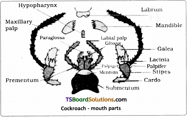
Question 3.
Is it possible to consider vegetative propagation’observed in plants like Bryophyllum, water hyacinth and ginger as a type of asexual reproduction? Give two / three reasons.
Answer:
a) Yes, reproductive leaves in Bryophyllurrt, offsets in water hyacinth and rhizome in ginger are vegetative propagules.

In asexual reproduction, a single individual (parent) is capable of producing offspring. Hence, the offspring produced are identical to each other and are also exact copies (clones) of their parent.

b) In production of vegetative propagules, there is no involvement of two parents, formation of gamete and fertilization.
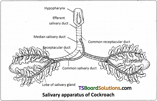
TS Inter 1st Year Botany Study Material Chapter 6 Modes of Reproduction

Question 4.
“Fertilisation is not an obligatory event for fruit production in certain plants.” Explain the statement.
Answer:
In some plants parthenocarpy is observed. Parthenocarpy means development of fruits without fertilisation. They are seedless. The development of an embryo from unfertilised egg cell is known as parthenogenesis. It is a form of asexual reproduction. Hence fertilisation is not an obligatory event for fruit production in certain plants. Eg : Guava, pineapple etc.
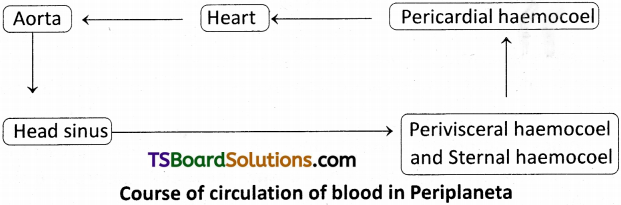
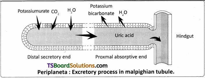
Question 5.
List the changes observed in angiosperm flower subsequent to pollination and fertilisation. [Mar. ’17 – A.P. : Mar. ’14, ’13]
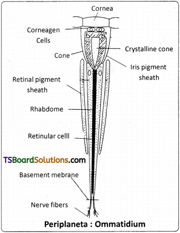
Answer:
Changes taking place in the angiospermic flower after fertilization are called post-fertilization changes.
TS Inter 1st Year Botany Study Material Chapter 6 Modes of Reproduction 1

  1. Sepals, petals, stamens, styles and stigma fall off.
  2. After fertilization ovary develops into fruit. It stores food materials.
  3. Fertilized ovules develop into seeds.
  4. Zygote changes into – Embryo
  5. Synergids & Antipodals – Degenerate
  6. Primary endosperm nucleus forms – Endosperm

Question 6.
Suggest a possible explanation why the seeds in pea pod are arranged in a row, whereas those in tomato are scattered in the juicy pulp.
Answer:

  1. Seeds develop from ovules after post-fertilization changes, while dry transforms into fruit.
  2. In pea plant, there is marginal placentation. Ovules are borne on a ridge (placenta) along the yentral suture of the ovary. Hence, seeds are present in row in pea pod.
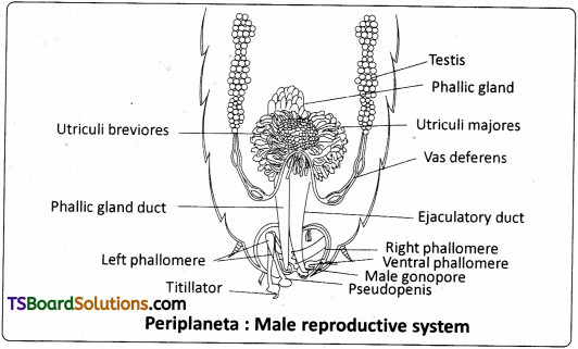
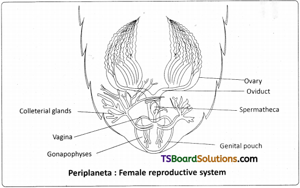
  3. In tomato, pariental placentation is present, the ovules develop on the inner wall of the ovary or on peripheral part. So seeds are scattered in juicy pulp formed from mesocarp and endocarp.

Question 7.
Justify the statement ‘Vegetative reproduction is also a type of asexual reproduction’. [May ’17]
Answer:

  1. Reproduction not involving the fusion of male and female gametes is called asexual reproduction.
  2. In Algae, moulds and mushrooms, the plant body break into smaller portions (fragmentation) and each fragment thing is formed and develops into a mature plant. Gemmae of liver worts are also vegetative reproductive structures.
  3. In flowering plants runner, stolon, sucker, offset rhizome, corm, tuber, bulb, bulbil, reproductive leaves are also vegetative propagules.
  4. In vegetative reproduction, there is no involvement of two parents, male and female gametes are not formed and are not fused. Offspring produced vegetatively by a plant are identical to one another and are exact copies (clones) of their parent.
  5. Hence, vegetative reproduction is also a type of asexual reproduction.

TS Inter 1st Year Botany Study Material Chapter 6 Modes of Reproduction

Question 8.
Define (a) Juvenile phase. (b) Reproductive phase. [Mar. – 2020]
Answer:
(a) Juvenile Phase :
All organisms have to reach a certain stage of growth and maturity in their life before they can reproduce sexually. This stage is known as Juvenile phase or Vegetative phase.

(b) Reproductive phase :
The end of juvenile phase is the beginning of reproductive phase. It can be seen easily in the higher plants when they come to flower.

Question 9.
Distinguish between asexual and sexual reproduction. Why is vegetative reproduction also considered as a type of asexual reproduction? [Mar. 15 – T.S.]
Answer:

  1. When an offspring is produced by a single parent with or without the involvement of gamete formation it is called asexual reproduction.
  2. Sexual reproduction involves the formation of the male and female gametes, either by the same individual or by different individuals of the opposite sex.
  3. Vegetative reproduction does not involve two parents, hence it is also considered as a type of asexual reproduction.

Question 10.
Identify each part in a flowering plant and write whether it is haploid (n) or diploid (2n). [Mar. – 2018]
(a) Ovary _____ (b) Anther _____ (c) Egg _____ (d) Pollen _____ (e) Male gamete _____ (f) Zygote _____
Answer:
a) Ovary – Diploid (2n)
b) Anther – Diploid (2n)
c) Egg – Haploid (n)
d) Pollen – Haploid (n)
e) Male gamete – Haploid (n)
f) Zygote – Diploid (2n)

Question 11.
Give a brief account on the phases of the life cycle of an angidsperm plant.
Answer:

  1. The life cycle of an angiospermic plant has two alternating phases namely the sporophytic and gametophytic phases.
  2. In angiosperms the plant body belongs to diploid (2n) sporophytic phase. This dominant phase of life cycle bears reproductive organs (flowers).
  3. The haploid (n) gametophytic phase of angiospermic plant is dervied from microspores (n) and megaspores (n).
  4. Microspores (pollen grains) are the meiotic products of microspore mother cells that develop in an anther.
  5. Megaspores are the meiotic products of megaspore mother cell that develop from the nucellus of the ovule.
  6. Micro and megaspores produce male and female gametophytes respectively.
  7. Male and female gametes formed respectively from male and female gametophytes are fused to form diploid (2n) zygote.
  8. Zygote after undergoing repeated mitotic divisions produce embryo in the seed.
  9. The embryo (2n) develops into a sporophytic plant during seed germination.

TS Inter 1st Year Botany Study Material Chapter 6 Modes of Reproduction 2

Long Answer Type Questions

Question 1.
Enumerate the differences between asexual and sexual reproduction. Describe the types of asexual reproduction exhibited by unicellular organisms.
Answer:
Differences between asexual and sexual reproduction :

Asexual ReproductionSexual Reproduction
1) Involves a single organism.1) Involves one or two organisms.
2) No production of gametes.2) Male and female gametes are formed.
3) No fusion of gametes.3) Involves fusion of male and female gametes.
4) Requires only mitotic divisions.4) Required meiotic followed by mitotic divisions.
5) Produces offspring that are identical to the parent.5) Offspring will have some characters from male parent and others from female parent. Some characters may not be present in either of the parents.
6) Chance of genetic variation is only through random mutation.6) More chance for genetic variation.
7) Not very useful for natural selection in evolution of species.7) Highly useful for natural selection in evolution of species.

Asexual reproduction by unicellular organisms :
It is by binary fission, budding and spore formation.
TS Inter 1st Year Botany Study Material Chapter 6 Modes of Reproduction 3

Binary fission :
Many unicellular organisms reproduce by binary fission. In this the parent cell divides into two equal halves and each one grows into new individuals. Eg : Euglena, bacteria etc. It is common in Protista and Monera.

Budding :
In yeast, asexual reproduction is done by budding. Small buds are produced that remain attached initially to the parent cell which eventually get separated and grows into new individual.
TS Inter 1st Year Botany Study Material Chapter 6 Modes of Reproduction 4

Spore :
In unicellular algae like Chlamydomonas asexual reproduction is done by spores. These spores are motile, hence called zoospores.
TS Inter 1st Year Botany Study Material Chapter 6 Modes of Reproduction 5

Question 2.
Although sexual reproduction is Song drawn, energy intensive complex forms of reproduction, many groups of organisms in kingdom plantae prefer this mode of reproduction. Give atleast three reasons for this.
Answer:

  1. Sexual reproduction involves formation of the male and female gametes, either by the same individual or by different individuals of the opposite sex. These gametes fuse to form the zygote which develops to forms the new organism.
  2. It is an elaborate, complex and slow process as compared to asexual reproduction.
  3. Sexual reproduction results in offspring that are not identical to the parents or amongst themselves due to fusion of male and female gametes.
  4. Sexual reproduction follow a regular sequence, and is characterized by the fusion (or fertilization) of the male and female gametes, the formation of zygote and embryogenesis. These sequential events may be grouped into 3 distinct stages namely, the pre-fertilization, fertilization and post-fertilization events.

A) Pre-fertilization events:
These include two events namely gametogenesis and gamete transfer that occur prior to the fusion of gametes. Gametogenesis refers to formation of two types (male and female) of gametes (haploid cells).

i) In some algae (eg. Cladophore) the two types of gametes may be so similar in appearance (isogametes) or may be two morphologidly distinct types (heterogametes) as in majority of sexually reproducing organisms. In plants that produce heterogametes (i.e., Runaria, Pteris and Cycas), the male gamete is called the antherozoid or sperm and the female gamete is called the egg.

ii) In some cases male and female gametes may be produced by same plant (bisexual), that has both male and female reproductive structures. In some other cases the male and female reproductive structures develop of two different plants of same species (unisexual). Bisexual condition can be denoted by terms such as homothallic (Fungi) and monoecious (plants). Similarly unisexual condition is described as heterothallic (Fungi) and dioecious (plants).

iii) In flowering plants, unisexual male flower being only stamen is called staminate and female flower bearing only pistils is called pistillate. In that case both male and female flowers may he present on same plant (monoecious cucurbits) or on two separate plants (dioecious – papaya and date palm).

iv) Organisms belonging to monera, fungi, algae and bryophytes have haploid plant body, they produce gametes by mitotic division. Whereas ptesido- phytes, gymnosperms and angiosperms having diploid plant body produce gametes through meiosis in meiocytes (gamete mother cells).

v) After the formation, male and female gametes must be physically brought together to facilitate fusion (fertilization). In majority of organisms (with exception of few fungi and algae), male gamete is motile and female gamete is stationary. In algae, bryophytes and pteridophytes water is the medium through which gamete transfer takes place.

vi) In seed plants pollen grains are the carriers of male gametes and ovule has the egg. in bisexual and self pollinating plants (Eg. Pea) pollen grains produced from anthers are transferred to stigma of same flower. But in cross pollinating plants (including dioecious) pollen from a flower reaches stigma of another flower).

vii) Pollen grains germinate on the stigma and pollen tubes carying the male gametes reach the ovule and discharge male gametes near the egg.

B) Fertilization :
It is the process of fusion of gametes and it is the most vital event of sexual reproduction. This process is also called as syngamy and result in the formation of a diploid zygote.
i) In majority of algae, syngamy occurs in the external medium (water) and is called external fertilization. But in many fungi and majority of plants (Bryophytes; Ptesidophytes, Gymnosperms and Angiosperms) syngamy (internal fertilization) occurs inside the body of the organism.

C) Post-fertilizaton events :
Events that occur after the formation of zygote are called as post-fertilization events. The diploid zygote is formed in all sexually reproducing organisms.

  1. In fungi and algae with haplontic life cycle zygote develops a thick wall that is resistant to desication and damage, undergoes a period of rest before germination. In those organisms zygote divides by meiosis immediately after keryogamy to form haploid spores that grow into haploid individuals.
  2. Zygote is the vital link that ensures continuity of species between organisms of one generation and the next.
  3. Every sexually reproducing organisms begins life as a single called zygote.
  4. Embryogenesis refers to the process of development of embryo from the zygote.
  5. During embryogenesis, zygote undergoes cell divisions (mitosis) and cell differention (formation of tissues and organs) to form an organism.
  6. In flowering plants, the zygote is formed inside the ovule, which transforms into seed. In angiosperms, seeds are present inside the fruit that formed from ovary.

Significance of sexual reproduction :

  1. Sexual reproduction enables the organisms to survive during unfavourable con¬ditions by producing resistant structures (thick walled zygote, seeds), So, even Algae and Fungi that have predominance of asexual/vegetative reproduction in life cycle shift to sexual process just before the onset of adverse conditions.
  2. Sexual reproduction involves fusion of gametes from two parents of opposite sex and hence responsible for genetic variation in offspring.
  3. Seeds are useful to surpass unfavourable conditions. In viviporous Mangroves seeds germinate on mother plant itself. This is a strategy to lower the environmental stress and ensuring successful establishment of plant let.

TS Inter 1st Year Botany Study Material Chapter 6 Modes of Reproduction

Question 3.
Describe the post-fertilisation changes in a flower. [Mar. ’17 – A.P]
Answer:
Changes taking place in the angiospermic flower after fertilization are called post-fertilization changes.

  1. Sepals, petals, stamens, styles and stigma fall off.
  2. In some members of Solanaceae, calyx remains persistent even after fertilisation and grows along with the fruit. In Asteraceae, persistent calyx, pappus helps in the fruits dispersal.
  3. After fertilization ovary develops into fruit. It stores food materials.
  4. Fertilized ovules develop into seeds.
  5. Part of ovule – Changes occurring after fertilization
    Funiculus – Stalk of the seed
    Outer integument – Testa (outer seed coat)
    Inner integument – Tegmen (inner seed coat)
    Micropyle – Seed pore
    Zygote – Embryo
    Synergids – Degenerate
    Antipodals – Degenerate
    Primary endosperm nucleus – Endosperm
    Scar of the ovule – Hilum (scar of the seed)

TS Inter 1st Year Botany Study Material Chapter 6 Modes of Reproduction 6
6) Endosperm is nutritive tissue useful for developing embryos. It is triploid in angiosperms and formed after fertilization.

Intext Question Answers

Question 1.
Why is reproduction essential for organisms?
Answer:
To ensure the continuity of species reproduction is essential. Reproduction will

  1. replace those species that die and
  2. allow an increase in total numbers of the species under suitable conditions.

Question 2.
Which is a better mode of reproduction sexual or asexual? Why?
Answer:
Sexual reproduction is the better mode of reproduction. It brings in diversity of characters in the new generation. It may give a better chance to adjust or adopt to changing environmental conditions, to tolerate diseases, to spread to new areas, to increase their population.

Question 3.
Why is the offspring formed by asexual reproduction referred to as clone?
Answer:
Offsprings formed by asexual method do not involve two parents. They are not only identical to one another but are also exact copies of their parent. Hence they are called clones.

TS Inter 1st Year Botany Study Material Chapter 6 Modes of Reproduction

Question 4.
How does the progeny formed from asexual reproduction differ from those formed by sexual reproduction?
Answer:
The progeny formed from asexual reproduction are genetically identical whereas those formed by sexual reproduction shows genetic variability.

Question 5.
What is vegetative propagation? Give two suitable examples.
Answer:
When the body breaks or gets separated into smaller portions, each fragment can develop into individual body. Eg: Algae, mould, mushrooms, ginger, turmeric, etc.

Question 6.
Higher organisms have resorted to sexual reproduction in spite of its complexity. Why?
Answer:

  1. In sexual reproduction variants are found in the offspring and their survival rate was enhanced.
  2. It gives better chance to adjust to changing environment.

Question 7.
Explain why meiosis and gametogenesis are always interlinked.
Answer:
Gametes are formed from meiocytes only after meiosis. This is called gametogenesis. Thus meiosis leads to the formation of gametes.

Question 8.
Define external fertilisation. Mention its disadvantages.
Answer:
In most aquatic organisms, such as algae, syngamy occurs in the external medium i. e., outside the body of the organism. This type of gametic fusion is called external fertilisation. Its disadvantage is they have to release a large number of gametes into the surrounding water in order to enhance the chances of syngamy.

Question 9.
Differentiate between a zoospore and a zygote.
Answer:
Zoospore is.a motile asexual spore that uses a flagellum for locomotion, also called a swarmspore, these spores are created by some algae, bacteria and fungi to propagate themselves.

Zygote is diploid, formed during sexual reproduction, by the fusion of male and female gametes.

Activity

Question 10.
Examine a few flowers of any cucurbit plant and try to identify the staminate and pistillate flowers. Do you know any other plant that ears unisexual flowers?
Answer:
Cucurbit plant show unisexual flowers. It bears only stamens in male flowers and only carpels in female flowers.

In coconut, male and female flowers are present on the same plant.

In Borasus, male flowers are present in male plant and female flowers are present in female plant.

TS Inter 1st Year Botany Study Material Chapter 6 Modes of Reproduction

Question 11.
What is a bisexual flower? Collect five bisexual flowers from your neighboured and with the help of your teacher find out their common and scientific names.
Answer:
The flower which has both androecium and gynoecium is called a bisexual flower. Collect different flowers as you collect for the herbarium.