Telangana TSBIE TS Inter 1st Year Zoology Study Material 7th Lesson Type Study of Periplaneta Americana (Cockroach) Textbook Questions and Answers.
TS Inter 1st Year Zoology Study Material 7th Lesson Type Study of Periplaneta Americana (Cockroach)
Very Short Answer Type Questions
Question 1.
Why do you call cockroach a pest? (U).
Answer:
Cockroach is commonly found in kitchen and contaminates our food with its excreta. It can transmit a number of bacterial diseases by contaminating food. Hence it is called a pest.
Question 2.
Name the terga of thoracic segments of cockroach. (K)
Answer:
The terga of thoracic segments are one large pronotum covering prothorax. The terga on the mesothorax and metathorax are called mesonotum and metanotum.
Question 3.
What are the structures with which cockroach walks on smooth surfaces and on rough surfaces respectively? (U)
Answer:
The claws and the arolium help in locomotion on rough surfaces whereas plantulae are useful on smooth surfaces.
Question 4.
Name the chitinous tubes that support the wings of cockroach. (K)
Answer:
The wings of cockroach contain a network of hollow veins or nervures.
Question 5.
What is tegmen? What is its function? (KJ
Answer:
The fore wings are thick, opaque and leathery. They donot help in flight, but cover and protect the hind wings when they are not in use. They are called tegmina (singular: tegmen).
![]()
Question 6.
Why is the head in cockroach called hypognathous? (U)
Answer:
The head of cockroach is called hypognathous because it lies hanging almost at right angles to the body with the posterior wider part upwards and the mouth parts directed downwards.
Question 7.
How is a tripod formed with reference to locomotion in cockroach? (U)
Answer:
Each tripod is formed by fore leg and hind leg of one side and the middle leg of the other side in cockroach.
Question 8.
Name the muscles that help in elevating and depressing the wings of a cockroach. (K)
Answer:
Wings are elevated by the contraction of dorsoventral muscles. Contraction of the dorso – longitudinal muscles depresses the wings.
Question 9.
Name the different blood sinuses in cockroach. (K)
Answer:
The blood sinuses are a) Pericardial haemocoel or the dorsal sinus b) Perivisceral haemocoel or the middle sinus, c) Sternal haemocoel or ventral sinus.
Question 10.
How are the fat bodies similar to the liver of the vertebrates? (A)
Answer:
The haemocoel of cockroach contains many large sized fat bodies called corpora adiposa. These are similar to the liver of the vertebrates in certain function like storing of food, secrete lipids, store uric acid and contain symbiotic bacteria.
Question 11.
What are the three regions of the alimentary canal in cockroach? (K)
Answer:
The three regions are foregut or stomodaeum, midgut or mesenteron and hindgut or proctodaeum.
Question 12.
How many denticulate plates are present in the gizzard of cockroach? (K)
Answer:
The chitinous inner lining of the gizzard of cockroach has six powerful teeth, which form an efficient grinding apparatus.
![]()
Question 13.
Which part of the gut secretes the peritrophic membrane in cockroach? (U)
Answer:
The ‘bolus’ of food in the mesenteron is enveloped by a chitinous and porous membrane called peritrophic membrane, which is secreted by the funnel like stomodeal valve of the gizzard.
Question 14.
Which parts of cockroach help in locating the food? (U)
Answer:
Cockroach locates the food by the olfactory sensillae of antenna, labial palps and maxillary palps.
Question 15.
In which part of the gut of cockroach, water is reabsorbed? (K)
Answer:
The undigested food is passed into the ileum, colon and then reaches the rectum, where water is reabsorbed by rectal papillae.
Question 16.
Write the names of mouthparts in cockroach that help in biting and tasting the food. (K).
Answer:
Mandibles help in biting and chewing of food. Labrum helps in tasting the food.
Question 17.
What are alary muscles? [K)
Answer:
There is a series of paired triangular muscles, called “Alary muscles.” Every segment has one pair of these muscles. These are attached to the pericardial septum by their broad bases and to the terga by their pointed ends.
Question 18.
What is haemocoel? (K)
Answer:
The body cavity of an arthropod or a mollusk filled with haemolymph : derived from the blastocoels of the embryo, also called the “Primary body cavity.”
![]()
Question 19.
The three sinuses in a cockroach are not equal in size. Why? (U)
Answer:
The middle sinus is very large as it contains most of the viscera. The dorsal and ventral sinuses are small as they have only heart and nerve cord, respectively.
Question 20.
Why is the blood of Periplaneta called haemolymph? (U)
Answer:
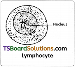
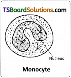
The blood of periplaneta is colourless and is called haemolymph. It consists of a fluid called plasma and free blood corpuscles or haemocytes which are phagocytic.
Question 21.
What is the function of haemocytes found in the blood of Periplaneta? (K)
Answer:
Haemocytes are phagocytic. They are large in size and can ingest foreign particles such as bacteria. Hence defensive in function.
Question 22.
Why does not the blood of Periplaneta help in respiration ? (A)
Answer:
There is no respiratory pigment in the blood and so it plays no major role in respiration.
Question 23.
Write important functions of blood in Periplaneta. (K)
Answer:
Blood functions
- It absorbs digested food from alimentary canal and distributes it to the rest of the body.
- It brings nitrogenous wastes from all parts of the body to the excretory organs for their elimination.
- Phagocytes of blood are defensive in function.
- It transports secretions of the ductless glands to the target organs.
Question 24.
The blood of Periplaneta is not red. Which pigment, do you think, is absent in it? (U)
Answer:
The blood of periplaneta is not red. There is no respiratory pigment called haemoglobin in the blood and so it plays no major role in respiration.
Question 25.
How many spiracles are present in cockroach? Mention their locations. (K)
Answer:
There are ten pairs of openings called stigmata or spiracles. The first two pairs of spiracles are present in the thoracic segments (2nd and 3rd). Remaining eight pairs are present in the first eight abdominal segments.
![]()
Question 26.
What are trichomes? Write their functions. (K)
Answer:
All spiracles bear small hair like structures called ‘trichomes’ to filter the dust particles.
Question 27.
Why is the respiratory system of cockroach called polypneustic and holopneustic system? (U)
Answer:
The spiracles of cockroach are polypneustic (as they are more than 3 pairs) and holopneustic (as all of them are functional).
Question 28.
Name the chitinous ring that encircles the spiracle of cockroach. (K)
Answer:
All spiracles are valvular and each of them is surrounded by a chitinous ring called peritreme.
Question 29.
What is intima? (K)
Answer:
Trachea in cockroach is made up of three layers, outer basement membrane, a middle one cell thick epithelium and an inner layer of cuticle called intima. The intima is produced into spiral thickenings called taenidia.
Question 30.
Name the protein that lines the tracheole of the cockroach. (K)
Answer:
Tracheoles are formed of a protein called trachein.
![]()
Question 31.
During inspiration which spiracles are kept open and which are kept closed? (K)
Answer:
During inspiration the thoracic spiracles are kept open and the abdominal spiracles are kept closed.
Question 32.
Which factors regulate the opening of the spiracles? (U)
Answer:
Opening and closing or spiracles is influenced by C02 tension in haemolymph and oxygen tension in the tracheae.
Question 33.
Inspiration in cockroach is a passive process and expiration is an active process. Justify. (U)
Answer:
During inspiration air is drawn in due to the relaxation of the muscles, the process is a “passive process”. Expiration involves the contraction of muscles, hence is described as ‘active process.’
Question 34.
The nitrogenous wastes in Periplaneta are removed from the body through alimentary canal. Why? (U)
Answer:
The removal of nitrogenous waste material through the alimentary canal helps in complete reabsorption of water from the wastes and formation of dry uric acid. It is an adaptation for conservation of water.
Question 35.
How does the cuticle of a cockroach help in excretion? (A)
Answer:
Some nitrogenous waste materials are deposited on the cuticle and eliminated during moulting.
Question 36.
How do fat bodies help in excretion? (A)
Answer:
Fat bodies absorb and store uric acid throughout the life. This is called storage excretion as they remain stored in the cells of the corpora adiposa.
Question 37.
What is ‘storage excretion’? (U)
Answer:
Fat bodies absorb and store uric acid throughout the life. This is called storage excretion as they remain stored in the cells of the corpora adiposa.
![]()
Question 38.
In which part of the alimentary canal of Periplaneta more water is reabsorbed? (K)
Answer:
The part of the alimentary canal of Periplaneta more water is reabsorbed in rectum containing rectal papillae.
Question 39.
List out the organs associated with excretion in Periplaneta. (K)
Answer:
The organs associated with excretion are
1) Malpighian tubules of anterior end of hind gut 2) Fat bodies 3) Uricose glands 4) Cuticle.
Question 40.
Which part of malpighian tubules extract water, salts and nitrogenous wastes from the haemolymph? (K)
Answer:
The distal portion of malpighian tubules containing glandular cells absorb salts, water and nitrogenous wastes from the haemolymph.
Question 41.
Which structure of cockroach acts as sensory and endocrine centre? (K)
Answer:
Brain or cerebral ganglia is mainly a sensory and an endocrine centre.
Question 42.
Distinguish between scolopidia and sensillae. (U)
Answer:
- Sensillae are the units of cuticular receptors and chemoreceptors.
- Scolopidia are the subcuticular units of mechanoreceptors of chordo-tonal organs.
Question 43.
How is the ommatidium of cockroach different from that of a diurnal insect? (A]
Answer:
In cockroach a nocturnal insect ommatidia form superposition image in which overlapping of images occur and it is a blurred image.
In diurnal insects the image is called apposition image as it is formed by the juxtaposition of small parts of the visual field. This type of vision is called mosaic vision.
![]()
Question 44.
How many segmental ganglia are present on the ventral nerve cord of cockroach? (K)
Answer:
On the ventral nerve cord segmental ganglia show 3 + 6 arrangement. 3 thoracic ganglia and 6 abdominal ganglia in first 7 segments except 5th segment.
Question 45.
Which of the abdominal ganglia is the largest and why? (U)
Answer:
The last or the 6th abdominal ganglion is the largest of all the abdominal ganglia. It is formed by the fusion of the ganglia of the 7th, 8th, 9th and 10th abdominal segments.
Question 46.
Name the structural and functional unit of compound eye of cockroach. How many such units are present in a single compound eye ? (K)
Answer:
Structural and functional unit of compound eye is ommatidium. In a single compound eye of cockroach 2000 ommatidia are present.
Question 47.
Why is the brain called the principal sensory centre in cockroach? (U)
Answer:
In brain protocerebrum receives sensory impulses from compound eyes through optic nerves, deutero cerebrum receives sensory impulses from antennae through antennel nerves and tritocerebrum receives sensory impulses from the labrum. Hence brain in principally sensory in nature.
Question 48.
Which parts of an ommatidium constitute dioptric region? (K)
Answer:
The region containing the cornea and crystalline cone constitute the dioptrical or focussing region of the ommatidium. ,
Question 49.
Distinguish between apposition image and superposition image. (U)
Answer:
In cockroach a nocturnal insect ommatidia form superposition image in which overlapping of images occur and it is a blurred image. In diurnal inserts the image is called apposition image as it is formed by the juxtaposition of small parts of the visual field. This type of vision is called mosaic vision.
Question 50.
List out the characters that help in understanding the difference between male and female cockroaches. (K)
Answer:
The female is different from the male in respect of short and broad abdomen, presence of brood pouches and absence of anal styles.
Question 51.
What is the function of mushroom gland in cockroach? (K)
Answer:
A characteristic mushroom shaped gland is present in the 6th and 7th abdominal segments which functions as an accessory reproductive gland.
Question 52.
Compare the utriculi majores and utriculi breviores of the mushroom gland functionally. (U)
Answer:
Secretion of utriculi majores forms the inner layer of the spermatophore while that of utriculi breviores nourishes the sperms.
![]()
Question 53.
How many ovarioles are present in a single ovary of Periplaneta and what are the two parts of a single ovariole? (U)
Answer:
Each ovary consists of eight tubules called ovarioles. Each ovariole consists of a tapering anterior filament called germarium and a posterior wider vitellarium.
Question 54.
What are phallomeres? (K)
Answer:
Surrounding the male genital opening there are chitinous and asymmetrical structures called phallic organs or gonapophyses or phallomeres which help in copulation. These are the male external genitalia.
Question 55.
What are gonapophyses? (K)
Answer:
Three pairs of plate like chitinous structures called gonapophyses are present around the female genital aperture. These gonapophyses guide the ova into ootheca as ovipositors. These are female external genitalia.
Question 56.
How is colleterial gland helpful in reproduction of Periplaneta? (A)
Answer:
Secretion of the two collateral glands forms a hard egg case called ootheca around the eggs.
![]()
Question 57.
What is paurometabolous development ? (U)
Answer:
Cockroach is paurometabolous, which means the development is gradual through nymphal stages.
Short Answer Type Questions
Question 1.
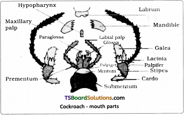
Draw a neat labelled diagram of the mouth parts of cockroach. (S)
Answer:
Question 2.
Describe the physiology of digestion in cockroach. (K)
Answer:
Digestion :
After swallowing, the food passes through the pharynx and oesophagus, and reaches the crop. In the crop, food is mixed with digestive juices that are regurgitated into it through the grooves of the gizzard. Hence, most of the food is digested in the crop. The partly digested food is filtered by the bristles of the gizzard and later it passes through the stomodeal valve into the ventriculus.
The enzyme amylase of the salivary juice converts starches into disaccharides. Invertase or sucrase digests sucrose into glucose and fructose. Maltase converts maltose into glucose. The enzyme lipase digests lipids into fatty acids and glycerol. Proteases digest proteins into amino acids. Cellulose of the food is digested by the enzyme cellulase secreted by the microorganisms present in the hindgut of cockroach. Cellulose in converted into glucose.
In the ventriculus, the digested food is absorbed. The undigested food is passed into the ileum, colon, and then reaches the rectum, where water is reabsorbed by rectal papillae. Then the remaining material is finally defaecated as dry pellets, through the anus.
Question 3.
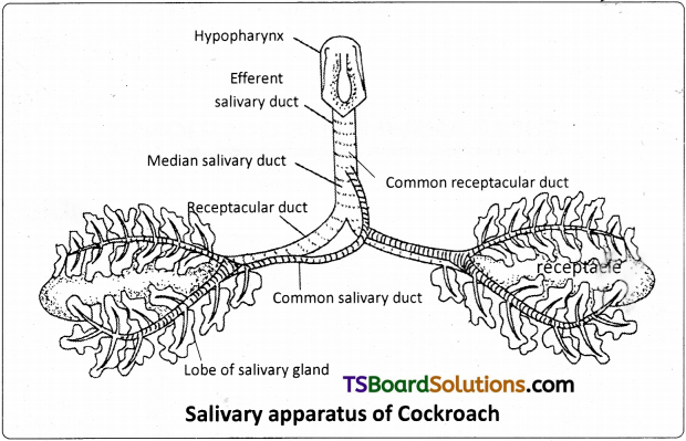
Draw a neat labelled diagram of the salivaary apparatus of cock roach. (S)
Answer:
Question 4.
What is haemocoel? Describe it with reference to Periplaneta. (U)
Answer:
The blood filled body cavity of an organism is called Haemocoel.
Haemocoel in Periplaneta :
The haemocoel of cockroach is divided in to three sinuses by two muscular, horizontal membranes, called dorsal diaphragm or ‘pericardial septum’ and ventral diaphragm. Both the diaphragms have pores. There is a series of paired triangular muscles, called alary muscles. Every segment has one pair of these muscles situated on the lateral sides of the body.
These are attached to the pericardial septum by their broad bases and to the terga by their pointed ends or apices. The three sinuses of the haemocoel are known as pericardial haemocoel or the ‘dorsal sinus’, the perivisceral haemocoel or the ‘middle sinus’ and sternal haemocoel or ‘venral sinus’ or ‘perineural sinus’. The middle sinus is very large as it contains most of the viscera. The dorsal and ventral sinuses are small as they have only heart and nerve cord, respectively.
![]()
Question 5.
Describe the structure and function of the heart in Periplaneta. (K)
Answer:
Heart :
The heart lies in the pericardial haemocoel or dorsal sinus. It is a long, muscular, contractile tube found along the mid dorsal line, beneath the terga of the thorax and abdomen. It consists of 13 chambers. Every chamber opens into the other present in front of it. Three of the thirteen chambers are situated in the thorax and ten in the abdomen. Its posterior end is closed while the anterior end is continued forward as the anterior aorta. At the posterior side of each chamber, except the last, there is a pair of small apertures called ‘ostia’ one on each side. Ostia have valves which allow the blood to pass only into the heart from the dorsal sinus.
Question 6.
Describe the process of blood circulation in Periplaneta. (K)
Answer:
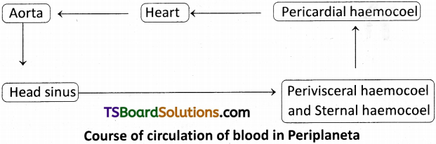
Circulation of blood :
The blood flows forward in the heart by the contractions of its chambers. At the anterior end of the heart, the blood flows into the aorta and from there it enters the sinus of the head. From the head sinus, the blood flows into the perivisceral and sternal sinuses. On contraction of the alary muscles the pericardial septum is pulled down. This increases the volume of the pericardial sinus.
Hence blood flows from the perivisceral sinus into the pericardial sinus through the appertures of the pericardial septum. On relaxation of the alary muscles, the pericardial septum moves upwards to its original position. This forces the blood, to enter the chambers of the heart through the ostia from the pericardial sinus.
Question 7.
How do contraction and relaxation of alary muscles help in circulation? (A)
Answer:
On contraction of the alary muscles the pericardial septum is pulled down. This increases the volume of the pericardial sinus. Hence blood flows from the perivisceral sinus into the pericardial sinus through the appertures of the pericardial septum. On relaxation of the alary muscles, the pericardial septum moves upwards to its original position. This forces the blood, to enter the chambers of the heart through the ostia from the pericardial sinus.
Question 8.
Describe the structure of trachea of cockroach. (K)
Answer:
Structure of Trachea :
The wall of the tracheae is made of three layers. They are an outer basement membrane, a middle one cell thick epithelium and an inner layer of cuticle called intima. It has protein / chitin layer and epicuticle towards lumen. The intima is produced into spiral thickenings called taenidia. In taenidia, protein / chitin layer is differentiated as exocuticle. The taenidia keep the tracheae always open.
![]()
Question 9.
Explain the structure of malpighian tubules. (U)
Answer:
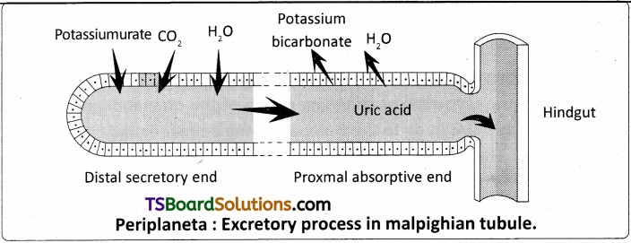
Malpighian tubule :
The malpighian tubules are long, unbranched yellowish tubules, attached at the extreme anterior end of the hindgut, lying freely in the haemolymph, but do not open into it, being blind at the free ends. They are 100 – 150 in number arranged in 6 – 8 bundles, each bundle having 15-25 tubules. Marcello Malpighi, described these tubules and called them vasa varicosa Meckel called them Malpighian tubules. Each tubule is lined by a single layer of glandular epithelium with a brush border on the inner surface. The ‘distal portion’ of the tubule is secretory and the ‘proximal part’ is absorptive in nature.
Question 10.
What are different excretory organs in Periplaneta? Describe the process of excretion in detail. (K)
Answer:
The structures associated with excretory function are
a) Malpighian tubules b) Fat bodies c) Uricose glands d) Cuticle.
Malpighian tubules :
The glandular cells of the malpighian tubules absorb water, salts, CO2 and nitrogenous wastes from the haemohymph and secrete them into the lumen of the tubules. The cells of the proximal part of the tubules reabsorb water and certain inorganic salts. By the contraction of the tubules urine is pushed into the ileum. More water is reabsorbed from it, when it moves in to the rectum and almost solid uric acid is excreted along with faecal matter.
The removal of nitrogenous waste material through the alimentary canal helps in complete reabsorption of water from the wastes and formation of dry uric acid. It is an adaptation for conservation of water as it is very important in terrestrial organisms.
Fat bodies :
Fat body is a lobed white structure. Urate cells present in these bodies are associated with excretion in a way. These cells absorb and store uric acid throughout the life. This is called storage excretion as they remain stored in the cells of the corpora adiposa.
Uricose glands :
Uric acid is stored in uricose gland or utriculi majores of the mushroom gland in male cockroach. It is discharged during copulation.
Cuticle :
Some nitrogenous waste materials are deposited on the cuticle and eliminated during moulting.
Question 11.
How does Periplaneta conserve water? Explain it with the help of excretion in it. (A)
Answer:
The removal of nitrogenous waste material through the alimentary canal helps in complete reabsorption of water from the wastes and formation of dry uric acid. It is an adaptation for conservation of water as it is very important in terristrial organisms.
Fat bodies :
Fat body is a lobed white structure. Urate cells present in these bodies are associated with excretion in a way. These cells absorb and store uric acid throughout the life. This is called storage excretion as they remain stored in the cells of the corpora adiposa.
![]()
Question 12.
Describe the structure of an ommatidium and label its parts. (K)
Answer:
Structure of an Ommatidium :
Each typical ommatidium is an elongated sub unit of the compound eye, consisting of the following parts.
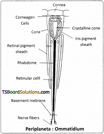
1. Cornea :
It is the outermost part of an ommatidium and corresponds to a ‘hexagonal facet’ of the compound eye. It is a biconvex, transparent part of the cuticle and allows light to pass through it. Cornea is secreted by specialized cells of epidermis. Cornea is the ‘refractive* 1 2 3 region of ommatidium.
2. Corneagen cells or lenticular cells :
These are two transparent specialized epidermal cells that secrete cornea. These cells later form the ‘primary pigment sheath’ or iris pigment sheath.
3. Vitrellae or cone cells (Semper cells) :
These are four transparent more or less conical cells that lie below the corneagen cells. They surround the transparent crystalline cone. Crystalline cone is secreted by the cone cells.
4. Crystalline cone :
It is the transparent conical structure that is secreted by the vitrellae and is surrounded by them. Light absorbing dark primary pigment cells surround the vitrellae. The region containing the cornea and crystalline cone constitute the dioptrical or focussing region of the ommatidium. Crystalline cone focuses the light on to the next part of the ommatidium.
5. Retinulae :
These are the innermost and elongated cell of an ommatidium. They are seven in number. They rest on the basement membrane. Each cell bears microvilli towards the inner surface. Microvilli of each retinular cell collectively form a rhabdomere that contains photoreceptor pigments. These rhabdomeres fuse along the axis of the ommatidium to form the rhabdome in the centre. Retinulae are the nerve cells from which sensory nerve fibres leave as the optic nerve to the protocerebrum. They are the photoreceptor cells of the ommatidium. Rhabdome and retinulae form the retinal or receptor region. Receptor region is surrounded by seven secondary pigment cells, which absorb light and serve to isolate each ommatidium from the rest (retinal pigment sheath).
Question 13.
Draw a neat and labelled diagram of ommatidium. (S)
Answer:
Question 14.
How can you identify the male and female cockroaches? Explain it describing the chief features of the external and internal genetalia. (U)
Answer:
The sexual dimorphism is evident both externally and internally. The female is different from the male in respect of short and broad abdomen, presence of brood pouches and absence of anal styles.
Male :
Internally male reproductive system contains a pair of testes, vas deferens, ductus ejaculatorius, mushroom shaped gland, conglobate gland.
Surrounding the male genital opening there are chitinous and asymmetrical structures called phallic organs or phallomeres which help in copulation. These are the male external genitalia.
Female :
Internally female reproductive system consists of a pair of ovaries, a pair of oviducts, vagina, spermathecae, spermathecal papilla and colleteral glands.
Three pairs of plate like chitinous structures called gonopophyses are present around the female genital aperture. These gonopophyses guide the ova into ootheca as ovipositors. These are the female external genitalia.
Question 15.
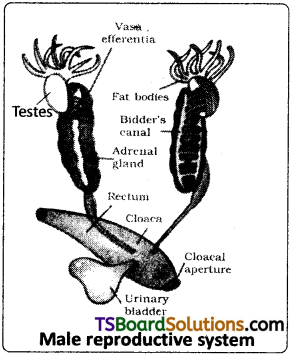
Describe the male reproductive system of cockroach. (K)
Answer:
Male Reproductive System :
The male reproductive system .consists of a pair of testes. These are elongated and lobed structures lying one on each lateral side in the fourth to sixth abdominal segments. They are embedded in the fat bodies. From the posterior end of each testis, there starts a thin duct, the vas deferens. The two vasa deferntia run backwards and inwards to open into a wide median duct, the ductus ejaculatorius in the seventh segment.
A characteristic mushroom shaped gland is present in the 6th and 7th abdominal segments which functions as an accessory reproductive gland. The gland consists of two types of tubules, i) long slendertubules, the utriculi majores or ‘peripheral tubules’ ii) Short tubules, the utriculi breviores. Secretion of utriculi majores forms the inner layer of the spermatophore while that of utriculi breviores nourishes the sperms. These tubules open into the anterior part of the ejaculatory duct.
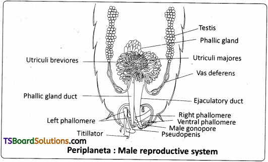
The seminal vesicles are present on the ventral surface cf the ejaculatory duct. These sacs store the sperms in the form of bundles called spermatophores. The ejaculatory duct is a muscular tube that extends posteriorly and opens at the gonopore or the ‘male genital pore’. The duct of phallic or conglobate gland also opens near the gonopore. Its function is still not known. Surrounding the male genital opening there are chitinous and asymmetrical structures called phallic organs or gonapophyses or phallomeres which help in copulation. These are the male external genitalia.
![]()
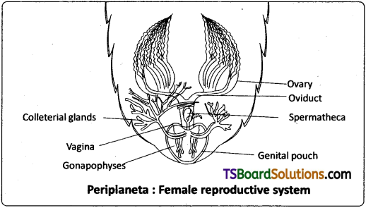
Question 16.
Describe the female reproductive system of cockroach. (K)
Answer:
Female Reproductive System :
The female reproductive system of Periplaneta consists of a pair of ovaries, a pair of oviducts, vagina, spermathecae, spermathecal papilla, and colleterial glands.
Ovaries :
A pair of large ovaries lies laterally in 2 to 6 abdominal segments. They are light yellow in colour surrounded by fat bodies. Each ovary consists of eight tubules called ovarian tubules or ovarioles. Each ovariole consists of a tapering anterior filament called germarium and a posterior wider vitellarium. The germarium contains various stages of developing ova, and the vitellarium contains mature ova with yolk. The tapering ends of the ovarioles of each ovary unite to form a single thread which attaches to the dorsal body wall. The ovarioles, at their posterior end unite to form a short wide oviduct.
The oviducts unite to form a very short median vagina. The vertical opening of the vagina is called female genital pore. It opens into a large genital pouch on the eighth sternum. A spermatheca or ‘receptaculium seminis’. consisting of a left-sac like and a right filamentous caecum, is present in the 6th segment which opens by a median aperture on a small spermathecal papilla in the dorsal wall of the genital pouch on the ninth sternum. In a fertile female, the spermatheca contains spermatophores, obtained during copulation.
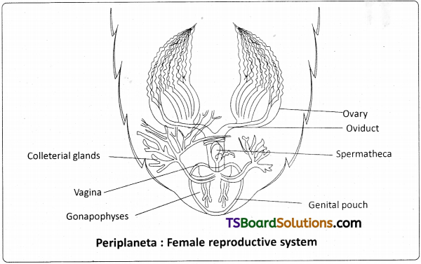
A pair of branched colleterial glands is present behind the ovaries. These glands open into the genital pouch separately, just above the spermathecal aperture. Secretion of the two collaterial glands forms a hard egg case called ootheca around the eggs.
Genital pouch is formed by 7th, 8th, and 9th abdominal sterna. The sternum of the seventh segment is boat shaped and forms the floor and side walls of the genital pouch. The sterna of the eighth and ninth segments, which are tucked into the seventh segment, constitute the anterior wall and the roof of the genital pouch, respectively. The genital pouch has two chambers the anterior ‘gynatrium’ or genital chamber and posterior ‘vestibulum’ or oothecal chamber.
Three pairs of plate like chitinous structures called gonapophyses are present around the female genital aperture. These gonapophyses guide the ova into ootheca as ovipositors. These are the female external genitalia.
Essay Answer Type Questions
Question 1.
Descirbe the structure of the head of cockroach, with the help of a neat labelled diagram. (K & S)
Answer:
Heat :
The head of cockroach is small and triangular. It is called hypognathous because it lies hanging almost at right angles to the body with the posterior wider part upwards, and the mouthparts directed downwards.
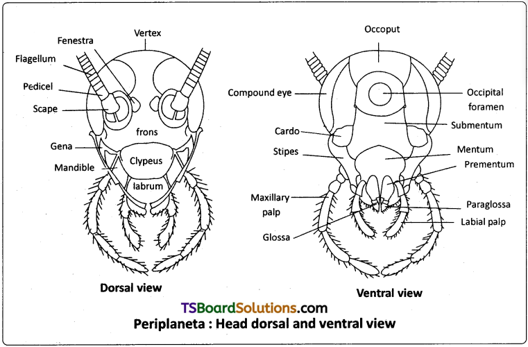
The head of cockroach is formed by the fusion of six embryonic segments. It is movably attached to the thorax by a short neck or cervicum. it is covered by a number of sclerites which fuse to form a capsule. The top of the head between the eyes is called vertex. The vertex has two sclerites called ‘epicranial plates’ connected by an ‘epicranial suture’. Below the vertex, the sclerites covering the’ head in front are a large frons, a narrow rectangular clypeus and a movable labrum. Covering the sides of the head, below the compound eyes are.the ‘cheek sclerities’ or ‘genae. At the back of the head capsule there is a large opening called occipital foramen.
It is bordered by a sclerite called occiput. The occipital foramen forms a passage for the oesophagus, aorta; nerve cord and tracheae. At the base of each antenna, a small whitish speck called fenestra or ‘ocellar spot’ or ‘simple eye’ is present. Appendages are absent in the first and third segments of the head. The second segment bears a pair of long, slenife^ and segmented antennae, one on each side of the head. The antennae are tactile and olfactory in function. The fourth segment bears a pair of mandibles. The fifth segment has a pair of ‘first maxillae’. The sixth segment bears a pair of ‘second maxillae’, which fuse to form the labium (also called ‘lower lip’)
![]()
Question 2.
Describe the abdomen of cockroach. (K)
Answer:
Abdomen :
The abdomen consists of ten segments. Each segment is covered by the dorsal tergum, the ventral sternum and the two lateral pleura or pleurites. There are ten terga but only nine sterna as the tenth sternum is absent. The eighth tergum in the male and both eighth and ninth terga in the female are not visible as they are overlapped by the seventh tergum. The tenth tergum extends beyond the posterior end of the body and has a deep notch/groove in the middle of its free end. In the male nine sterna are visible whereas in the female, only seven sterna are visible. The seventh, eighth and ninth sterna together form a brood pouch. The brood pouch has two parts the anterior genital chamber or gynatrium and posterior oothecal chamber.
The posterior end of the abdomen has a pair of anal cerci, a pair of anal styles and gonapophyses in the males. Anal cerci are jointed and arise from the lateral sides of the tenth tergum and are found in both the sexes. The anal styles are without joints and arise from the ninth sternum (seen only in the males). The gonapophyses are small chitinous processes arising from the ninth sternum in the males and eighth and ninth sterna in the females. They are the external genital organs. The anus is at the posterior end of the abdomen. The genital aperture in male is present just below the anus on one of the gonapophyses and in female it is located on the eighth sternum.
![]()
Question 3.
Describe the digestive system of cockroach with the help of a neat labelled diagram. (K & S)
Answer:
The digestive system of cockroach consists of an alimentary canal and the associated glands. The preoral cavity, surrounded by the mouth parts, is present in front of the mouth. The hypopharynx divides it into two chambers called cibarium (anterior) and salivarium (posterior)
Alimentary canal :
The alimentary canal of cockroach is a long tube and is coiled at some places. It extends between the mouth and the anus. It is divided into three regions, namely, foregut or stomodaeum, midgut or mesenteron and hindgut or proctodaeum. The foregut and hindgut are internally lined by ectoderm. The mesenteron is lined by the endodermal cells.
Foregut of stomodaeum :
The foregut includes pharynx, oesophagus, crop, and gizzard. It is internally lined by a chitinous cuticle. Mouth opens into the pharynx, which in turn leads into a narrow tubular oesophagus. The oesophagus opens behind into a thin walled distensible sac called crop. The crop serves as a reservoir for storing food. Its outer surface is covered by a network of tracheae.
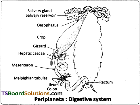
Behind the crop there is a thick walled muscular proventriculus, or gizzard. The chitinous inner lining of the gizzard has six powerful teeth, which form an efficient grinding apparatus. Behind each tooth is a hairy pad, which bears backwardly directed bristles. Among these plates, food is thoroughly ground into fine particles. These food particles are filtered by the bristles. The gizzard thus acts both as a grinding mill and also as a sieve. There is a membranous projection of the gizzard into the mesenteron in the form of a funnel called stomodeal valve. This valve prevents the entry (regurgitation) of food from the mesenteron back into the gizzard.
Midgut (mesenteron or ventriculus) :
The midgut is a short and narrow tube behind the gizzard. It is also called mesenteron or ventriculus. Between the ventriculus and the gizzard, arising from ventriculus, there are six to eight finger like diverticula called hepatic caecae. They are helpful in digestion and absorption of the digested t food materials. Ventriculus is functionally divided into an anterior secretory part and a posterior absorptive part.
The secretory part of the ventriculus has many gland cells and it secretes several enzymes. The ‘bolus’ of food in the mesenteron is enveloped by a chitinous and porous membrane called peritrophic membrane, which is secreted by the funnel like stomodeal valve of the gizzard.
Digested food is absorbed into the blood through the peritrophic membrane in the posterior absorptive region of the ventriculus. The peritrophic membrane protects the wall of the ventriculus from hard food particles in the food. The opening of the ventriculus into the hindgut is controlled by a sphincter muscle. It prevents entry of undigested food and uric acid from the hindgut into the midgut.
Hindgut or proctodaeum :
The hindgut is a long coiled tube, consisting of three regions namely ileum, colon and rectum, it is internally lined by chitinous cuticle. The ileum that lies behind the mesenteron is a short tube. Six bundles of fine yellow, blind tubules called malpighiari tubules open into the ileum near the junction of mesenteron and ileum. Malpighian tubules are excretory in function.
Ileum collects uric acid from the malpighian tubules and undigested food from the mesenteron. Ileum opens behind into a long coiled tube called colon. Colon leads into a short and wide rectum, which opens out through the anus. Rectum bears on its inner side six longitudinal chitinous folds called rectal papillae. They are concerned with the reabsorption of water from the undigested food.
Digestive glands :
The digestive glands associated with the alimentary canal of cockroach are salivary glands, hepatic caecae and glandular cells of the mesenteron. Salivary glands : There is a pair of salivary glands attached to the ventrolateral sides of the crop, one on each side. Each salivary gland has two lobes. Each lobe of salivary gland has many lobules called acini. Each acinus is a group of secretory cells called zymogen cells with a small ductule. The ductules of both the lobes of a salivary gland unite to form a common salivary duct on each side.
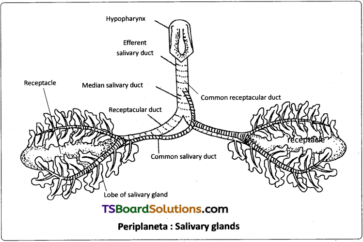
The two common salivary ducts are joined to form the median salivary duct. Between the two lobes of a salivary gland of each side is a sac called salivary receptacle that stores saliva. It leads into a receptacular duct, or ‘reservoir duct’. The receptacular ducts of both the sides are united to form a common receptacular duct, or ‘common reservoir duct’. The median salivary duct opens into the common receptacular duct. Later these two form an efferent salivary duct. The efferent salivary duct opens at the base of the hypopharynx. Acinar cells secrete saliva, which contains starch digesting enzymes such as amylase.
Question 4.
Describe the blood circulatory system of Periplaneta in detail and draw a neat labelled diagram of it. (K & S)
Answer:
Circulatory system of Periplaneta :
The circulatory system helps in the transportation of digested food, hormones etc., from one part to another in the body. Periplaneta has an open type of circulatory system as the blood, or haemohymph, flows freely within the body cavity or haemocoel. Blood vessels are poorly developed and open into spaces. Visceral organs located in the haemocoel are bathed in the blood. The three main parts associated with the blood circulatory system of Periplaneta are the haemocoel, heart, and blood.
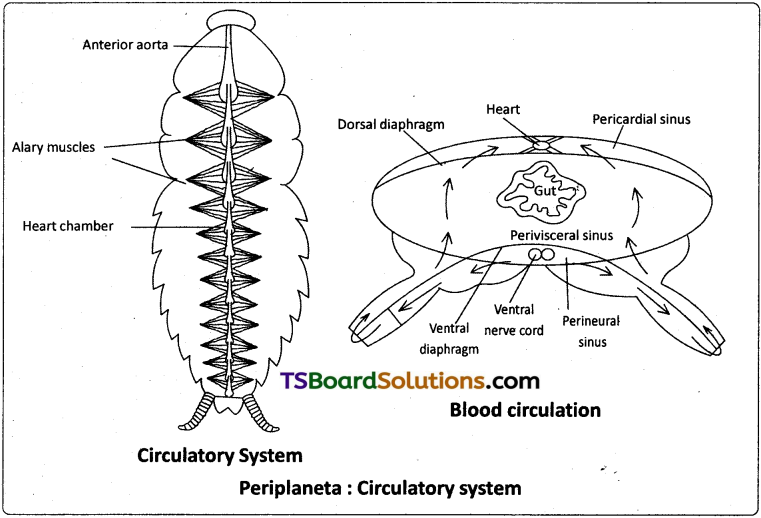
Haemocoel :
The haemocoel of cockroach is divided into three sinuses by two muscular, horizontal membranes, called dorsal diaphragm or ‘pericardial septum1 and ventral diaphragm. Both the diaphragms have pores. There is a series of paired triangular muscles, called alary muscles. Every segment has one pair of these muscles situated on the lateral sides of the body. These are attached to the pericardial septum by their broad bases and to the terga by their pointed ends or apices.
The three sinuses of the haemocoel are known as pericardial haemocoel or the ‘dorsal sinus’, the perivisceral haemocoel or the ‘middle sinus’ and sternal haemocoel or ‘vental sinus’ or ‘perineural sinus’. The middle sinus is very large as it contains most of the viscera. The dorsal and ventral sinuses are small as they have only heart and nerve cord, respectively.
Heart :
The heart lies in the pericardial haemocoel or dorsal sinus. It is along, muscular, contractile tube found along the mid dorsal line, beneath the terga of the thorax and abdomen. It consists of 13 chambers. Every chamber opens into the other present in front of it. Three of the thirteen chambers are situated in the thorax and ten in the abdomen. Its posterior end is closed while the anterior end is continued forward as the anterior aorta. At the posterior side of each chamber, except the labt, there a pair of small apertures called ostia’ one on each side. Ostia have valves which allow the blood to pass only into the heart from the dorsal sinus.
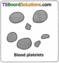
Blood :
The blood of Periplaneta is colourless and is called haemolymph. it consists of a fluid called plasma, and free blood corpuscles or haemocytes, which are ‘phagocytic1. The phagocytes are large in size and can’ingest1 foreign particles such as bacteria. There is no respiratory pigment in the blood and so it plays no major role in respiration. The important functions of the blood are :
- It absorbs digested food from the alimentary canal and distributes it to the rest of the body.
- It brings nitrogenous wastes from all parts of the body to the excretory organs for their elimination.
- It carries defensive phagocytes to the places of infection where they engulf the germs and disintegrating tissue parts.
- It transports secretions of the ductless glands to the target organs.
Circulation of blood :
The blood flows forward in the heart by the contractions of its chambers. At the anterior end of the heart, the blood flows into the aorta and from there it enters the sinus of the head. From the head sinus, the blood flows into the perivisceral and sternal sinuses. On contraction of the alary muscles, the pericardial septum is pulled down. This increases the volume of the pericardial sinus. Hence blood flows from the perivisceral sinus into the pericardial sinus through the appertures of the pericardial septum. On relaxation of the alary muscles, the pericardial septum moves upwards to its original position. This forces the blood, to enter the chambers of the heart through the ostia from the pericardial sinus.
Question 5.
The blood circulatory system of Periplaneta is of open type. Illustrate the statement describing the course of circulation in it. (U)
Answer:
The circulatory system helps in the transportation of digested food, hormones etc. from one part to another in the body. Periplaneta has an open type of circulatory system as blood or haemolymph flows freely with in the body cavity or haemocoel.
Circulation of blood :
The blood flows forward in the heart by the contraction of its chambers. At the anterior end of the heart, the blood flows into the aorta and from there it enters the sinus of the head. From the head sinus, the blood flows into the perivisceral and sternal sinuses. On contraction of the alary muscles the pericardial septum is pulled down. This increases the volume of the pericardial sinus. Hence blood flows from the perivisceral sinus into the pericardial sinus through the appertures of the pericardial septum. On relaxation of the alary muscles, the pericardial septum moves upwards to its original position. This forces the blood, to enter the chambers of the heart through the ostia from the pericardial sinus.
Question 6.
Describe the respiratory system of cockroach with the help of neat and labelled diagrams. (K & S)
Answer:
Respiratory System of Periplaneta :
Due to the absence of respiratory pigment, the blood of cockroach is colourless and it cannot carry oxygen to different tissues. Therefore a tracheal system is developed to carry the air directly to the tissues. The respiratory system of cockroach consists of stigmata, tracheae and tracheoles.
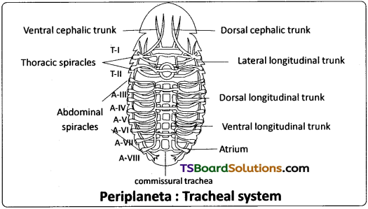
Stigmata or spiracles :
The tracheal system communicates with the exterior by ten pairs of openings called stigmata or spiracles. The first two pairs of spiracles are present in the thoracic segments, one pair in mesothorax and one pair in the metathorax. The remaining eight pairs are present in the first eight abdominal segments. Spiracles are located in the pleura of their respective segments. The respiratory system in insects is classified on the basis of number and nature of spiracles.
The spiracles of cockroach are polypneustic (as they are more than 3 pairs) and holopneustic (as all of them are functional). All spiracles are valvular and each of them is surrounded by a chitinous ring called peritreme. All spiracles bear small hair like structures called trichomes to filter the dust particles. Each spiracle opens into a small chamber called atrium.
Tracheae :
From the atrium of each thoracic spiracle several horizontal tracheae run inside. They join with each other in the thorax to form many tracheal trunks like dorsal cephalic, ventral cephalic trunks and their branches. These branches enter all organs of the head. The thoracic region also contains lateral longitudinal trunks. The abdominal spiracles lead into atria. From the atrium of each abdominal spiracle three tracheal tubes arise. All these tracheal tubes of one side open into three separate longitudinal tracheal trunks.
They are lateral, dorsal and ventral longitudinal trunks. Lateral longitudinal trunks are the longest tracheal trunks. The three pairs of longitudinal tracheal trunks of both the sides are interconnected by many commissural tracheae. From all the tracheal trunks several branches are given out, which enter different organs. All tracheal branches entering into an organ end in a special cell called tracheole cell.
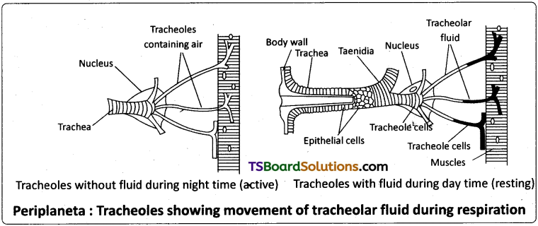
The wall of the tracheae is made of three layers. They are an outer basement membrane, a middle one cell thick epithelium and an inner layer of cuticle called intima. The intima is produced into spiral thickenings called taenidia. The taenidia keep the tracheae always open and prevent it from collapsing.
Tracheoles :
The terminal cell of trachea is called tracheoblast or tracheole cell. It has several intracellular tubular extensions called tracheoles. Tracheoles are devoid of intima and taenidia. They are formed of a protein called trachein. Tracheolar fluid is present inside the tracheoles. The level of the tracheolar fluid varies with the metabolic activity of the insect. It is more when the insect is inactive and completely reabsorbed into the tissues, when the insect is more active. Tracheoles penetrate the cell and are intimately associated with mitochondria (to supply oxygen to them).
Mechanism of respriation :
Respiration includes two events, viz., inspiration and expiration. The muscles helpful are dorsoventral muscles and ventral longitudinal musdes. Dorsoventral muscles are the principal muscles of respriation.
Inspiration :
Taking in of air is inspiration. lifts effected by the relaxation of the dorsoventral muscles and ventral longitudinal muscles. Due to the relaxation of the dorsoventral muscles, tergal plates are elevated and the volume of the body cavity increases. Due to the relaxation of the ventral longitudinal muscles, the telescoped segments come to the normal position. So the volume of the body cavity increases in the longitudinal axis. As air is drawn in due to the relaxation of the muscles, the process is a ‘passive’ process. During inspiration the thoracic spiracles are kept open and the abdominal spiracles are kept closed.
Expiration :
Sending out air from the body is called expiration. On contraction the dorsoventral muscles depress the tergal plates. Body cavity decreases in size and pressure increases. Due to the contraction of the ventral longitudinal muscles, the segments are telescoped and the volume of the body cavity decreases in the longitudinal axis increasing the pressure further. As this process involves the contraction of muscles, expiration is described as active process. During expiration thoracic spiracles are closed and abdominal spiracles are kept open.
Exchange of gases :
As air enters the tracheoles, oxygen from the air is taken into the cells and CO2 is released into haemolymph. The CO2 from the haemolymph mostly goes out through the inter-segmental membranes of the body wall. Cockroach and some other insects such as grasshoppers and beetles exhibit the phenomenon of discontinuous ventilation. In this mode of respiration continuous exchange of gases is interrupted by extended periods during which spiracles remain closed. The expulsion of CO2 from the body occurs in bursts, when the spiracles are open.
The exchange of gases depends on the metabolic rate and temperature. When air enters the tracheoles, oxygen diffuses faster into the tissues due to its high partial pressure. At the same time the carbon dioxide of tissues, instead of passing into the tracheal system, goes into the haemolymph. Carbon dioxide is carried more quickly into the haemolymph due to its greater solubility in it. This CO2 accumulates near the spiracles and diffuses into the artial chambers near the spiracles and goes o.ut in bursts through the abdominal spiracles. Opening and closing of spiracles is influenced by CO2 tension in haemolymph and oxygen tension in the trachea.
![]()
Question 7.
Describe the nervous system of Periplaneta and draw a labelled diagram of it. (K & S)
Answer:
The nervous sytem of cockroach consists of central nervous system, peripheral nervous system and autonomous nervous system.
Central Nervous System :
It consists of a nerve ring, and a ganglionated double ventral nerve cord.
Nerve ring :
The nerve ring, which is present around the oesophagus, is formed by the following.
Brain :
Brain lies above the oesophagus. The brain is mainly a sensory and an endocrine centre. Three lobes of the brain are protocerebrum,deutocerebrum and tritocerebrum. the protocerebrum receives sensory impulses from the compound eyes through optic nerves; deutocerebrum receives sensory impulses from antennae through antennal nerves; and tritocerebrum receives sensory impulses from the labrum. Hence brain is principally ‘sensory’ in nature.
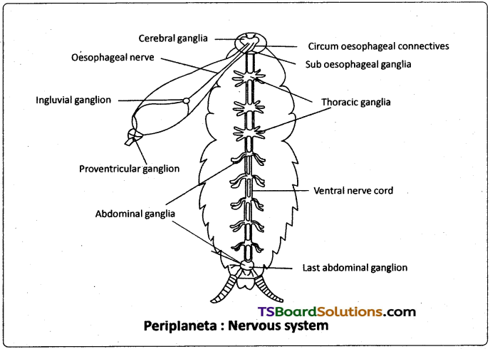
Sub-oesophageal ganglion :
It lies below the oesophagus. It is the motor center that controls the movements of mouthparts, legs and wings, it is formed by the fusion of paired ganglia of mandibular, maxillary and labial segments of the head. Circum-oesophageal connectives : A pair of circum-oesophageal connectives is present around the oesophagus, connecting the tritocerebrum with the sub – oesophageal ganglion/ sub oesophageal ganglia.
Ventral nerve cord :
The two ventral nerve cords are solid and ganglionated. They arise from the sub-oesophageal ganglion and extend upto the 7th abdominal segment. The two nerve cords remain separate except at the ganglia. Three thoracic ganglia are present, one in each thoracic segment. In addition, there are six abdominal ganglia. The first to the fourth abdominal segments have one abdominal ganglion each. The 5th abdominal segment has no ganglion . The serially 5th abdominal ganglion is present in the 6th segment. The serially 6th abdominal ganglion is present in the 7th segment. The last or the 6th abdominal ganglion is the largest of all the abdominal ganglia. It is formed by the fusion of the ganglia of the 7th, 8th, 9th, and 10th abdominal segments.
Peripheral Nervous System :
It consists of nerves arising from the central nervous system. It receives a pair of optic nerves, from the compound eyes, a pair of antennal nerves, from the antennae and a pair of labral nerves from the labrum. Motor neurons of the frontal nerve to the frontal ganglion join the sensory neurons of the labral nerve to form the larbro frontal nerve arising from the tritocerebrum. Sub – oesophageal ganglion gives off motor nerves to the mandibles, maxillae, labium, wings and legs. It is the principal ‘motor centre’ in the body. Thoracic ganglia supply nerves to the parts of their respective segments. Metathoracic ganglia send nerves to the first abdominal segment also.
Nerves from the first four abdominal ganglia supply to the organs of the segments 2 – 6 serially (the 1st to the 4th ganglia innervate segments 2nd to 5th respectively). The 5th ganglion present in the 6th segment innervates the organs of the 6th segment. All organs present in 7th to 10th segments receive nerves from the last abdominal ganglion (present in the 7th segment.) The organs include the reproductive organs, copulatory appendages besides anal cerci.
Autonomous Nervous System :
This system is also called stomatogastric nervous system or ‘visceral nervous system’. It controls the visceral organs, particularly the muscles of the alimentary canal, and the heart. Autonomous nervous system includes four ganglia, a frontal ganglion on the dorsal wall of the pharynx, in front of the brain, hypocerebral ganglion or occipital ganglion above the oesophagus, behind the brain, a visceral ganglion or ingluvial ganglion on the wall of the crop and a proventricular ganglion on the gizzard.
These ganglia contain the ‘somata’ of the post ganglionic motor neurons. Pregaglionic motor neurons of tritocerebrum go to the frontal ganglion as labrofrontal and frontal nerve. Frontal ganglion is connected to the hypocerebral ganglion by a ‘recurrent nerve’. Hypocerebral ganglion is connected to the visceral ganglion and in turn the visceral ganglion is connected to proventricular ganglion.
![]()
Question 8.
Describe the reproductive system of Periplaneta and draw neat and labelled diagrams of it. (K & S)
Answer:
Reproductive System of Periplaneta :
Periplaneta is dioecious, or unisexual, and both the sexes have well developed reproductive organs. The sexual dimorphism is evident both externally and internally. The female is different from the male in respect of short and broad abdomen, presence of brood pouches and absence of anal styles.
Male Reproductive System :
The male reproductive system consists of a pair of testes. These are elongated and lobed structures lying one on each lateral side in the fourth to sixth abdominal segments. They are embedded in the fat bodies. From the posterior end of each testis, there starts a thin duct, the vas deferens. The two vasa deferentia run backwards and inwards to open into a wide median duct, the ductus ejaculatorius ip the seventh segment. A characteristic mushroom shaped gland is present in the 6th and 7th abdominal segments which functions as an accessory reproductive gland. The gland consists of two types of tubules, i) long slender tubules. The utriculi majores or ‘peripheral tubules’ ii) Short tubules, the utriculi breviores. Secretion of utriculi majores forms the inner layer of the spermatophore while that of utriculi breviores nourishes the sperms. These tubules open into the anterior part of the ejaculatory duct.
The seminal vesicles are present on the ventral surface of the ejaculatory duct. These sacs store the sperms in the form of bundles called spermatophores. The ejaculatory duct is a muscular tube that extends posteriorly and opens at the gonopore or the ‘male genital ‘pore’. The duct of phallic or conglobate gland also opens near the gonopore. Its function is still not known. Surrounding the male genital opening there are chitinous and asymmetrical structures called phallic organs or gonapophyses or phallomeres which help in copulation. These are the male external genitalia.
Female Reproductive System :
The female reproductive system of Periplaneta consists of a pair of ovaries, a pair of oviducts, vagina, spermathecae, spermathecal papilla, and colleterial glands.
Ovaries :
A pair of large ovaries lies laterally in 2 to 6 abdominal segments. They are light yellow in colour surrounded by fat bodies. Each ovary consists of eight tubules called ovarian tubules or ovarioles. Each ovariole consists of a tapering anterior filament called germarium, and a posterior wider vitellarium. The germarium contains various stages of developing ova, and the vitellarium contains mature ova with yolk. The tapering ends of the ovarioles of each ovary unite to form a single thread which attaches to the dorsal body wall. The ovarioles, at their posterior end unite to form a short wide oviduct. The oviducts unite to form a very short median vagina.
The vertical opening of the vagina is called female genital pore. It opens into a large genital pouch on the eighth sternum. A spermatheca or receptaculum seminis, consisting of a left-sac like and a right filamentous caecum, is present in the 6th segment which opens by a median aperture on a small spermathecal papilla in the dorsal wall of the genital pouch on the ninth sternum. In a fertile female, the spermatheca contains spermatophores, obtained during copulation.
A pair of branched colleterial glands is present behind the ovaries. These glands open into the genital pouch separately, just above the spermathecal aperture. Secretion of the two collaterial glands forms a hard egg case called ootheca around the eggs.
Genital pouch is formed by 7th, 8th, and 9th abdominal sterna. The sternum of the seventh segment is boat shaped and forms the floor and side walls of the genital pouch. The sterna of the eighth and ninth segments, which are tucked into the seventh segment, constitute the anterior wall and the roof of the genital pouch, respectively. The genital pouch has two chambers the anterior ‘gynatrium’ or genital chamber and posterior ‘vestibuium’ or oothecal chamber.
Three pairs of plate like chitinous structures called gonapophyses are present around the female genital aperture. These gonapophyses guide the ova into ootheca as ovipositors. These are the female external genitalia.


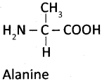
 is a zwitterionic form a neutral form due to equal positive and negative charges.